. 2023 Apr 20;30(4):383-393.e6.
doi: 10.1016/j.chembiol.2023.03.007. Epub 2023 Apr 3.

ITK degradation to block T cell receptor signaling and overcome therapeutic resistance in T cell lymphomas

Affiliations

ITK degradation to block T cell receptor signaling and overcome therapeutic resistance in T cell lymphomas

Baishan Jiang et al. Cell Chem Biol. .

Abstract

Interleukin (IL)-2-inducible T cell kinase (ITK) is essential for T cell receptor (TCR) signaling and plays an integral role in T cell proliferation and differentiation. Unlike the ITK homolog BTK, no inhibitors of ITK are currently US Food and Drug Administration (FDA) approved. In addition, recent studies have identified mutations within BTK that confer resistance to both covalent and non-covalent inhibitors. Here, as an alternative strategy, we report the development of BSJ-05-037, a potent and selective heterobifunctional degrader of ITK. BSJ-05-037 displayed enhanced anti-proliferative effects relative to its parent inhibitor BMS-509744, blocked the activation of NF-kB/GATA-3 signaling, and increased the sensitivity of T cell lymphoma cells to cytotoxic chemotherapy both in vitro and in vivo. In summary, targeted degradation of ITK is a novel approach to modulate TCR signal strength that could have broad application for the investigation and treatment of T cell-mediated diseases.

Keywords: GATA-3; ITK; PROTAC; T cell lymphoma; TCR signaling.


Conflict of interest statement

Declaration of interests N.S.G., D.M.W., B.S.J., and W.W. are inventors on a patent application related to the ITK degrader described in this manuscript. N.S.G. is a founder, scientific advisory board (SAB) member, and equity holder in Syros, C4, Allorion, Lighthorse, Voronoi, Inception, Matchpoint, CobroVentures, GSK, Shenandoah (board member), Larkspur (board member), and Soltego (board member). The Gray lab receives or has received research funding from Novartis, Takeda, Astellas, Taiho, Jansen, Kinogen, Arbella, Deerfield, Springworks, Interline, and Sanofi. D.M.W. is an employee of Merck and a founder, SAB member, and equity holder in Ajax and Travera. The Weinstock lab received research funding from AstraZeneca, Daiichi Sankyo, Secura, and Abcuro. E.S.F. is a founder, member of the SAB, and equity holder of Civetta Therapeutics, Jengu Therapeutics, Proximity Therapeutics, and Neomorph Inc.; SAB member and equity holder in Avilar Therapeutics and Photys Therapeutics; and a consultant to Astellas, Sanofi, Novartis, Deerfield, and EcoR1 capital. The Fischer laboratory receives or has received research funding from Novartis, Deerfield, Ajax, Interline, and Astellas. K.A.D. is a consultant to Kronos Bio and Neomorph Inc.

Figures

Figure 1.
Figure 1.. Design and Development of BSJ-05-037
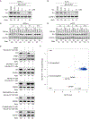
(A) Co-crystal structure of BMS-509744 (pink) bound to ITK (gray, PDB: 3MJ2) revealing solvent-exposed isopropylamine (circled) where linker was attached. (B) Chemical structure of BSJ-05-037 and BSJ-05-037-bump (negative control). (C) TREEspot visualization of the biochemical kinome selectivity profile of BSJ-05-037 (1 μM). ITK is highlighted in blue, while all other inhibited kinases are highlighted in red (Table S1).
Figure 2.
Figure 2.. BSJ-05-037 Induces Potent Degradation of ITK Dependent on CRBN, Neddylation, and the Proteasome
(A and B) Immunoblot for ITK and GAPDH in DERL-2 (A) and Hut78 (B) cells after 16-h treatment with DMSO or BSJ-05-037 at the concentrations indicated (data are representative of independent biological triplicate). (C and D) Immunoblot for ITK and GAPDH after treatment of DERL-2 (C) and Hut78 (D) cells with 250 nM of BMS-509744, BSJ-05-037-bump, or BSJ-05-037 at the time points indicated (data are representative of at least two independent biological replicates). (E) Immunoblot for ITK, CRBN, and GAPDH after 4-h treatment of Hut78 parental or CRBN-overexpressed cells with or without BSJ-05-037 (100 nM). Immunoblots for ITK and GAPDH after 16-h co-treatment of DERL-2 cells with DMSO, MLN-4924 (1 μM), MG132 (10 μM), BMS-509744 (2.5 μM), or pomalidomide (2.5 μM) and either BSJ-05-037 (250 nM or 1 μM) or DMSO (data are representative of at least two independent biological replicates). (F) Scatterplot depicts the change in relative protein abundance of BSJ-05-037 (100 nM, 5 h)-treated MOLT4 cells compared with DMSO vehicle control-treated cells. Protein abundance measurements were made using tandem mass tag quantitative mass spectrometry and significant changes were assessed by moderated t test as implemented in the limma package. The log2 fold change (log2 FC) is shown on the y axis and negative log10 p value (−log10 p value) on the x axis. See also Table S2.
Figure 3.
Figure 3.. BSJ-05-037 Induces Anti-proliferative Effects and Increased Sensitivity to Vincristine in TCLs
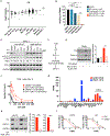
(A) Microarray analysis of ITK transcriptional levels among PTCL entities and in normal T cells. Mean of log2 intensity ≥ 8 is considered expression (indicated by the dashed line). (B) Cell viability assay in DERL-2 cells treated with BSJ-05-037-bump (1 μM), Thalidomide (10 μM), BMS-509744 (1 μM), and BSJ-05-037 or DMSO for 10 days. The number of viable cells was determined by both total cell counts and PI staining (measured by flow cytometry), and then normalized to DMSO control to determine relative cell viability. The experiment was performed in two independent biological replicates with technical triplicate. Data are presented as mean ± SEM. (C) Immunoblot for ITK, phospho-PLCγ1 (Y783), total PLCγ1, and GAPDH after 8-h treatment of DERL-2 cells with BSJ-05-037 or BMS-509744 at the concentrations indicated (data are representative of independent biological triplicate). (D) Schematic of T cell signaling, emphasizing ITK/NF-kB/GATA-3 signaling downstream of the TCR. (E and F) Cells were cultured in the presence or absence of Dynabeads Human T-Activator CD3/CD28. (E) Representative immunoblot (left) and quantitative analysis (right) of ITK and GATA-3 expression in T8ML1 cells upon exposure to DMSO or BSJ-05-037 (1 μM) for 24 hours (data are representative of independent biological triplicate). (F) Cell viability of T8ML1 cells after 3-d treatment with BSJ-05-037 (1 μM) and vincristine at the concentrations indicated. The experiment was performed in two independent biological replicates with technical triplicate. Data are presented as mean ± SD. (G) RNA expression levels of GATA3 and TBX21 by reads per kilobase of transcript, per million mapped reads (RPKM) in TCL cell lines. (H) Representative immunoblot (left) and quantitative analysis (right) for ITK, GATA-3, and GAPDH in Hut78 and H9 cells after 24-h treatment of DMSO or BSJ-05-037 (1 μM) (data are representative of independent biological triplicate). (I) Dose-response curves for Hut78 and H9 cells treated with vincristine at indicated dose range for 4 days in the presence or absence of BSJ-05-037 (1 μM). The experiment was performed in two independent biological replicates with technical triplicate. Data are presented as mean ± SD. **p<0.01; ***p<0.001.
Figure 4.
Figure 4.. BSJ-05-037 Induces Anti-proliferative Effects and Increased Sensitivity to Vincristine in TCLs
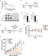
(A) Pharmacokinetic analysis of BSJ-05-037 after a single intraperitoneal dose at 10 mg/kg in C57BL/6 mice. Half-life = 1.9 (+/− 0.14), Cmax=3 μM. n=3 mice. (B) Workflow of CTCL Hut78 in vivo model. (C and D) Representative immunoblot (C) and quantitative analysis (D) of ITK and GATA-3 expression in xenografted tumor cells upon exposure to vehicle or BSJ-05-037 for 24 hours. (E) Workflow of CTCL H9 in vivo model. (F and G) Tracking of mouse body weight (F) and tumor volume (G) in H9 in vivo model (E) under treatment with vehicle control, BSJ-05-037 (50 mg/kg I.P., Q.O.D), vincristine (0.4 mg/kg I.V., weekly), or combination for 14 days. The dotted line indicates 5% body weight loss. ns, not statistically significant; *p<0.05; ***p<0.001.

Comment in

References

    1. Qi Q, Sahu N, and August A (2006). Tec kinase Itk forms membrane clusters specifically in the vicinity of recruiting receptors. J Biol Chem 281, 38529–38534. 10.1074/jbc.M609180200. - DOI - PubMed
    1. Bunnell SC, Diehn M, Yaffe MB, Findell PR, Cantley LC, and Berg LJ (2000). Biochemical interactions integrating Itk with the T cell receptor-initiated signaling cascade. J Biol Chem 275, 2219–2230. 10.1074/jbc.275.3.2219. - DOI - PubMed
    1. Weeks S, Harris R, and Karimi M (2021). Targeting ITK signaling for T cell-mediated diseases. iScience 24, 102842. 10.1016/j.isci.2021.102842. - DOI - PMC - PubMed
    1. Heyeck SD, Wilcox HM, Bunnell SC, and Berg LJ (1997). Lck phosphorylates the activation loop tyrosine of the Itk kinase domain and activates Itk kinase activity. J Biol Chem 272, 25401–25408. 10.1074/jbc.272.40.25401. - DOI - PubMed
    1. Wilcox HM, and Berg LJ (2003). Itk phosphorylation sites are required for functional activity in primary T cells. J Biol Chem 278, 37112–37121. 10.1074/jbc.M304811200. - DOI - PubMed

Publication types

Substances