. 2023 Apr;52(4):20230058.
doi: 10.1259/dmfr.20230058. Epub 2023 Apr 4.

Cinematic rendering to improve visualization of supplementary and ectopic teeth using CT datasets

Affiliations

Cinematic rendering to improve visualization of supplementary and ectopic teeth using CT datasets

Ines Willershausen et al. Dentomaxillofac Radiol. 2023 Apr.

Abstract

Objectives: Ectopic, impacted, and supplementary teeth are the number one reason for cross-sectional imaging in pediatric dentistry. The accurate post-processing of acquired data sets is crucial to obtain precise, yet also intuitively understandable three-dimensional (3D) models, which facilitate clinical decision-making and improve treatment outcomes. Cinematic rendering (CR) is anovel visualization technique using physically based volume rendering to create photorealistic images from DICOM data. The aim of the present study was to tailor pre-existing CR reconstruction parameters for use in dental imaging with the example of the diagnostic 3D visualization of ectopic, impacted, and supplementary teeth.

Methods: CR was employed for the volumetric image visualization of midface CT data sets. Predefined reconstruction parameters were specifically modified to visualize the presented dental pathologies, dentulous jaw, and isolated teeth. The 3D spatial relationship of the teeth, as well as their structural relationship with the antagonizing dentition, could immediately be investigated and highlighted by separate, interactive 3D visualization after segmentation through windowing.

Results: To the best of our knowledge, CR has not been implemented for the visualization of supplementary and ectopic teeth segmented from the surrounding bone because the software has not yet provided appropriate customized reconstruction parameters for dental imaging. When employing our new, modified reconstruction parameters, its application presents a fast approach to obtain realistic visualizations of both dental and osseous structures.

Conclusions: CR enables dentists and oral surgeons to gain an improved 3D understanding of anatomical structures, allowing for more intuitive treatment planning and patient communication.

Keywords: 3D visualization; CBCT; CT; dental anatomy; dental radiology; volume rendering.


Conflict of interest statement

Conflicts of Interest: The authors disclose no conflict of interest related to the present article. With regard to activities not related to this article, Fabian Necker is research student at Siemens Healthineers GmbH, Erlangen, Germany.

Figures

Figure 1.
Figure 1.
Displays the post-processing pipeline of volumetric data for 3D visualization by means of cinematic rendering. 3D, three-dimensional; DICOM, Digital Imaging and Communications in Medicine; GPU, graphics processing unit; PACS, picture archiving and communications system.
Figure 2.
Figure 2.
An 11.7-year-old girl with a horizontally impacted canine (white arrows, Figure 1a–d) and a persisting deciduous 63 canine (white arrowheads, Figure 1a–d). (a) A panoramic radiograph is displayed. (b) Semi-transparent reconstruction parameters are utilized to visualize both bone and teeth, as well as the different dental tissues. (c, d) The individual dentition and bone tissue with a soft kernel show a photorealistic visualization in frontal and lateral view.
Figure 3.
Figure 3.
An 11.2-year-old boy with a mesiodens and a further supplementary tooth (white arrows; 3D, f) within the hard palate. (a) A panoramic radiograph is displayed. (b) Semi-transparent reconstruction parameters are displayed. (c, d) The bone reconstruction parameters with a soft kernel can lead to artifacts in regions with teeth near each other (yellow circle, in d). (e, f) The same situation is shown with a hard kernel, allowing a better differentiation between the roots of the mesiodens and of tooth 21 (yellow circle in f).
Figure 4.
Figure 4.
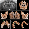
A 12.4-year-old boy with cleidocranial dysostosis and multiple supernumerary and impacted teeth. (a) The panoramic image is displayed. (b-e) The bone present with a soft kernel in the frontal, lateral, and palatal view is illustrated. (c-e) Tooth 11 (blue arrowhead), tooth 21 (green arrowhead), and tooth 13 (yellow arrowhead) are displayed, as well as their respective supernumerary teeth 11 (blue arrow), 21 (green arrow), and 13 (yellow arrow).

References

    1. Hodges RJ, Atchison KA, White SC. Impact of cone-beam computed tomography on orthodontic diagnosis and treatment planning. Am J Orthod Dentofacial Orthop 2013; 143: 665–74. doi: 10.1016/j.ajodo.2012.12.011 - DOI - PubMed
    1. Yeung AWK, Jacobs R, Bornstein MM. Novel low-dose protocols using cone beam computed tomography in dental medicine: A review focusing on indications, limitations, and future possibilities. Clin Oral Investig 2019; 23: 2573–81. doi: 10.1007/s00784-019-02907-y - DOI - PubMed
    1. Murray D, Whyte A. Dental panoramic tomography: What the general radiologist needs to know. Clin Radiol 2002; 57: 1–7. doi: 10.1053/crad.2001.0826 - DOI - PubMed
    1. Sams CM, Dietsche EW, Swenson DW, DuPont GJ, Ayyala RS. Pediatric panoramic radiography: Techniques, artifacts, and interpretation. Radiographics 2021; 41: 595–608. doi: 10.1148/rg.2021200112 - DOI - PubMed
    1. Caiado GM, Evangelista K, Freire M do CM, Almeida FT, Pacheco-Pereira C, Flores-Mir C, et al. . Orthodontists’ criteria for prescribing cone-beam computed tomography-a multi-country survey. Clin Oral Investig 2022; 26: 1625–36. doi: 10.1007/s00784-021-04135-9 - DOI - PMC - PubMed