. 2023 May;42(5):1374-1387.
doi: 10.1109/TMI.2022.3228075. Epub 2023 May 2.

Free-Breathing Liver Fat, R₂* and B₀ Field Mapping Using Multi-Echo Radial FLASH and Regularized Model-Based Reconstruction

Free-Breathing Liver Fat, R₂* and B₀ Field Mapping Using Multi-Echo Radial FLASH and Regularized Model-Based Reconstruction

Zhengguo Tan et al. IEEE Trans Med Imaging. 2023 May.

Abstract

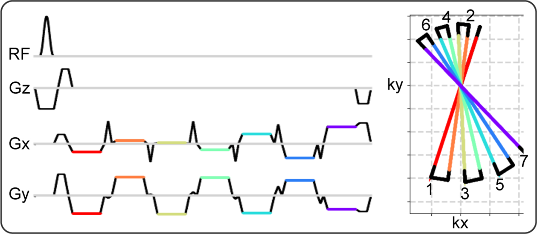
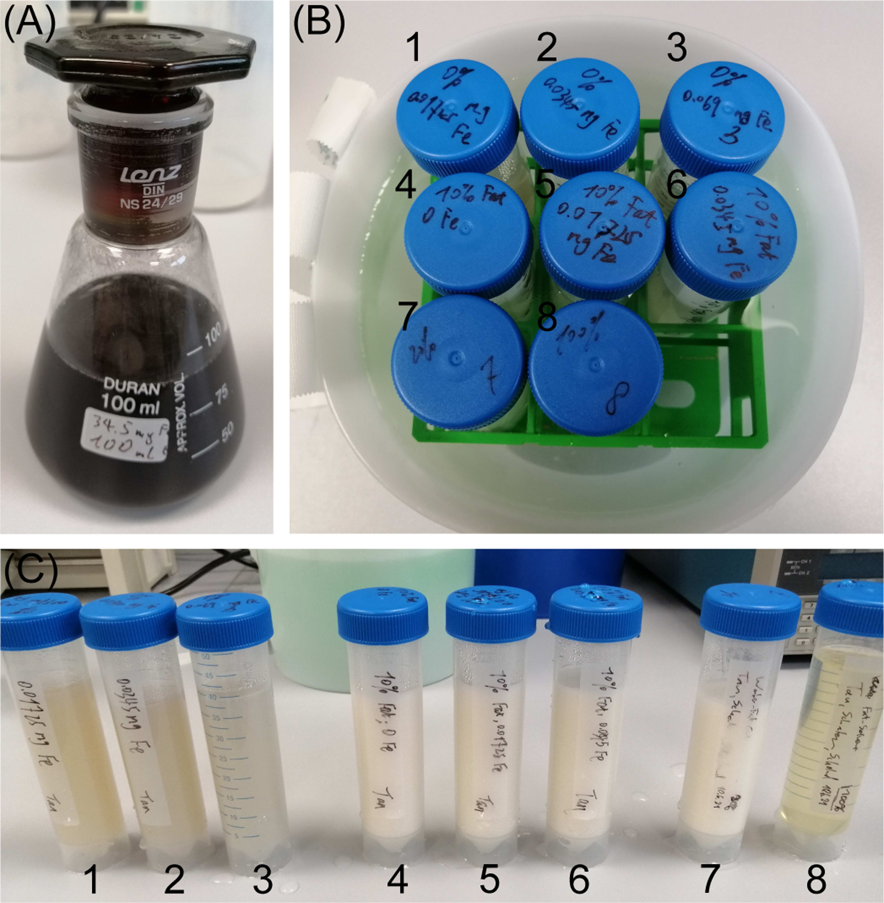
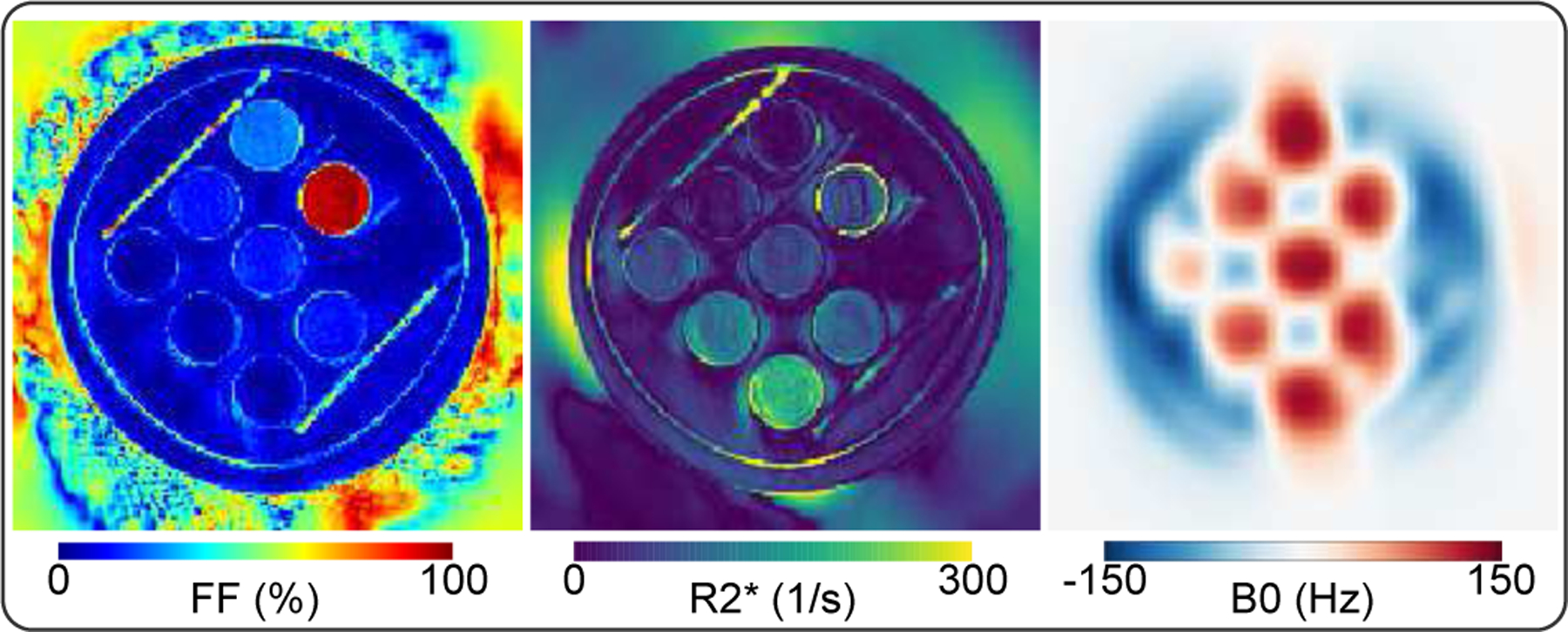
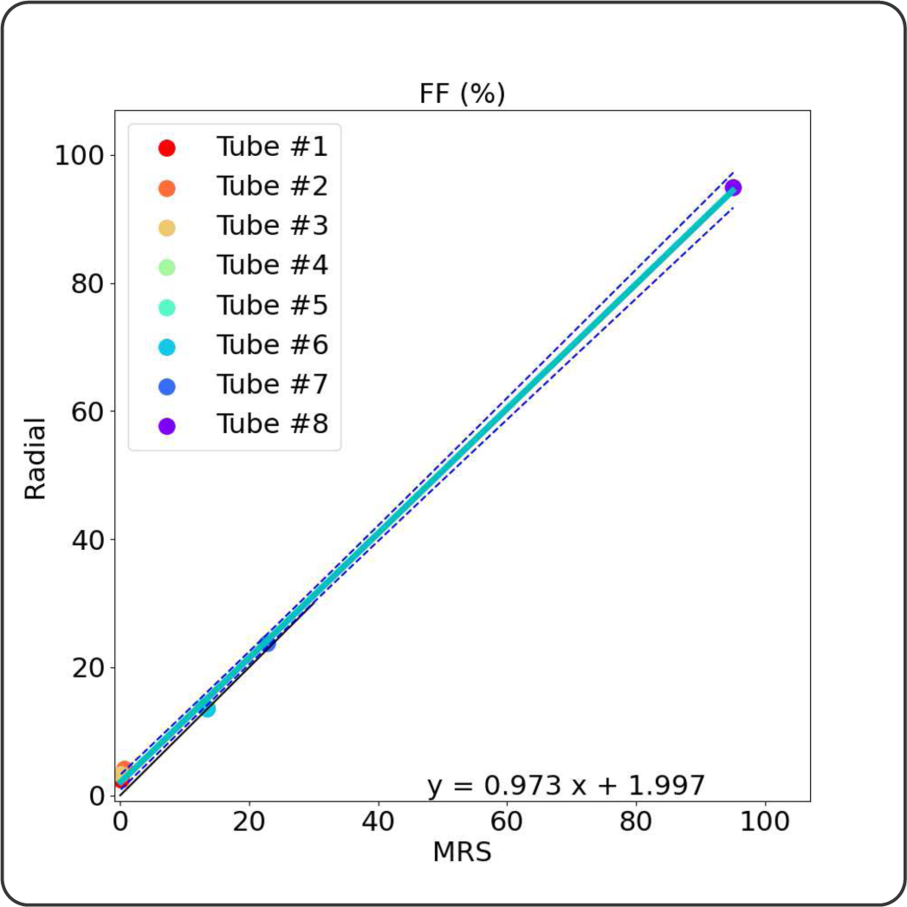
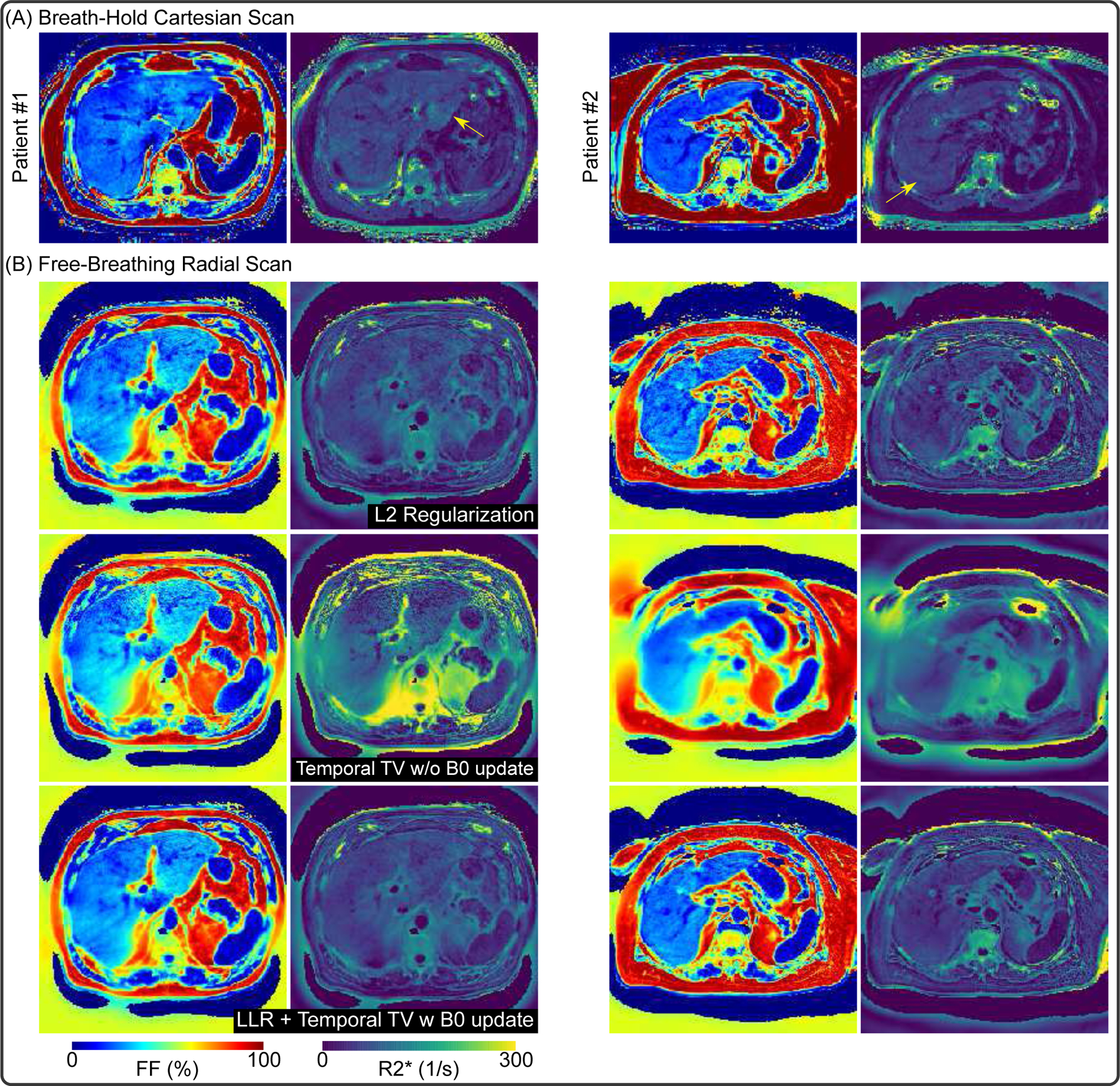
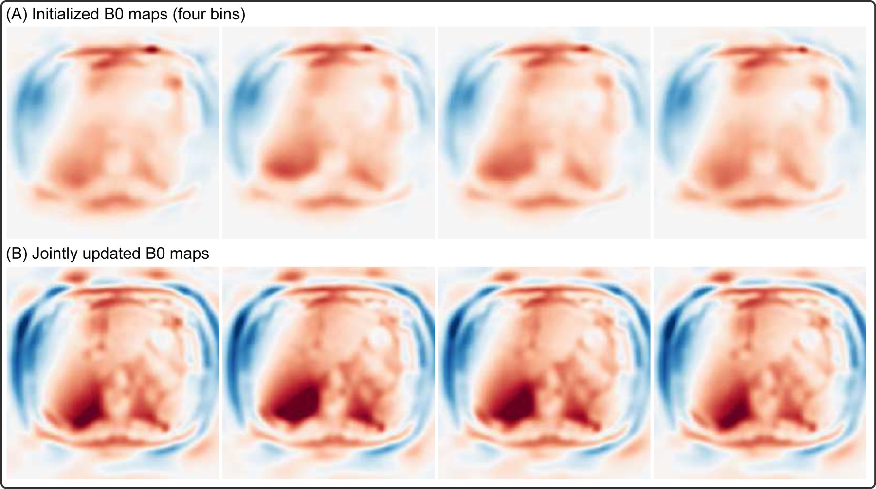
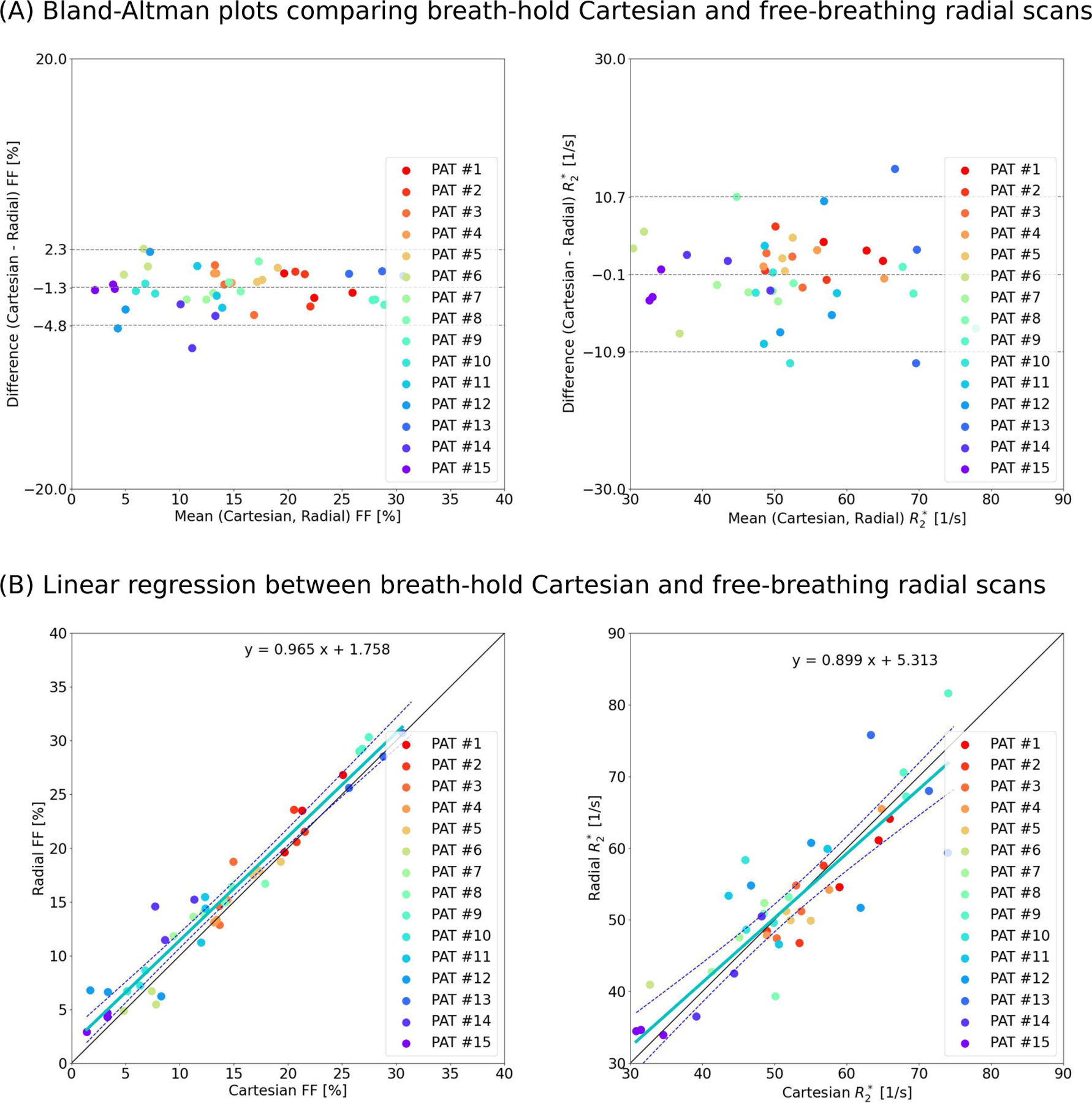
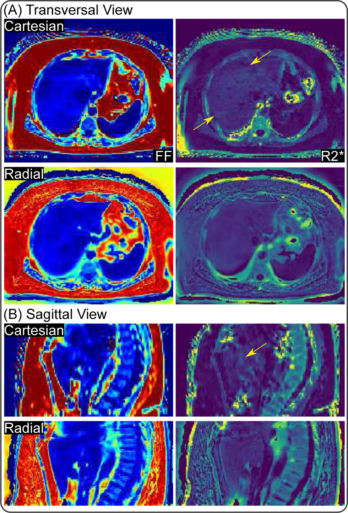
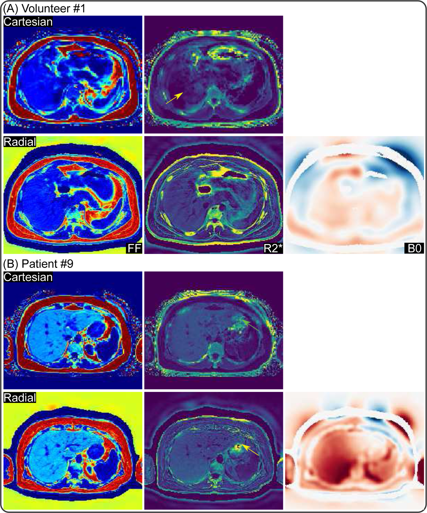
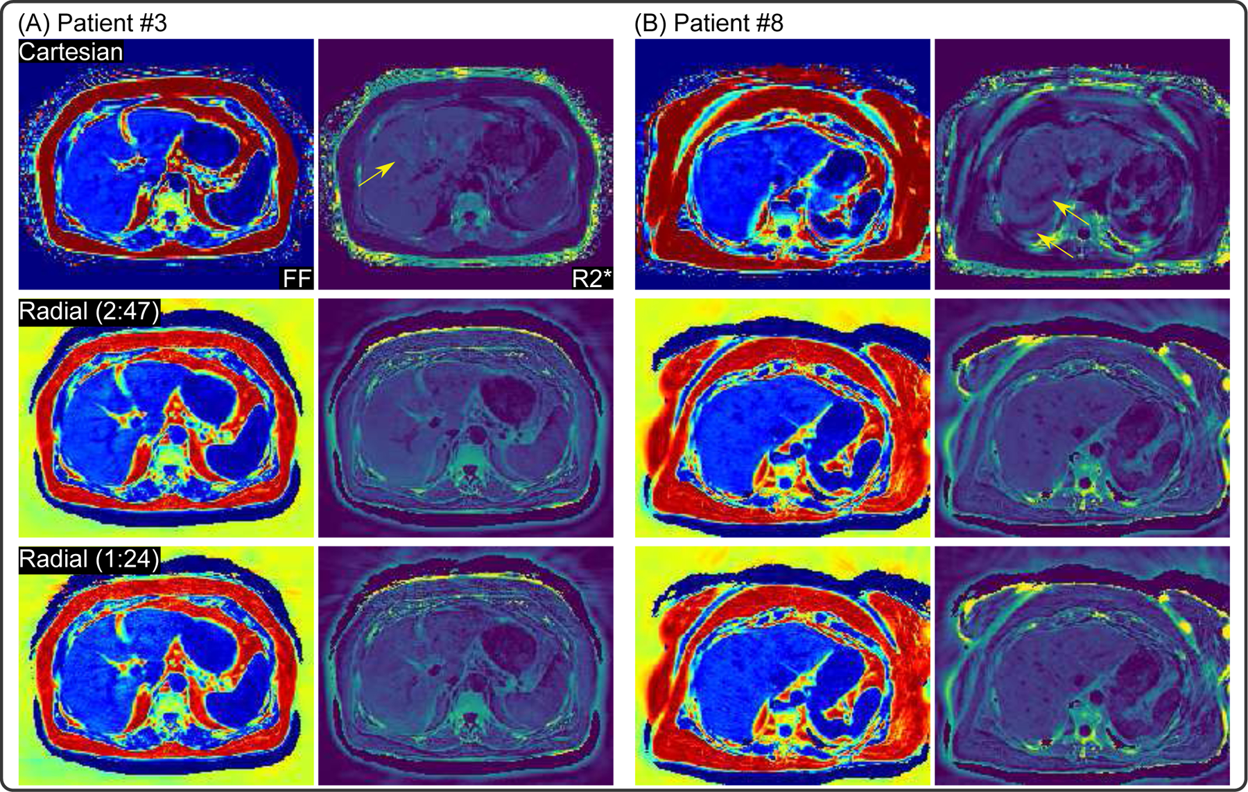
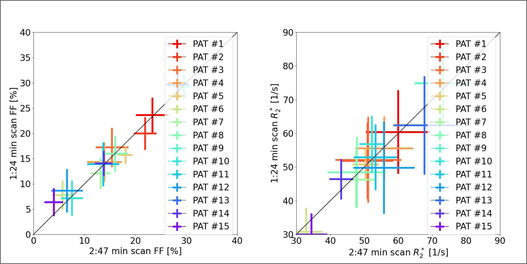
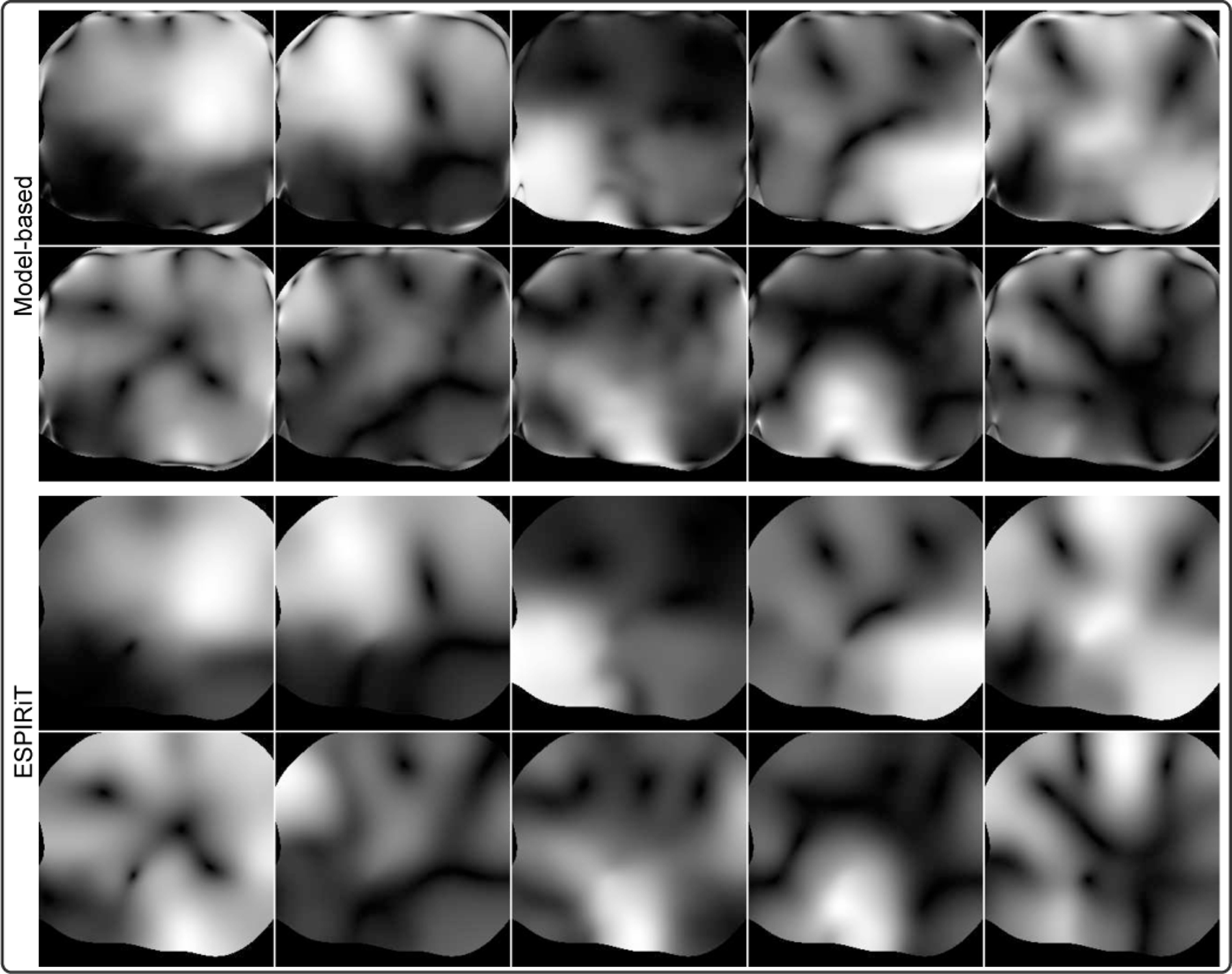
This work introduced a stack-of-radial multi-echo asymmetric-echo MRI sequence for free-breathing liver volumetric acquisition. Regularized model-based reconstruction was implemented in Berkeley Advanced Reconstruction Toolbox (BART) to jointly estimate all physical parameter maps (water, fat, R2 , and B0 field inhomogeneity maps) and coil sensitivity maps from self-gated k -space data. Specifically, locally low rank and temporal total variation regularization were employed directly on physical parameter maps. The proposed free-breathing radial technique was tested on a water/fat & iron phantom, a young volunteer, and obesity/diabetes/hepatic steatosis patients. Quantitative fat fraction and R2 accuracy were confirmed by comparing our technique with the reference breath-hold Cartesian scan. The multi-echo radial sampling sequence achieves fast k -space coverage and is robust to motion. Moreover, the proposed motion-resolved model-based reconstruction allows for free-breathing liver fat and R2 quantification in multiple motion states. Overall, our proposed technique offers a convenient tool for non-invasive liver assessment with no breath holding requirement.


Figures

Fig. 1.
Fig. 1.
(Left) One representative repetition time TR block of the proposed multi-echo asymmetric-echo radial sequence. (Right) The corresponding k-space trajectory. The echoes are color coded, indicating the period when ADC is switched on, while the dark solid lines indicate either the ramp or the blip gradients.
Fig. 2.
Fig. 2.
Photos of the constructed water/fat & iron phantom. (A) 34.5 mg iron nano particle diluted in 100 mL distilled water. (B) Phantom layout with eight tubes as listed in (C).
Fig. 3.
Fig. 3.
Multi-echo radial FLASH acquisition and model-based reconstruction results of the water/fat & iron phantom built in-house. Displayed images are FF, R2, and B0 maps, respectively.
Fig. 4.
Fig. 4.
Quantitative analysis of FF values for the eights tubes via linear regression between the standard MR Spectroscopy (MRS) and the proposed multi-echo radial acquisition with model-based reconstruction.
Fig. 5.
Fig. 5.
Comparison of (A) the reference breath-hold Cartesian scan and (B) the proposed free-breathing radial scan on Patients #1 and #2. Furthermore, for the radial data in (B), we compared three different regularizations: L2 regularization, temporal TV regularization without B0 update, and the proposed spatial LLR and temporal TV regularization with B0 update.
Fig. 6.
Fig. 6.
(A) Initial B0 maps of four respiratory bins for the model-based reconstruction without B0 update in Fig. 5. (B) Jointly updated B0 maps of four respiratory bins. Joint update using seven echoes helps to recover more details in the B0 maps.
Fig. 7.
Fig. 7.
(A) One coronal-view FF and R2 map from breath-hold Cartesian scan of Patient #1. (B) Plots of self-gating signal and phase portraits. (C) Four coronal-view FF and R2 maps from free-breathing radial scan with self-gated motion-resolved model-based reconstruction. The R2 map in (A) suffers from hyper intensities in the liver dome region (yellow ellipse), whereas the proposed radial scan shows consistent R2 values.
Fig. 8.
Fig. 8.
Quantitative analysis of reconstructed FF and R2 maps for all subjects comparing the reference breath-hold Cartesian scan and the proposed free-breathing radial scan, respectively.
Fig. 9.
Fig. 9.
(A) Transversal view and (B) sagittal view of reconstructed FF and R2 maps in Patient #6 comparing the reference breath-hold Cartesian scan and the proposed radial scan, respectively. This patient shows definite obesity symptom (see Table III), but has no fatty liver. In fact, the FF values of this patient is the lowest among all subjects (see also Fig. 8). Similar to the results of Patient #1 in Fig. 5, the Cartesian scan suffers from fold-in artifacts in the R2 maps (yellow arrows), while the proposed radial scan shows consistent R2 values.
Fig. 10.
Fig. 10.
(A) Volunteer #1 and (B) Patient #9 with elevated FF values, but no clear symptom of obesity. In particular, Patient #9 was diagnosed with hepatic steatosis by the standard liver biopsy and ultrasound.
Fig. 11.
Fig. 11.
Comparison of FF and R2 maps for Patients (A) #3 and (B) #8 among the reference Cartesian scan, the free-breathing radial 2:47 min scan, and the free-breathing radial scan with retrospective two-fold undersampling, corresponding to a scan time of 1:24 min. Note the ripple-like artifact in the R2 map from the Cartesian scans due to incomplete breath hold (yellow arrows in the 1st row).
Fig. 12.
Fig. 12.
Quantitative analysis of the reconstructed FF and R2 maps from radial scans: actual acquisition (2:47 min) and retrospectively undersampled acquisition (1:24 min), respectively.
Fig. 13.
Fig. 13.
Magnitude of coil sensitivity maps for Patient #1 from (top) the proposed model-based method and (bottom) the ESPIRiT method.

References

    1. Caussy C, Reeder SB, Sirlin CB, and Loomba R, “Noninvasive, quantitative assessment of liver fat by MRI-PDFF as an endpoint in NASH trials,” Hepatology, vol. 68, pp. 763–772, 2018. - PMC - PubMed
    1. Hu HH, Branca RT, Hernando D, Karampinos DC, Machann J, McKenzie CA, Wu HH, Yokoo T, and Velan SS, “Magnetic resonance imaging of obesity and metabolic disorders: Summary from the 2019 ISMRM Workshop,” Magn. Reson. Med, vol. 83, pp. 1565–1576, 2020. - PubMed
    1. Wood JC, “Impact of iron assessment by MRI,” Hematology, vol. 2011, pp. 443–450, 2011. - PubMed
    1. Hernando D, Levin YS, Sirlin CB, and Reeder SB, “Quantification of liver iron with MRI: State of the art and remaining challenge,” J. Magn. Reson. Imaging, vol. 40, pp. 1003–1021, 2014. - PMC - PubMed
    1. Dixon WT, “Simple proton spectroscopic imaging,” Radiology, vol. 153, pp. 189–194, 1984. - PubMed

Publication types