. 2023 May 10;43(19):3439-3455.
doi: 10.1523/JNEUROSCI.1355-22.2023. Epub 2023 Apr 4.

Interleukin (IL)-1 Receptor Signaling Is Required for Complete Taste Bud Regeneration and the Recovery of Neural Taste Responses following Axotomy

Affiliations

Interleukin (IL)-1 Receptor Signaling Is Required for Complete Taste Bud Regeneration and the Recovery of Neural Taste Responses following Axotomy

Guangkuo Dong et al. J Neurosci. .

Abstract

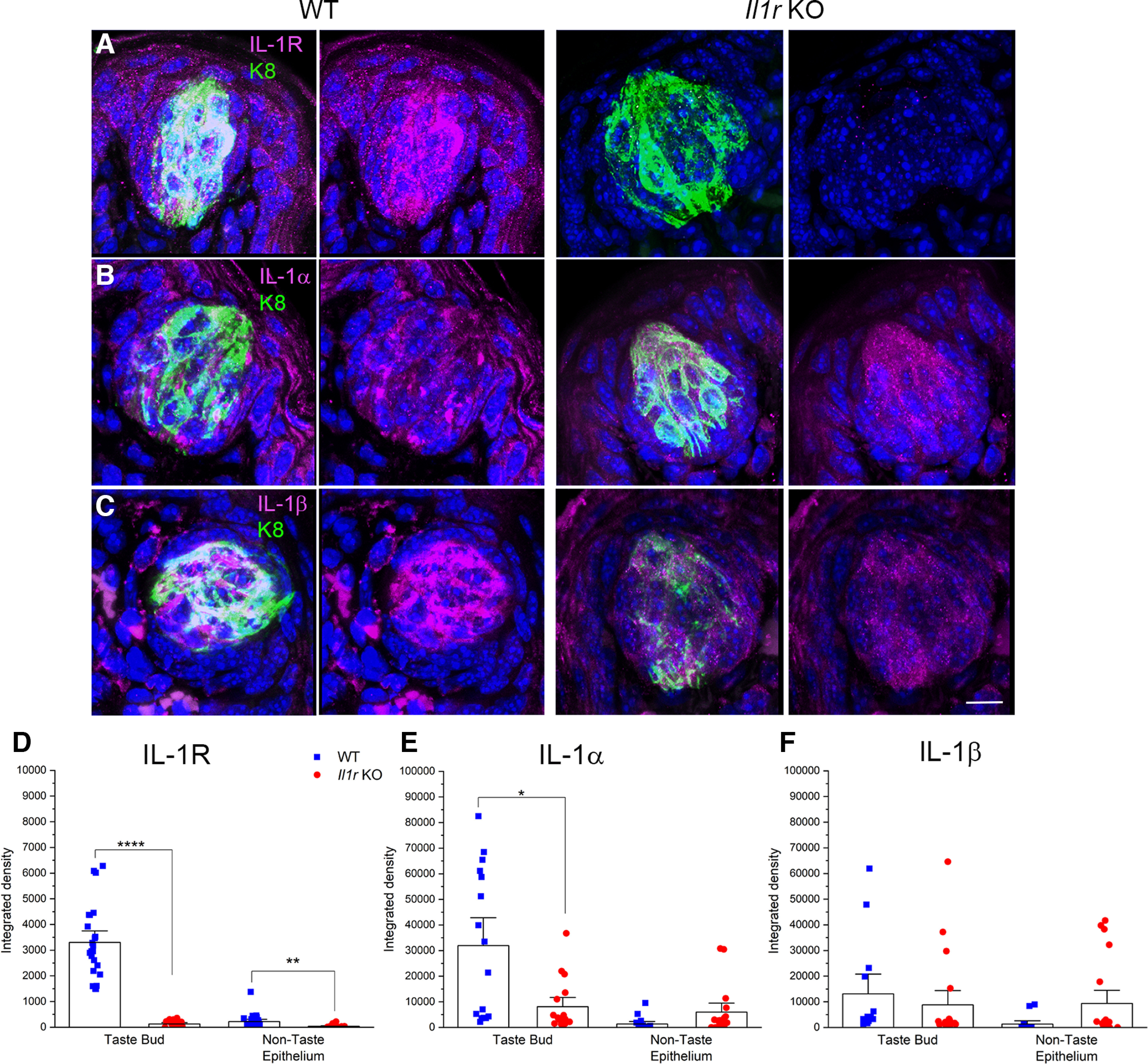
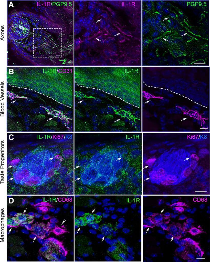
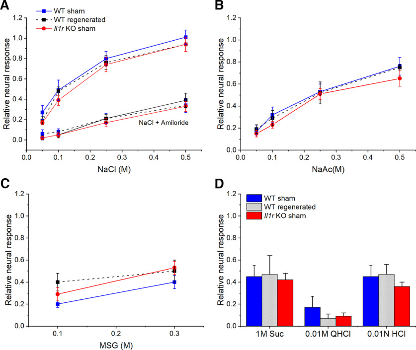
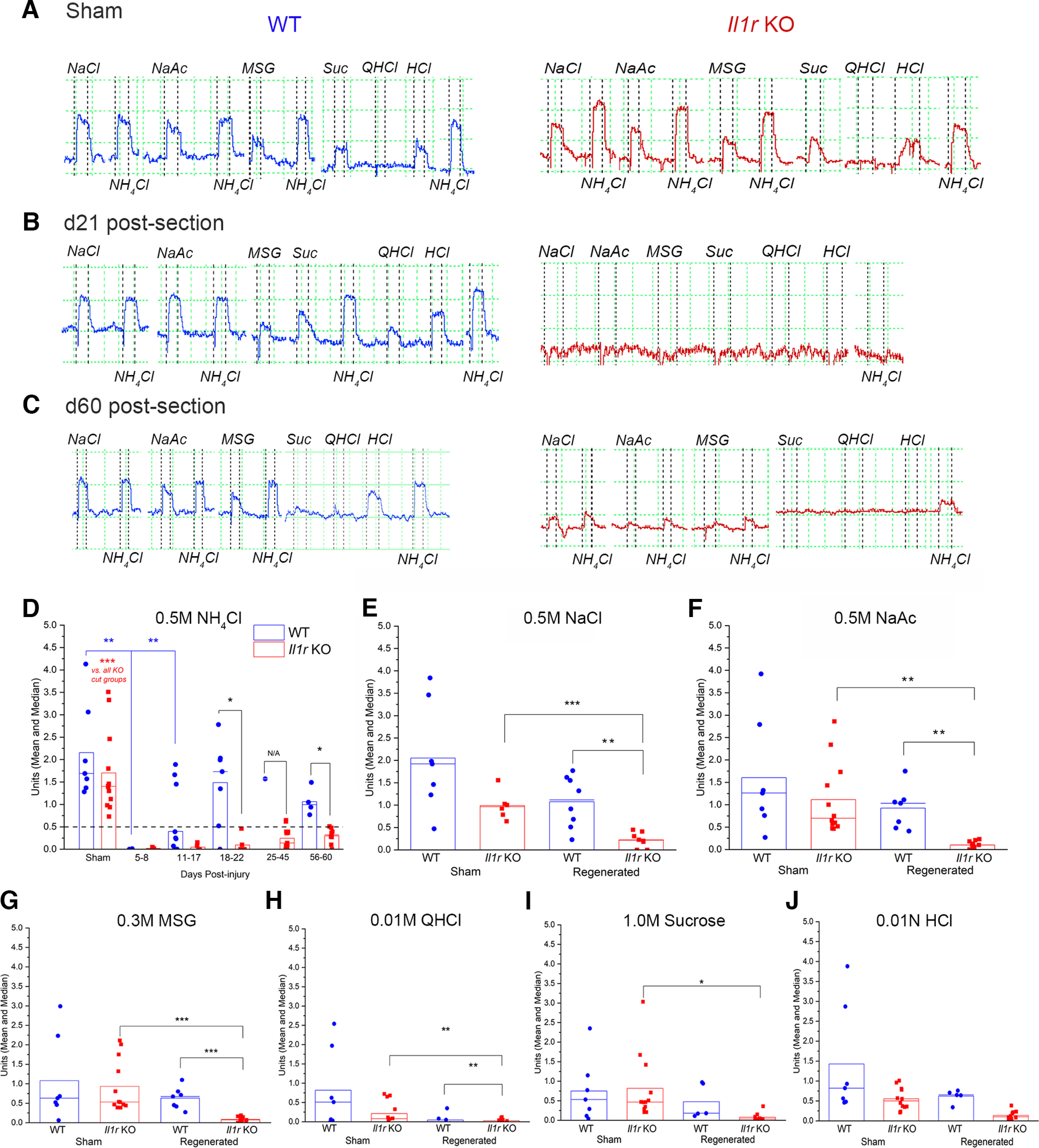
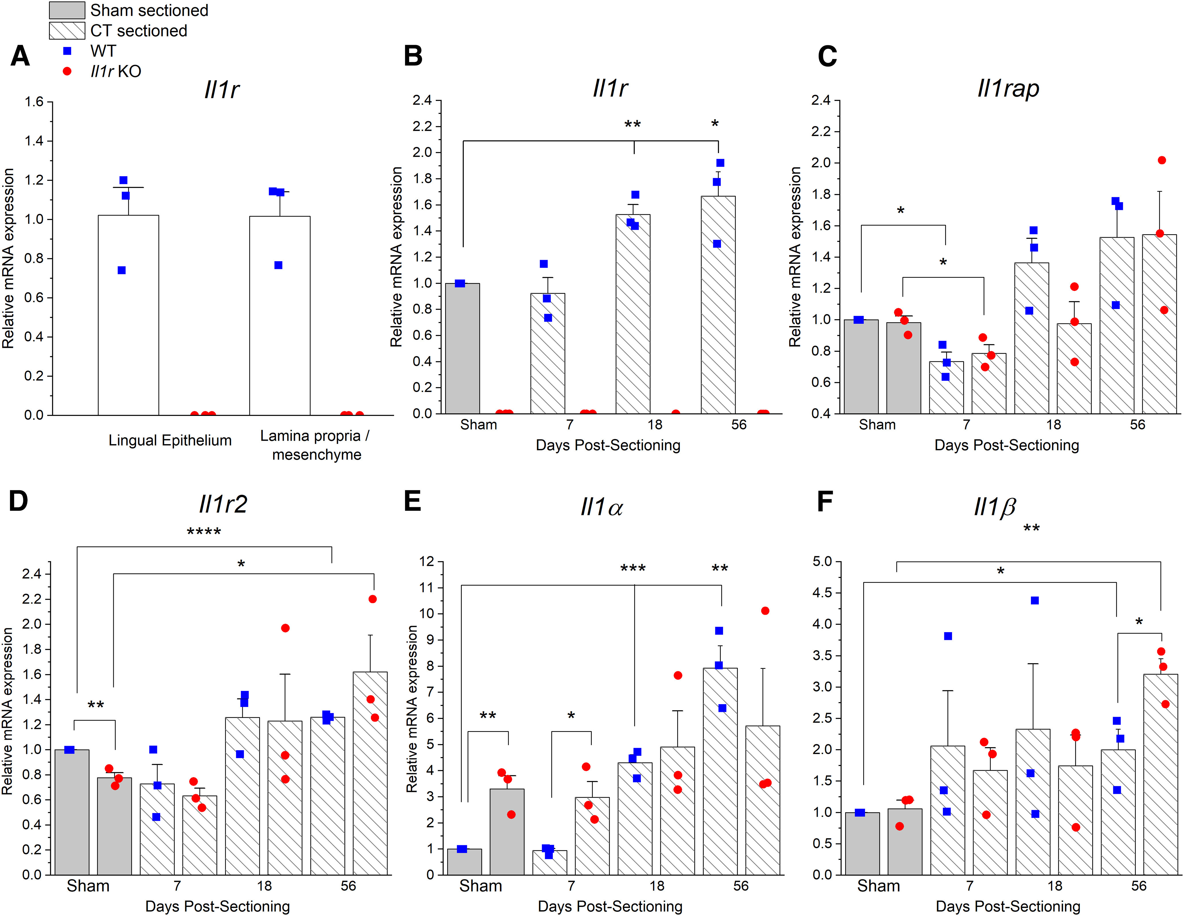
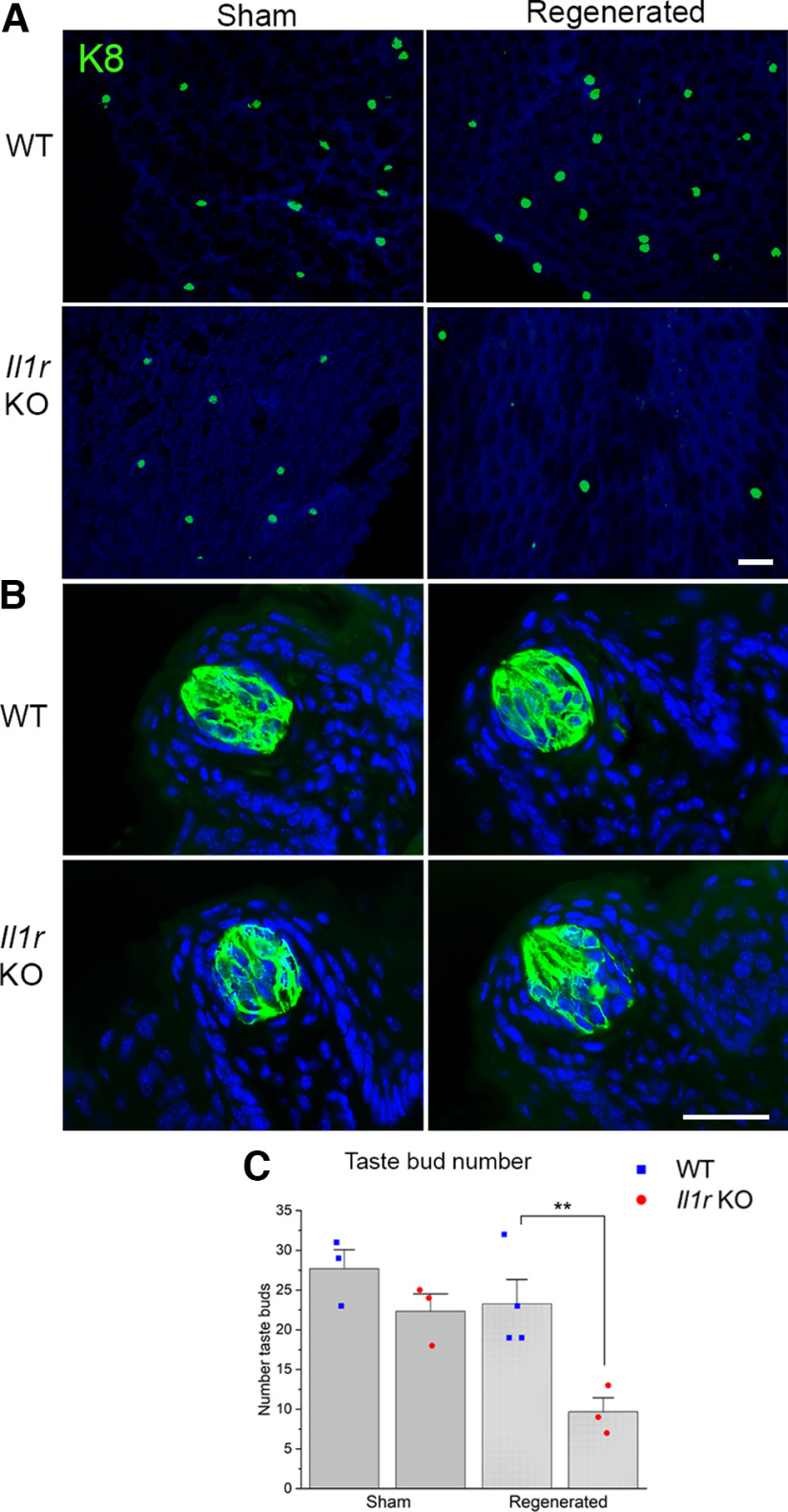
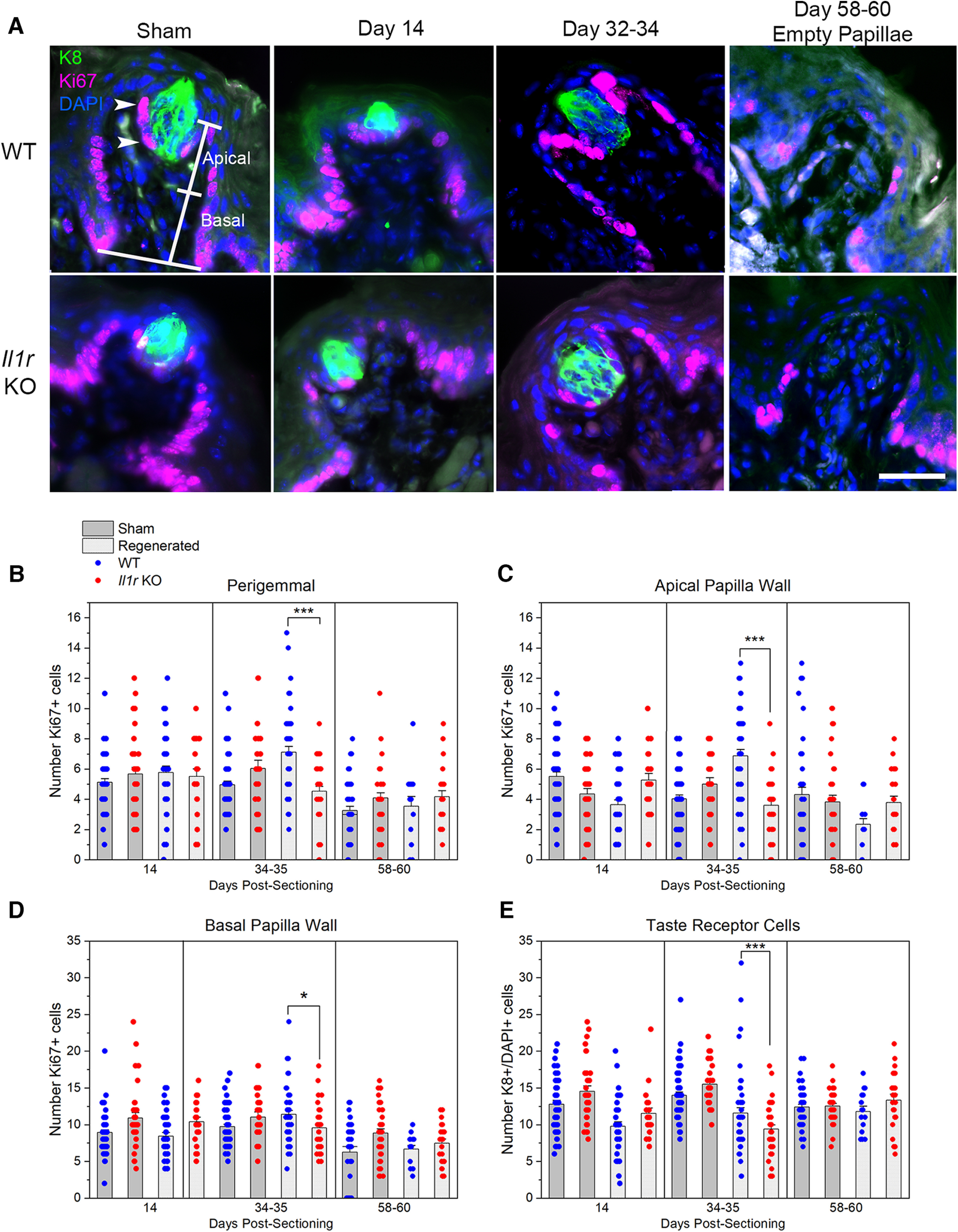
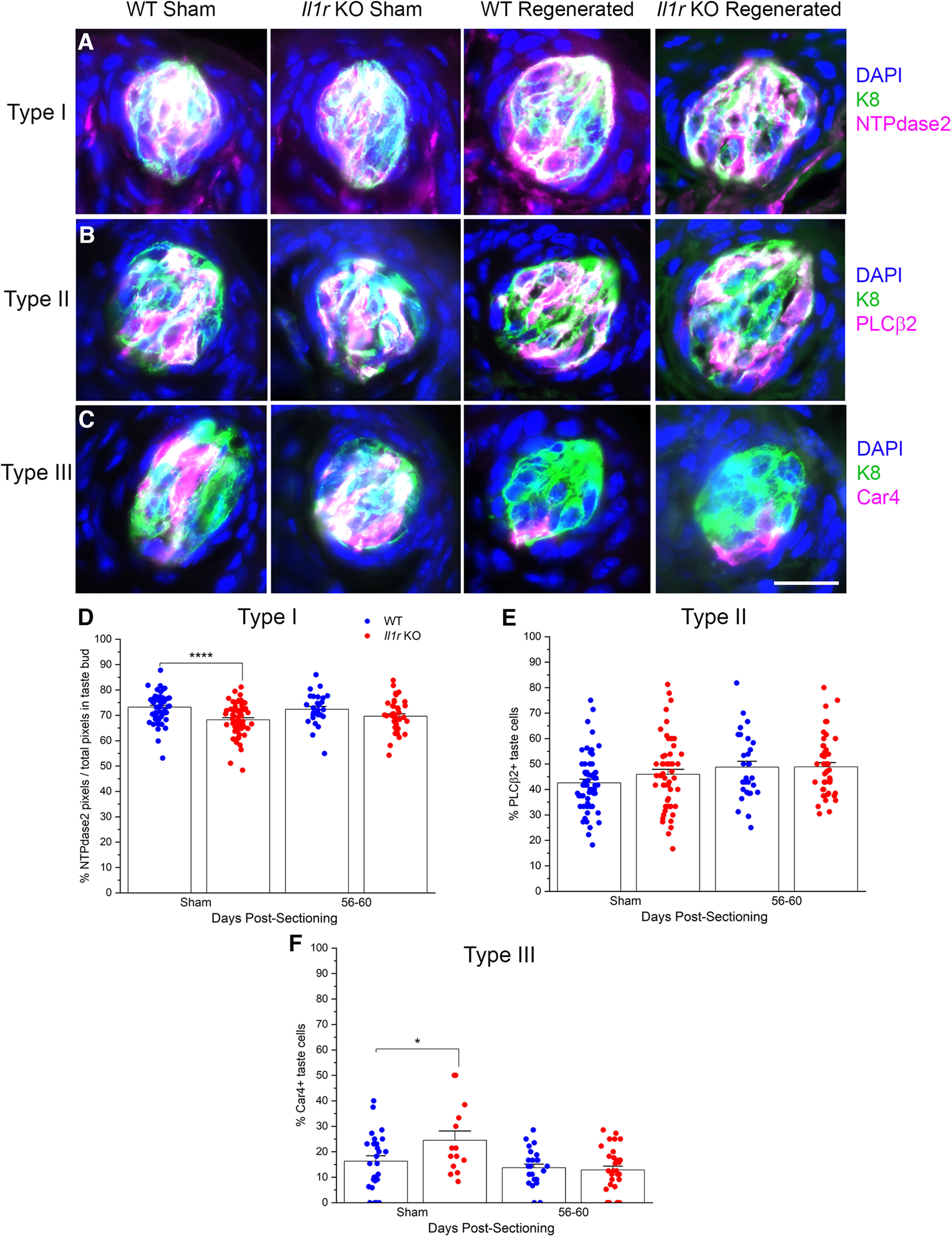
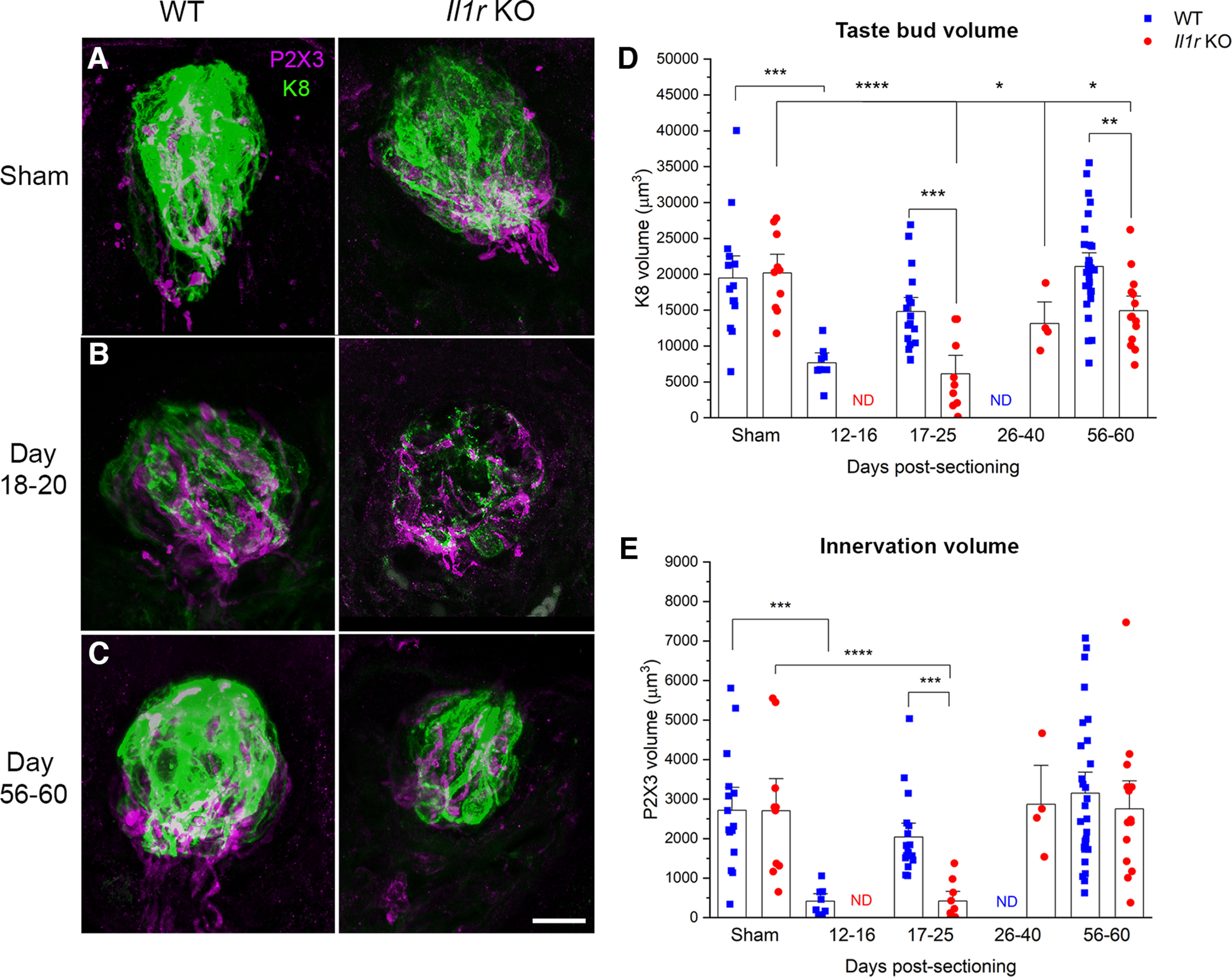
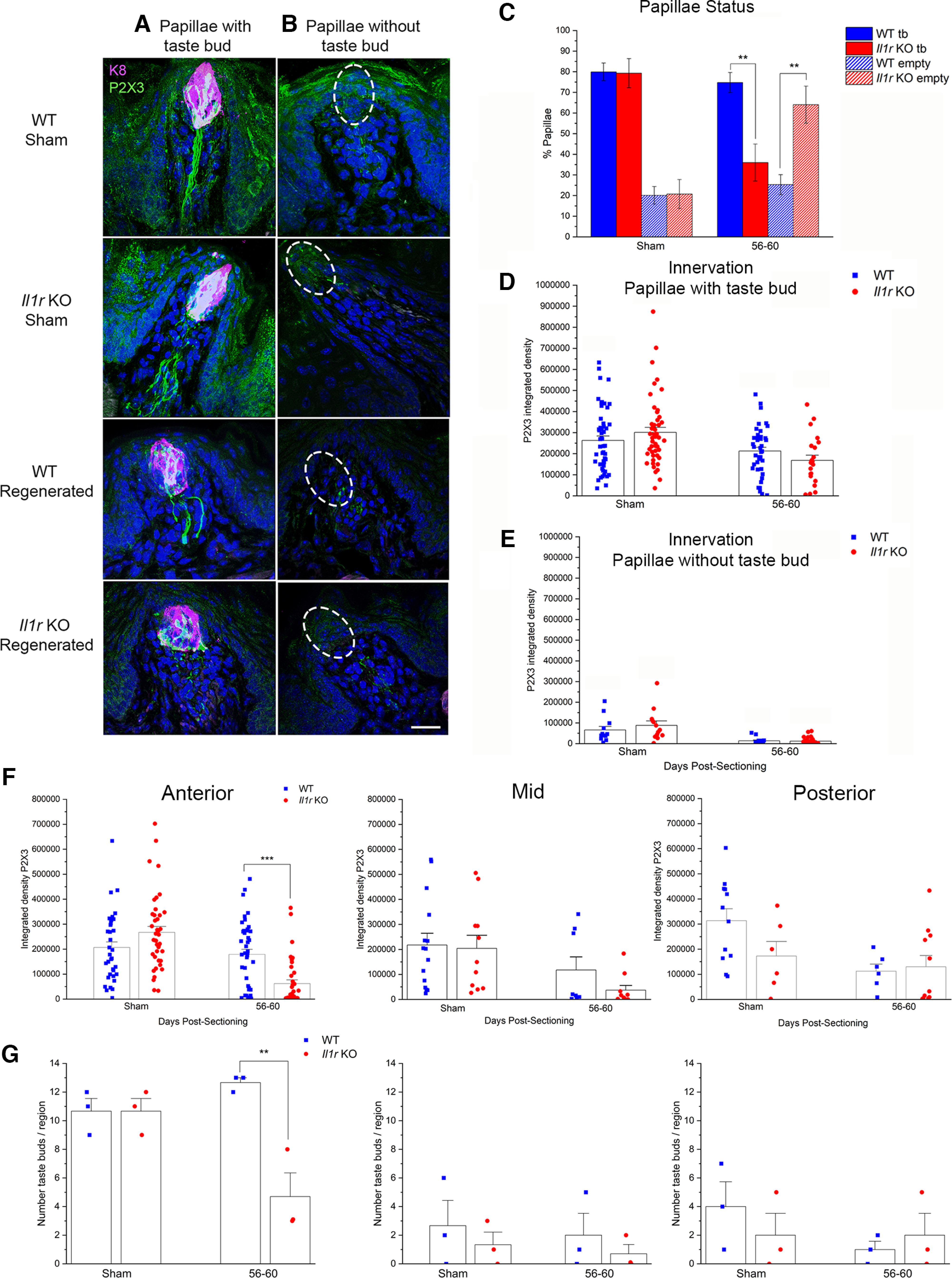
Experimental or traumatic nerve injury causes the degeneration of associated taste buds. Unlike most sensory systems, the sectioned nerve and associated taste buds can then regenerate, restoring neural responses to tastants. It was previously unknown whether injury-induced immune factors mediate this process. The proinflammatory cytokines, interleukin (IL)-1α and IL-1β, and their requisite receptor are strongly expressed by anterior taste buds innervated by the chorda tympani nerve. We tested taste bud regeneration and functional recovery in mice lacking the IL-1 receptor. After axotomy, the chorda tympani nerve regenerated but was initially unresponsive to tastants in both WT and Il1r KO mice. In the absence of Il1r signaling, however, neural taste responses remained minimal even >8 weeks after injury in both male and female mice, whereas normal taste function recovered by 3 weeks in WT mice. Failed recovery was because of a 57.8% decrease in regenerated taste buds in Il1r KO compared with WT axotomized mice. Il1a gene expression was chronically dysregulated, and the subset of regenerated taste buds were reinnervated more slowly and never reached full volume as progenitor cell proliferation lagged in KO mice. Il1r signaling is thus required for complete taste bud regeneration and the recovery of normal taste transmission, likely by impairing taste progenitor cell proliferation. This is the first identification of a cytokine response that promotes taste recovery. The remarkable plasticity of the taste system makes it ideal for identifying injury-induced mechanisms mediating successful regeneration and recovery.SIGNIFICANCE STATEMENT Taste plays a critical role in nutrition and quality of life. The adult taste system is highly plastic and able to regenerate following the disappearance of most taste buds after experimental nerve injury. Several growth factors needed for taste bud regeneration have been identified, but we demonstrate the first cytokine pathway required for the recovery of taste function. In the absence of IL-1 cytokine signaling, taste bud regeneration is incomplete, preventing the transmission of taste activity to the brain. These results open a new direction in revealing injury-specific mechanisms that could be harnessed to promote the recovery of taste perception after trauma or disease.

Keywords: chorda tympani nerve; cytokine; degeneration; electrophysiology; inflammation; taste receptor cell.


Figures

Figure 1.
Figure 1.
Taste buds express IL-1R and its ligands. (A) IL-1R, (B) IL-1α, and (C) IL-1β expression detected by immunofluorescence in keratin K8+ fungiform taste buds from WT (n = 4) and Il1r KO (n = 4) mice. D-F, Mean ± SEM integrated density of cytokine expression. D, IL-1R expression was higher in WT taste buds compared with KO taste buds, and in the surrounding WT versus KO lingual epithelium. E, IL-1α expression was also significantly higher in WT compared with Il1r KO taste buds. F, IL-1β levels were similar in WT and KO taste and nontaste epithelium. *p < 0.05. **p < 0.01. ****p < 0.0001. Scale bar, 10 µm.
Figure 2.
Figure 2.
IL-1R expression in the anterior taste epithelium and mesenchyme. A, IL-1R is colocalized with PGP9.5+ nerve fibers innervating fungiform taste buds and (B) blood vessels in the lamina propria. Dashed white lines indicate the basal lingual epithelium. IL-1R is also expressed by (C) perigemmal taste progenitor cells identified by K8 and Ki67, and (D) a subset of CD68+ macrophages. Scale bars, 10 µm.
Figure 3.
Figure 3.
Mean ± SEM relative CT nerve responses to tastants were similar in surgical and strain control groups. CT responses were normalized to 0.5 m NH4Cl responses. We recorded from sham-sectioned WT mice (n = 7), sham-sectioned Il1r KO (n = 12) mice, and WT mice at day 22-60 post-sectioning (n = 7). There were significant effects of stimulus when multiple concentrations of (A) NaCl, (B) NaAc, and (C) MSG were tested as described in the text. However, groups did not significantly differ in responses to stimulus series or (D) 1.0 m sucrose (Suc), 0.01 m quinine (QHCl), or 0.01 m HCl, demonstrating that Il1r KO did not affect CT responses in the absence of injury.
Figure 4.
Figure 4.
The regenerated CT does not recover taste responsivity in Il1r KO mice. Response traces were recorded from the CT nerve of (A) WT (blue) and Il1r KO (red) mice. Responses to 0.5 m NH4Cl over time indicate the stability of the recording. Light green dashed vertical lines indicate 0.5 s. Heavy vertical black dashed lines indicate stimulus application and rinse. Horizontal lines indicate comparison of response magnitudes. Robust responses to multiple taste qualities were recorded from both strains (A) 59 (WT) or 56 (KO) days after sham sectioning. Representative (B) 21 d post-sectioning, the regenerated nerve of a WT mouse responded robustly to all tastants, while the regenerated CT of the KO mouse was unresponsive. C, At 60 d post-sectioning, the regenerated CT of a WT mouse was similarly responsive while responses to even the strongest stimulus, 0.5 m NH4Cl, were minimal in the KO mouse. The baseline for this Il1r KO mouse increased slightly over time, although taste-elicited responses remained low. D-J, Absolute response magnitudes are plotted for individual mice along with the mean (bar) and median (horizontal line) responses to the highest concentration of each taste stimulus tested. For reference, the dashed horizontal line in D indicates the arbitrary cutoff at which we typically consider a recording preparation usable. D, Absolute responses to 0.5 m NH4Cl recorded at distinct post-sectioning periods. Responses from sham-sectioned mice recorded from day 20-59 post-injury were not different over time in either strain, and are collapsed here. WT responses were significantly reduced at day 5-8 and 11-17 post-sectioning compared with shams but recovered by day 18-22. Responses from the regenerated CT of Il1r KO mice were significantly smaller at each post-injury period compared with KO shams and at day 56-60 compared with WT mice. Responses from the regenerated nerve to the salts (E) 0.5 m NaCl, (F) 0.5 m NaCl, (G) an umami stimulus, 0.3 m MSG, and (H) a bitter stimulus, 0.01 m QHCl, follow a similar trend and were significantly reduced in Il1r KO mice at day 56-60. I, Neural responses to 1.0 M sucrose were significantly reduced in Il1r KO axotomized versus sham-sectioned mice at this period. J, CT responses to the acid, HCl, followed a similar trend but were not significantly different. N = 4-21 mice/surgical group/strain/post-injury period, with the exception of n = 1 WT mouse at day 35. *p < 0.05. **p < 0.01. NA, statistical comparison between strains not applicable at this period.
Figure 5.
Figure 5.
Il1 family gene expression dynamics are distinctly altered by CT nerve sectioning in WT and Il1r KO mice. A, In the absence of injury, Il1r expression was confirmed in peeled lingual epithelium and the mesenchyme/lamina propria of WT but not Il1r KO mice. We analyzed Il1 family mRNA expression in the right/regenerated side of the tongue using qRT-PCR. Expression levels were normalized to gapdh expression, and analyzed at day 7 in sham-sectioned mice. B, Il1r significantly increased at day 18 and 56 post-sectioning in WT mice. C, Il1rap expression was reduced at day 7 post-injury but then rebounded in WT and Il1r KO mice. D, Il1r2 expression significantly increased at day 56-60 post-injury in both strains. E, Il1α was upregulated in shams and at day 7 after axotomy in KO mice but chronically elevated only in WT mice. F, Il1β increased at day 56-60 in both strains but was elevated in KO mice at this period. N = 3/group/treatment period. *p < 0.05. **p < 0.01. ***p < 0.001. ****p < 0.0001.
Figure 6.
Figure 6.
Fewer taste buds regenerate following axotomy in Il1r KO mice. A, A low-magnification view of K8+ taste buds on the anterior lingual epithelium in Il1r KO compared with WT mice at day 58-60 after CT nerve or sham sectioning. B, K8+ taste buds were counted in serial sagittal sections to determine (C) the mean (±SEM) total number of taste buds on the right Sham/regenerated side of the anterior tongue. The number of taste buds at day 5 post-sectioning was similar between strains but significantly reduced in KO mice at day 56-60. Scale bars: A, 100 µm; B, 35 µm. N = 3 or 4 mice/group. *p < 0.05.
Figure 7.
Figure 7.
Il1r KO alters taste cell proliferation dynamics after nerve injury. We analyzed Ki67+ proliferating cells and K8+/DAPI+ taste receptor cells in the center profile of taste buds at day 14, 34-35, and 56-60 after sham or CT sectioning. A, Ki67+ (red) perigemmal cells are shown adjacent to K8+ taste buds (green; arrowheads) and in the apical (top half) and basal wall (bottom half) of fungiform papillae as indicated by white lines. B, The mean (±SEM) number of Ki67+ perigemmal cells was significantly reduced in Il1r KO mice at day 32-34 post-sectioning. C, In the apical and (D) basal papilla wall, Ki67+ cells were significantly reduced in Il1r KO compared with WT mice at day 32-34. E, The number of K8+/DAPI+ taste cells was also significantly lower in KO mice at day 32-34 post-sectioning. N = 4 mice and 22-50 taste buds/group. *p < 0.05. ***p < 0.001. Scale bar, 35 µm.
Figure 8.
Figure 8.
Il1r KO taste buds that regenerate are repopulated by all taste cell types. Taste buds immunostained with markers for (A) Type I, (B) Type II, or (C) Type III taste receptor cells colocalized with K8 after sham or CT sectioning (day 56-60). D, Type I cells were assessed by measuring determining the percentage of NTPdase2+ pixels/total pixels in the center profile of the K8+ taste bud. The density of NTPdase2 staining significantly decreased in Il1r KO compared with WT taste buds after sham sectioning but not after CT sectioning. The percentage of (E) Type II (PLCβ2+/K8+/DAPI+) taste cells with the center taste bud profile was similar across groups in posttests. F, The percentage of Type III (Car4+/K8+/DAPI+) taste cells/taste bud profile was significantly increased in sham-sectioned Il1r KO mice. Scale bar, 20 µm. *p < 0.05. ****p < 0.0001.
Figure 9.
Figure 9.
Taste buds that regenerate are innervated more slowly and fail to reach full volume in Il1r KO mice. Representative images of reconstructed K8+ taste buds (green) and P2X3+ CT nerve fibers (magenta) from WT and KO mice (A) 7 d after sham-sectioning, (B) 18-20 d post-injury, or (C) 56-60 d post-injury. D, The mean (±SEM) volume of K8 labeling was significantly reduced in WT mice at day 12-16 after axotomy but recovered to sham-like size by day 17-25. In KO mice, K8 volume was significantly decreased at all post-injury periods compared with day 7 shams, and smaller than WT at day 17-25 and 56-60 post-injury. E, The volume of P2X3 label within taste buds was significantly decreased in WT mice at day 12-16 but recovered by day 17-25. At day 17-25 post-injury, innervation volumes were significantly reduced in KO mice compared with day 7 sham-sectioned KO and WT mice. Scale bar, 10 µm. *p < 0.05. **p < 0.01. ***p < 0.001. ****p < 0.0001. ND, Not determined.
Figure 10.
Figure 10.
Papillae without taste buds were more frequent in Il1r KO mice and sparsely innervated in both strains. At day 56-60 after CT or sham sectioning, (A) papillae housing K8+ taste buds received more P2X3 innervation compared with (B) papillae without a taste bud. Dashed white lines indicate where a taste bud would typically be located in dorsal papilla. C, The mean (±SEM) percentage of papillae containing a taste bud was significantly reduced in parallel with a significant increase in empty papillae in Il1r KO mice after axotomy. The integrated density of P2X3+ nerve fibers (mean + SEM) of papillae (D) containing a taste bud or (E) without a taste bud was similar across strain and surgical treatment. We also performed regional analyses in papillae with a taste bud. F, CT innervation was significantly reduced in papillae located on the anterior region in Il1r KO compared with WT mice after axotomy. Innervation density in papillae located in the mid and posterior regions of the fungiform field was statistically similar across mouse strain or surgical treatment. G, The number of taste buds in the anterior field was also significantly reduced in KO compared with WT mice at day 56-60 post-axotomy. Fewer taste buds were located in the mid or posterior regions, and numbers were similar regardless of strain and surgical treatment. Scale bar, 20 µm. **p < 0.001. ***p < 0.0001.

Similar articles

Cited by

References

    1. Bastien D, Lacroix S (2014) Cytokine pathways regulating glial and leukocyte function after spinal cord and peripheral nerve injury. Exp Neurol 258:62–77. 10.1016/j.expneurol.2014.04.006 - DOI - PubMed
    1. Beidler LM, Smallman RL (1965) Renewal of cells within taste buds. J Cell Biol 27:263–272. 10.1083/jcb.27.2.263 - DOI - PMC - PubMed
    1. Berling K, Mannstrom P, Ulfendahl M, Danckwardt Lilliestrom N, von Unge M (2015) The chorda tympani degenerates during chronic otitis media: an electron microscopy study. Acta Otolaryngol 135:542–548. 10.3109/00016489.2014.999875 - DOI - PubMed
    1. Cain P, Frank ME, Barry MA (1996) Recovery of chorda tympani nerve function following injury. Exp Neurol 141:337–346. 10.1006/exnr.1996.0169 - DOI - PubMed
    1. Castillo-Azofeifa D, Losacco JT, Salcedo E, Golden EJ, Finger TE, Barlow LA (2017) Sonic hedgehog from both nerves and epithelium is a key trophic factor for taste bud maintenance. Development 144:3054–3065. 10.1242/dev.150342 - DOI - PMC - PubMed

Publication types

LinkOut - more resources