. 2023 Aug;44(8):1665-1675.
doi: 10.1038/s41401-023-01080-z. Epub 2023 Apr 4.

GPR84 regulates pulmonary inflammation by modulating neutrophil functions

Affiliations

GPR84 regulates pulmonary inflammation by modulating neutrophil functions

Si-Wei Wang et al. Acta Pharmacol Sin. 2023 Aug.

Abstract

Acute lung injury (ALI) is an acute, progressive hypoxic respiratory failure that could develop into acute respiratory distress syndrome (ARDS) with very high mortality rate. ALI is believed to be caused by uncontrolled inflammation, and multiple types of immune cells, especially neutrophils, are critically involved in the development of ALI. The treatment for ALI/ARDS is very limited, a better understanding of the pathogenesis and new therapies are urgently needed. Here we discover that GPR84, a medium chain fatty acid receptor, plays critical roles in ALI development by regulating neutrophil functions. GPR84 is highly upregulated in the cells isolated from the bronchoalveolar lavage fluid of LPS-induced ALI mice. GPR84 deficiency or blockage significantly ameliorated ALI mice lung inflammation by reducing neutrophils infiltration and oxidative stress. Further studies reveal that activation of GPR84 strongly induced reactive oxygen species production from neutrophils by stimulating Lyn, AKT and ERK1/2 activation and the assembly of the NADPH oxidase. These results reveal an important role of GPR84 in neutrophil functions and lung inflammation and strongly suggest that GPR84 is a potential drug target for ALI.

Keywords: GPR84; ROS; acute lung injury; antagonist; inflammation; neutrophil.


Conflict of interest statement

YMZ, employee of Burgeon Therapeutics Co. Ltd, participated in the synthesis of BGT-004. All authors declare that they have no competing interests.

Figures
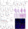
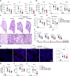
Fig. 1
Fig. 1. LPS-induced ALI is ameliorated in GPR84–/– mice.
The numbers of cells in the BALF (a) and Real-time qPCR analysis of GPR84 mRNA levels in BALF cells (b) at 0, 6, 12, 24 and 48 h after LPS treatment, *P < 0.05, **P < 0.01, ***P < 0.001 versus 0 h. c Real-time qPCR analysis of IL1β, IL6, and TNFα mRNA levels in the lungs of WT and GPR84–/– mice treated with LPS for 24 h. Gene expressions were normalized to GAPDH in the same sample and then normalized to WT control. d The concentrations of IL1β, IL6 and TNFα in BALF of WT and GPR84–/– mice treated with LPS for 24 h. Representative images of H&E stained lung sections (e) and statistical analysis (f) of histological scores. Solid arrow denotes hemorrhage and solid arrow tips point at alveolar septal thickening. Scale bars = 100 μm. g Content of BALF total protein in WT and GPR84–/– mice. h The cell numbers per mouse of total cells, CD45+ immune cells, CD45+ CD11b+ myeloid cells, CD45+ CD11b+ Ly6G+ neutrophils and CD45+ CD11b+ Ly6G- F4/80+ macrophages in the BALF of WT and GPR84-/- mice treated with LPS for 24 h. *P < 0.05, **P < 0.01, n = 8. Representative pictures (i) and statistical analysis (j) of immunofluorescence staining of MPO+ neutrophils (red) in lung sections of ALI mice. Nuclei were stained with Hoechst 33258 (blue). Scale bars = 50 μm. k, l Concentration of MPO and H2O2 in the lungs of WT and GPR84–/– ALI mice. All data are presented as means ± SEM. *P < 0.05, **P < 0.01, ***P < 0.001.
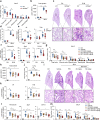
Fig. 2
Fig. 2. GPR84 antagonist BGT-004 alleviates LPS-induced ALI.
ALI mice were orally administered vehicle or BGT-004 (25 and 50 mg/kg) 0.5 h before induction and gavaged 12 h later. Mice were sacrificed 24 h after LPS treatment for following analysis. a Real-time qPCR analysis of IL1β, IL6, and TNFα mRNA levels in lung tissues. Gene expressions were normalized to GAPDH in the same sample and then normalized to WT control. b IL1β, IL6 and TNFα concentrations in the BALF. Representative pictures of H&E staining of the lung sections (c) and statistical analysis (d) of the histological scores. Solid arrow indicates hemorrhage and solid arrow tips denote septal thickening. Scale bars = 100 μm. e Content of total protein in the BALF. f The cell numbers per mouse of total cells, CD45+ immune cells, CD45+ CD11b+ myeloid cells, CD45+ CD11b+ Ly6G+ neutrophils and CD45+ CD11b+ Ly6G- F4/80+ macrophages in BALF, n = 8. Immunofluorescence staining (g) and statistical analysis (h) of MPO+ neutrophils (red) in lung sections and nuclei were stained with Hoechst 33258 (blue). Scale bars = 50 μm. The levels of MPO (i) and H2O2 (j) in lung tissue. All data are presented as means ± SEM, *P < 0.05, **P < 0.01, ***P < 0.001.
Fig. 3
Fig. 3. Activation of GPR84 promotes neutrophil chemotaxis, ROS production and degranulation.
a Chemotaxis of WT and GPR84–/– BM neutrophils in response to 6-OAU (10 μM). b Chemotaxis of BM neutrophils toward 6-OAU (10 μM) in the presence of BGT-004 (0.1, 1 or 10 μM). The chemotaxis index was assessed by counting cells in the lower chamber of the transwell and then normalized to control of the WT group. c ROS signal of unprimed WT BM neutrophils stimulated with 6-OAU (3 μM). d mRNA levels of GPR84 in BM neutrophils primed with LPS for 3 h. Gene expressions were normalized to GAPDH in the same sample and then normalized to control. e Dose-dependent stimulation of ROS release from LPS-primed WT BM neutrophils by 6-OAU. f Dose-dependent inhibition of 6-OAU (3 μM) stimulated ROS release from LPS-primed WT BM neutrophils by BGT-004. ROS production of LPS-primed WT and GPR84–/– BM neutrophils (g) or BALF neutrophils from ALI mice (h) after 6-OAU (10 μM) stimulation. Representative FACS plots (i) and statistical analysis (j) of CD63+ BM neutrophils after 6-OAU stimulation. Representative FACS plots (k) and statistical analysis (l) of CD63+ cells in BM neutrophils stimulated with 6-OAU (1 μM) in the presence of various concentrations of BGT-004. All data are presented as means ± SEM, *P < 0.05, **P < 0.01, ***P < 0.001.
Fig. 4
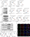
Fig. 4. GPR84 regulates ROS production via Lyn, AKT and ERK1/2 phosphorylation.
BM neutrophils were used for following experiments.a 6-OAU (3 μM)-induced ROS production in LPS-primed neutrophils in the presence of various concentrations of Lyn inhibitor PP2, AKT inhibitor MK2206 and ERK1/2 inhibitor PD0325901. Representative Western blot (b) and statistical analysis (c) of Lyn, AKT and ERK1/2 phosphorylation in LPS-primed neutrophils stimulated with 6-OAU (0.3, 3 or 30 μM). Representative Western blot (d) and statistical analysis (e) of Lyn, AKT and ERK1/2 phosphorylation stimulated by 6-OAU (3 μM) in the presence of BGT-004 in LPS-primed neutrophils. Representative Western blot analysis (f) and quantitation (g) of Lyn, AKT and ERK1/2 phosphorylation in LPS-primed WT and GPR84–/– BM neutrophils treated with 6-OAU (3 μM). All blots were representative results of three independent treatments and densitometry analysis was performed as the ratio between the phosphoprotein versus the same total protein and then normalized to control. h Representative immunofluorescence staining of cell membrane (WGA-labeled, green), p47PHOX (red) and nuclei (blue) in LPS-primed WT and GPR84-/- neutrophils stimulated with 6-OAU (3 μM). Scale bars = 10 μm. All data are presented as means ± SEM, *P < 0.05, **P < 0.01, ***P < 0.001.
Fig. 5
Fig. 5. Knockout or blockade of GPR84 reduces BLM-induced lung injury.
af Lung injury was induced in WT and GPR84-/- mice with intratracheal instillation of 2 U/kg BLM and the animals were sacrificed 7 days later for following analysis. a The cell numbers per mouse of total cells, CD45+ immune cells, CD45+CD11b+ myeloid cells, CD45+ CD11b+ Ly6G+ neutrophils and CD45+ CD11b+ Ly6G- F4/80+ macrophages in the BALF. b Total protein levels in the BALF. Representative images of H&E staining of the lung sections (c) and the statistical analysis of the histological scores (d). Solid arrows denote immune cells infiltration and solid arrow tips shows the hyaline accumulation. Scale bars = 100 μm. mRNA levels of proinflammatory cytokines IL6 and TNFα (e) and profibrotic factors Fibronectin, Spp1 and Col1α (f) in the lung tissues. Gene expressions were normalized to GAPDH in the same sample and then normalized to WT control. gl BLM-treated mice were gavaged with vehicle, PFD (300 mg/kg, bid) or BGT-004 (25 and 50 mg/kg, bid) and sacrificed 7 days after BLM treatment for further analysis. g FACS analysis of the cell numbers per mouse of the total cells, CD45+ immune cells, CD45+CD11b+ myeloid cells, CD45+ CD11b+ Ly6G+ neutrophils and CD45+ CD11b+ Ly6G- F4/80+ macrophages in the BALF. h Total protein levels in the BALF. Representative images of H&E staining of the lung sections (i) and the statistical analysis of the histological scores (j). Solid arrow tips indicate the hyaline formation and solid arrows point at immune cells accumulation. Scale bars = 100 μm. mRNA levels of proinflammatory cytokines (k) and profibrotic factors (l) in the lung tissues. Gene expressions were normalized to GAPDH in the same sample and then normalized to control. (m) Mice were orally administered vehicle, PFD (300 mg/kg, bid) or BGT-004 (25 and 50 mg/kg, bid) 0.5 h before BLM instillation and gavaged again 12 h later. BALF was collected 24 h after BLM treatment. Cell numbers per mouse of the total cells, CD45+ immune cells, CD45+CD11b+ myeloid cells, CD45+ CD11b+ Ly6G+ neutrophils and CD45+ CD11b+ Ly6G- F4/80+ macrophages were analyzed by FACS. All data are presented as means ± SEM, *P < 0.05, **P < 0.01, ***P < 0.001.
Fig. 6
Fig. 6. Mechanism of GPR84-induced ROS production in neutrophils.
Activation of GPR84 by its ligands induces the activation of Lyn. Activated Lyn phosphorylates the downstream AKT and ERK1/2. These two kinases then phosphorylate and induce membrane translocation of p47PHOX, a critical step in forming the functional NADPH oxidase. Whether and how GPR84 regulates the other subunits of the NADPH oxidase remain to be elucidated.

Similar articles

Cited by

References

    1. Wheeler AP, Bernard GR. Acute lung injury and the acute respiratory distress syndrome: a clinical review. Lancet. 2007;369:1553–64. doi: 10.1016/S0140-6736(07)60604-7. - DOI - PubMed
    1. Butt Y, Kurdowska A, Allen TC. Acute lung injury: A clinical and molecular Review. Arch Pathol Lab Med. 2016;140:345–50. doi: 10.5858/arpa.2015-0519-RA. - DOI - PubMed
    1. Maiese A, Baronti A, Manetti AC, Di Paolo M, Turillazzi E, Frati P, et al. Death after the administration of COVID-19 vaccines approved by EMA: has a causal relationship been demonstrated? Vaccines (Basel) 2011;10:10–308. - PMC - PubMed
    1. Matthay MA, Zemans RL. The acute respiratory distress syndrome: pathogenesis and treatment. Annu Rev Pathol. 2011;6:147–63. doi: 10.1146/annurev-pathol-011110-130158. - DOI - PMC - PubMed
    1. Huang X, Xiu H, Zhang S, Zhang G. The role of macrophages in the pathogenesis of ALI/ARDS. Mediat Inflamm. 2018;2018:1264913. doi: 10.1155/2018/1264913. - DOI - PMC - PubMed

Substances