. 2023 Jul;30(7):1029-1039.
doi: 10.1038/s41417-023-00610-5. Epub 2023 Apr 4.

Improving poxvirus-mediated antitumor immune responses by deleting viral cGAMP-specific nuclease

Affiliations

Improving poxvirus-mediated antitumor immune responses by deleting viral cGAMP-specific nuclease

Stephanie Riederer et al. Cancer Gene Ther. 2023 Jul.

Abstract

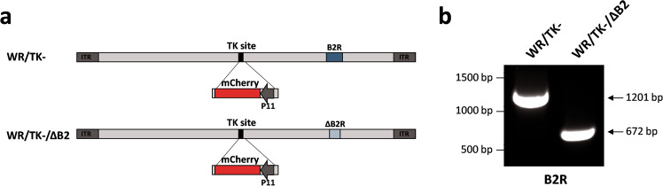
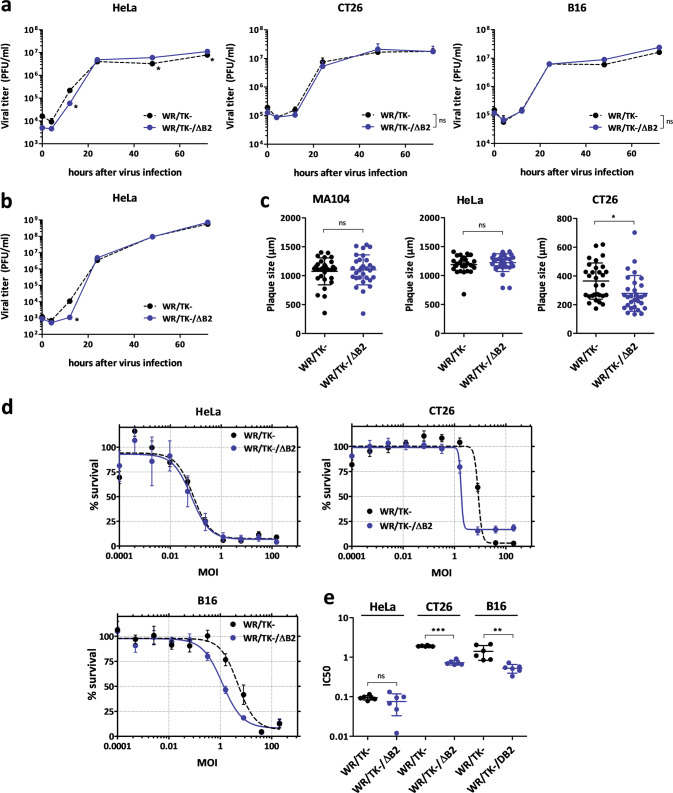
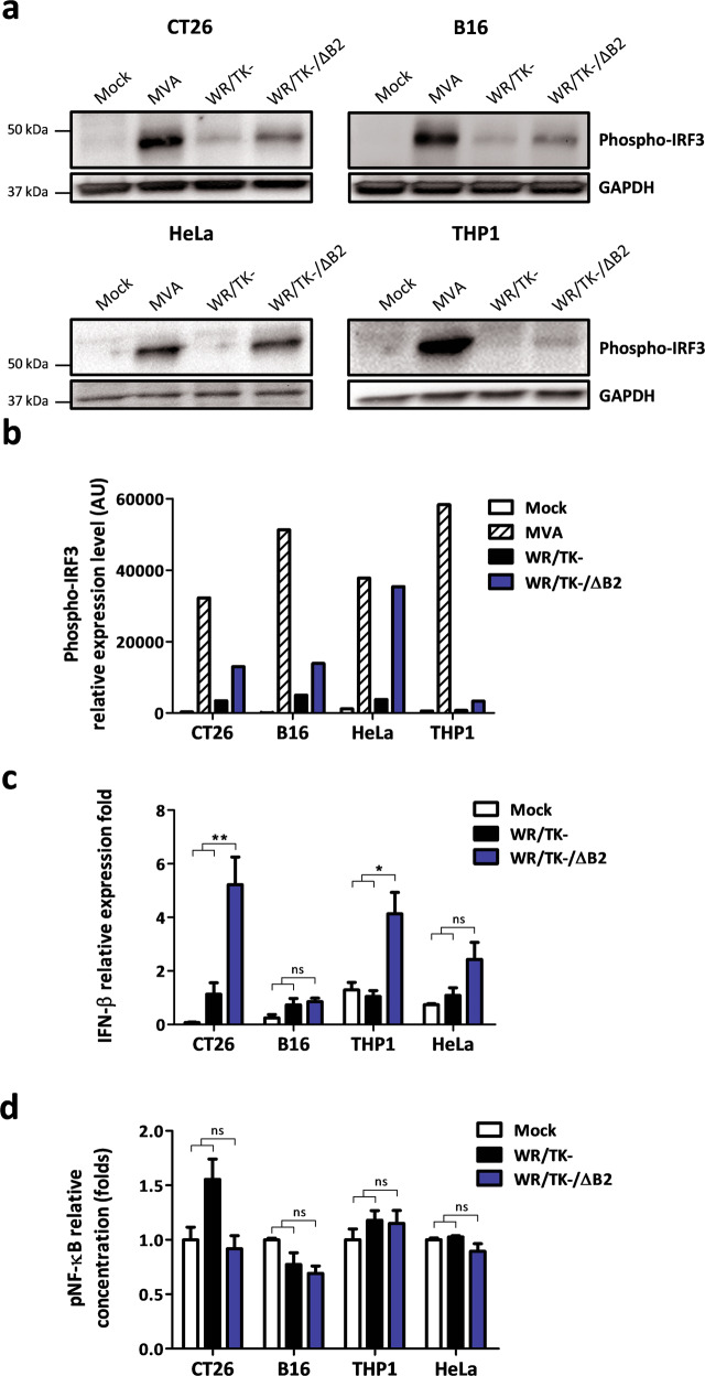
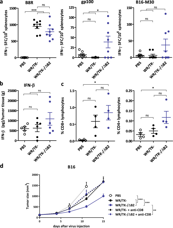
cGAMP-specific nucleases (poxins) are a recently described family of proteins dedicated to obstructing cyclic GMP-AMP synthase signaling (cGAS), an important sensor triggered by cytoplasmic viral replication that activates type I interferon (IFN) production. The B2R gene of vaccinia viruses (VACV) codes for one of these nucleases. Here, we evaluated the effects of inactivating the VACV B2 nuclease in the context of an oncolytic VACV. VACV are widely used as anti-cancer vectors due to their capacity to activate immune responses directed against tumor antigens. We aimed to elicit robust antitumor immunity by preventing viral inactivation of the cGAS/STING/IRF3 pathway after infection of cancer cells. Activation of such a pathway is associated with a dominant T helper 1 (Th1) cell differentiation of the response, which benefits antitumor outcomes. Deletion of the B2R gene resulted in enhanced IRF3 phosphorylation and type I IFN expression after infection of tumor cells, while effective VACV replication remained unimpaired, both in vitro and in vivo. In syngeneic mouse tumor models, the absence of the VACV cGAMP-specific nuclease translated into improved antitumor activity, which was associated with antitumor immunity directed against tumor epitopes.


Conflict of interest statement

The authors declare no competing interests.

Figures

Fig. 1
Fig. 1. Generation of an oncolytic VACV with a deletion in the B2R gene.
a Schematic diagram of WR/TK- and WR/TK- incorporating a deletion in the B2R gene (WR/TK-/ΔB2). To monitor viral replication in tumors, an expression cassette encoding the mCherry fluorescent protein under the control of the viral P11 promoter was inserted into the thymidine kinase (J2R) site in the virus genome. b PCR analysis to confirm the deletion in the B2R gene. Expected PCR products are: B2R = 1201 bp, ∆B2R = 672 bp.
Fig. 2
Fig. 2. cGAMP nuclease impairment does not have a major impact on viral replication and cancer cell killing capacity.
a, b Robust replication of the B2R-deleted virus. Human and mouse tumor cells were infected with a multiplicity of infection (MOI) of 5 (a) or 0.05 (b) and at indicated time points, samples were collected and viral titers were determined by plaque assay. Viral yield was evaluated in quadruplicate in two independent experiments. c Plaque size. The cell monolayers were infected at a MOI of 0.05 and, 72 h post infection, cells were dyed with crystal violet before measuring the diameter of the plaques. The diameter size (µm) of 25 representative plaques per group and mean ± SD are represented on the dot plot. (d, e) Comparative cytotoxicity in human and mouse tumor cell lines. Tumor cells were infected with the indicated viruses at doses ranging from 150 to 0.0001 PFU/cell. After 72 h, the % of cells killed (d) and IC50 values (e) were evaluated (3 different replicates from two independent experiments +SD). *p < 0.05 vs. WR/TK–; **p < 0.01 vs. WR/TK−; **p < 0.001 vs. WR/TK−; ns not significant.
Fig. 3
Fig. 3. Deletion of B2R in oncolytic VACV leads to IRF3 pathway activation.
a, b Phosphorylation of IRF3 in human and mouse cancer cells by candidate oncolytic VACV with a deletion of the B2R gene. The indicated cell lines were infected at a MOI of 10 and 5 h after infection, cells were lysed and Western blot analyses were performed using a monoclonal antibody against phospho-IRF3. Mouse phospho-IRF3 and human phosphor-IRF3 have a molecular weight of 45 and 55 kDa, respectively. The replication-deficient VACV MVA served as a positive control and GAPDH detection as a loading control. Two independent experiments were performed and representative Western blot (a) and associated densitometric analysis (b) are shown. c Relative expression of IFN-β measured by RT-qPCR. The indicated mouse and human cancer cells were infected at a MOI of 4 with Mock, WR/TK- or WR/TK-/ΔB2. Total RNA was obtained 6 h after infection. The qPCR data were normalized to the GAPDH gene in HeLa and THP1 cells or β-actin in CT26 and B16 cells by the 2−ΔΔCq method. d Activation of NF-κB pathway after infection with WR/TK- and WR/TK-/ΔB2. An ELISA assay was utilized to determine concentrations of pNF-κB in extracts of CT26, B16, THP-1, and HeLa infected at a MOI of 10. Analyses were performed 5 h after infection. Data were obtained in quadruplicate and are plotted as fold change versus Mock + SD. ns not significant; *p < 0.05; **p < 0.01.
Fig. 4
Fig. 4. Replication of the cGAMP nuclease mutant VACV is unimpaired in tumor models in vivo.
5 × 105 CT26 cells were subcutaneously implanted on the flank of 8-week-old BALB/C mice (n = 5–6). At day 0, a dose of 1 × 107 PFU of the indicated virus was intratumorally injected and 4 days later, mice were sacrificed and the tumors were harvested. a Images of representative tumors showing mCherry fluorescence intensity. b mCherry fluorescence from the tumors of each group was quantified using a MacroImaging system. Fluorescence intensity (arbitrary units) of individual tumors ±SD is shown. c Viral titers determined by plaque assays after tumor homogenization. Titers obtained from each independent tumor and means +SD are plotted on the graph. ns not significant; *p < 0.05.
Fig. 5
Fig. 5. Oncolytic VACV with a deleted cGAMP nuclease shows increased antitumor activity in mouse tumor models.
a Scheme of the treatment schedule. 5 × 105 B16 (b, c) or CT26 (d, e) tumor cells were subcutaneously implanted at day −9 in the flank of 8-week-old C57Bl/6 (B16 tumors) or BALB/C (CT26 tumors) mice, and viruses were intratumorally administrated at days 0 and 4 at a dose of 1 × 107 PFU/injection. PBS injected mice were used as the control group. To monitor tumor growth, the tumors were measured 2–3 times per week until they reached the termination criteria (≥750 mm3; see Materials and methods). Tumor volume (b, d) and overall survival (c, e) are plotted for 9–14 mice per group +SEM. Data shown in (b) for the control groups PBS and WR/TK– is the same as published in [23] as both studies were done in the same experiment following the principle of the 3Rs. *p < 0.05; **p < 0.01, ***p < 0.001.
Fig. 6
Fig. 6. Deletion of the cGAMP nuclease elicits antitumor T-cell responses directed against tumor antigens.
a Cellular immune response evaluated by IFN-γ ELISpot assay. C57Bl/6 mice harboring B16 tumors were treated as indicated in Fig. 5 and 8 days after virus administration splenocytes were prepared, stimulated in vitro with the indicated peptides, and analyzed for IFN-γ producing cells by ELISPOT. Individual values of IFN-γ spot forming cells (SPC)/106 splenocytes in 8 mice/group and means ± SD are plotted on the graphs. b Intratumoral IFN-β concentrations after VACV administration. BALB/C mice harboring CT26 tumors were injected intratumorally with a single dose of 1 × 107 PFU and 4 days later, tumors were harvested and homogenized. IFN-β concentrations were measured using an ELISA assay. c Treatment with WR/TK-/ΔB2 increases the number of CD8+ and CD4+ lymphocytes infiltrating the tumor. C57Bl/6 mice with subcutaneous B16 tumors were treated as before (Fig. 5) and tumors were harvested at day 8 after virus administration and evaluated for lymphocyte populations by flow cytometry. Percentage of CD8+ and CD4+ live lymphocytes are plotted. d CD8+ cells are essential for the antitumor efficacy mediated by WR/TK−/ΔB2. C57Bl/6 mice with subcutaneous B16 tumors were injected with CD8-depleting antibodies or isotype control and treated as before (Fig. 5). Tumor volume is plotted for 8–11 mice per group +SEM. ns not significant; *p < 0,05; **p < 0.01; ***p < 0.001.

References

    1. Guo ZS, Lu B, Guo Z, Giehl E, Feist M, Dai E, et al. Vaccinia virus-mediated cancer immunotherapy: cancer vaccines and oncolytics. J Immunother Cancer. 2019;7:6. doi: 10.1186/s40425-018-0495-7. - DOI - PMC - PubMed
    1. Zuo S, Wei M, He B, Chen A, Wang S, Kong L, et al. Enhanced antitumor efficacy of a novel oncolytic vaccinia virus encoding a fully monoclonal antibody against T-cell immunoglobulin and ITIM domain (TIGIT) EBioMedicine. 2021;64:103240. doi: 10.1016/j.ebiom.2021.103240. - DOI - PMC - PubMed
    1. Van Hoecke L, Riederer S, Saelens X, Sutter G, Rojas JJ. Recombinant viruses delivering the necroptosis mediator MLKL induce a potent antitumor immunity in mice. Oncoimmunology. 2020;9:1802968. doi: 10.1080/2162402X.2020.1802968. - DOI - PMC - PubMed
    1. Shi T, Song X, Wang Y, Liu F, Wei J. Combining Oncolytic Viruses With Cancer Immunotherapy: Establishing a New Generation of Cancer Treatment. Front Immunol. 2020;11:683. doi: 10.3389/fimmu.2020.00683. - DOI - PMC - PubMed
    1. Pelin A, Boulton S, Tamming LA, Bell JC, Singaravelu R. Engineering vaccinia virus as an immunotherapeutic battleship to overcome tumor heterogeneity. Expert Opin Biol Ther. 2020;20:1083–97. doi: 10.1080/14712598.2020.1757066. - DOI - PubMed

Publication types

Substances