

SENP3 facilitates M1 macrophage polarization via the HIF-1α/PKM2 axis in lipopolysaccharide-induced acute lung injury
- PMID: 37016838
- PMCID: PMC10164277
- DOI: 10.1177/17534259231166212
SENP3 facilitates M1 macrophage polarization via the HIF-1α/PKM2 axis in lipopolysaccharide-induced acute lung injury
Abstract
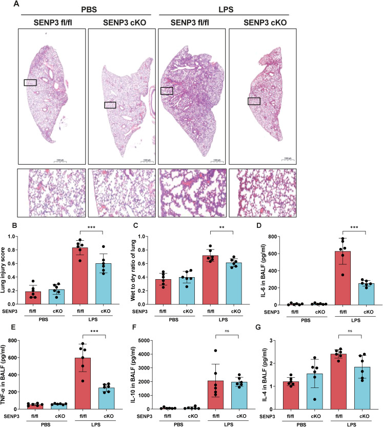
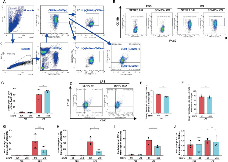
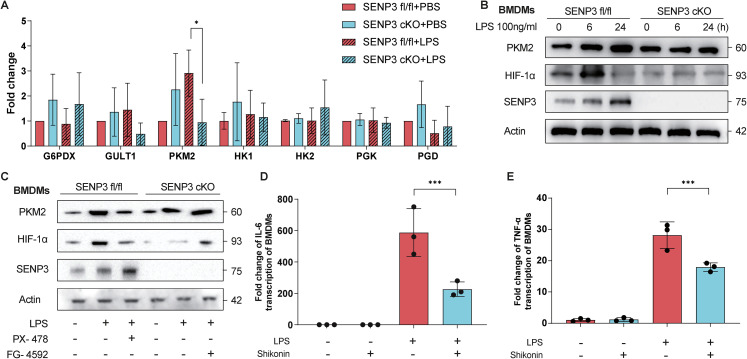
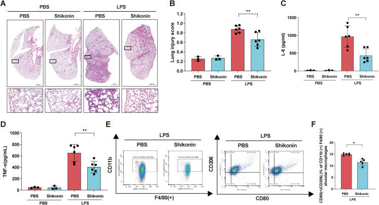
M1/M2 macrophage polarization plays a pivotal role in the development of acute lung injury (ALI). The hypoxia-inducible factor-1α/pyruvate kinase M2 (HIF-1α/PKM2) axis, which functions upstream of macrophage polarization, has been implicated in this process. The function of HIF-1α is known to be tightly regulated by SUMOylation. Upregulation of SUMO-specific peptidase 3 (SENP3), a deSUMOylation enzyme, is induced by reactive oxygen species (ROS), which are abundantly produced during ALI. To explore the links between SENP3, macrophage polarization, and lung injury, we used mice with Senp3 conditional knockout in myeloid cells. In the lipopolysaccharide (LPS)-induced ALI model, we found that in vitro and in vivo SENP3 deficiency markedly inhibited M1 polarization and production of pro-inflammatory cytokines and alleviated lung injury. Further, we demonstrated that SENP3 deficiency suppressed the LPS-induced inflammatory response through PKM2 in a HIF-1α-dependent manner. Moreover, mice injected with LPS after PKM2 inhibitor (shikonin) treatment displayed inhibition of M1 macrophage polarization and reduced lung injury. In summary, this work revealed that SENP3 promotes M1 macrophage polarization and production of proinflammatory cytokines via the HIF-1α/PKM2 axis, contributing to lung injury; thus, SENP3 may represent a potential therapeutic target for ALI treatment.
Keywords: HIF-1α; Macrophage; PKM2; SENP3; SUMOylation; acute lung injury.
Conflict of interest statement
The author(s) declared no potential conflicts of interest with respect to the research, authorship, and/or publication of this article.
Figures
Similar articles
-
Hypoxia inducible factor-1α is an important regulator of macrophage biology.Heliyon. 2023 Jun 9;9(6):e17167. doi: 10.1016/j.heliyon.2023.e17167. eCollection 2023 Jun. Heliyon. 2023. PMID: 37484306 Free PMC article. Review.
-
Norisoboldine Attenuates Sepsis-Induced Acute Lung Injury by Modulating Macrophage Polarization via PKM2/HIF-1α/PGC-1α Pathway.Biol Pharm Bull. 2021;44(10):1536-1547. doi: 10.1248/bpb.b21-00457. Biol Pharm Bull. 2021. PMID: 34602563
-
SENP3 in monocytes/macrophages up-regulates tissue factor and mediates lipopolysaccharide-induced acute lung injury by enhancing JNK phosphorylation.J Cell Mol Med. 2020 May;24(10):5454-5462. doi: 10.1111/jcmm.15199. Epub 2020 Mar 31. J Cell Mol Med. 2020. PMID: 32237051 Free PMC article.
-
DeSUMOylation of MKK7 kinase by the SUMO2/3 protease SENP3 potentiates lipopolysaccharide-induced inflammatory signaling in macrophages.J Biol Chem. 2018 Mar 16;293(11):3965-3980. doi: 10.1074/jbc.M117.816769. Epub 2018 Jan 19. J Biol Chem. 2018. PMID: 29352108 Free PMC article.
-
The role of natural products targeting macrophage polarization in sepsis-induced lung injury.Chin Med. 2025 Feb 5;20(1):19. doi: 10.1186/s13020-025-01067-4. Chin Med. 2025. PMID: 39910395 Free PMC article. Review.
Cited by
-
Laminarin Alleviates Acute Lung Injury Induced by LPS Through Inhibition of M1 Macrophage Polarisation.J Cell Mol Med. 2025 Mar;29(5):e70440. doi: 10.1111/jcmm.70440. J Cell Mol Med. 2025. PMID: 40045157 Free PMC article.
-
Molecular mechanisms of Sepsis attacking the immune system and solid organs.Front Med (Lausanne). 2024 Aug 29;11:1429370. doi: 10.3389/fmed.2024.1429370. eCollection 2024. Front Med (Lausanne). 2024. PMID: 39267971 Free PMC article. Review.
-
Hypoxia inducible factor-1α is an important regulator of macrophage biology.Heliyon. 2023 Jun 9;9(6):e17167. doi: 10.1016/j.heliyon.2023.e17167. eCollection 2023 Jun. Heliyon. 2023. PMID: 37484306 Free PMC article. Review.
-
The impact of glucose metabolism on inflammatory processes in sepsis-induced acute lung injury.Front Immunol. 2024 Dec 6;15:1508985. doi: 10.3389/fimmu.2024.1508985. eCollection 2024. Front Immunol. 2024. PMID: 39712019 Free PMC article. Review.
-
Post-translational modifications and bronchopulmonary dysplasia.Front Pediatr. 2025 Jan 3;12:1426030. doi: 10.3389/fped.2024.1426030. eCollection 2024. Front Pediatr. 2025. PMID: 39830627 Free PMC article. Review.
References
Publication types
MeSH terms
Substances
LinkOut - more resources
Full Text Sources
Molecular Biology Databases
Miscellaneous
