. 2023 Jun 26;35(7):2678-2693.
doi: 10.1093/plcell/koad099.

The OPAQUE1/DISCORDIA2 myosin XI is required for phragmoplast guidance during asymmetric cell division in maize

Affiliations

The OPAQUE1/DISCORDIA2 myosin XI is required for phragmoplast guidance during asymmetric cell division in maize

Qiong Nan et al. Plant Cell. .

Abstract

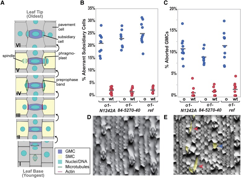
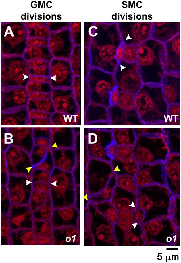
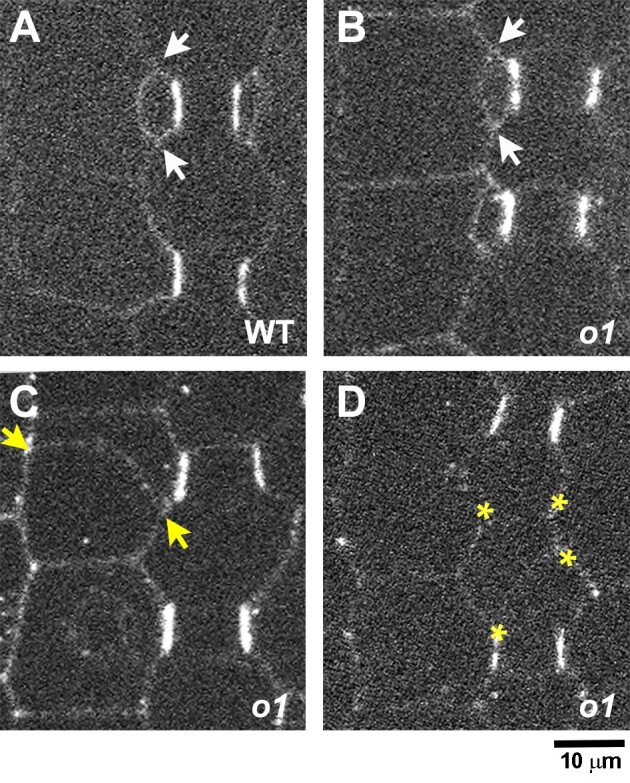
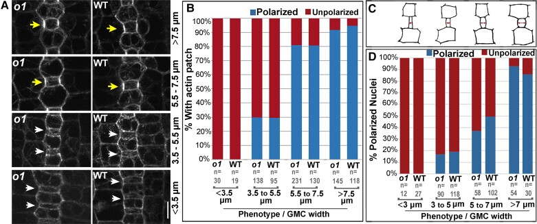
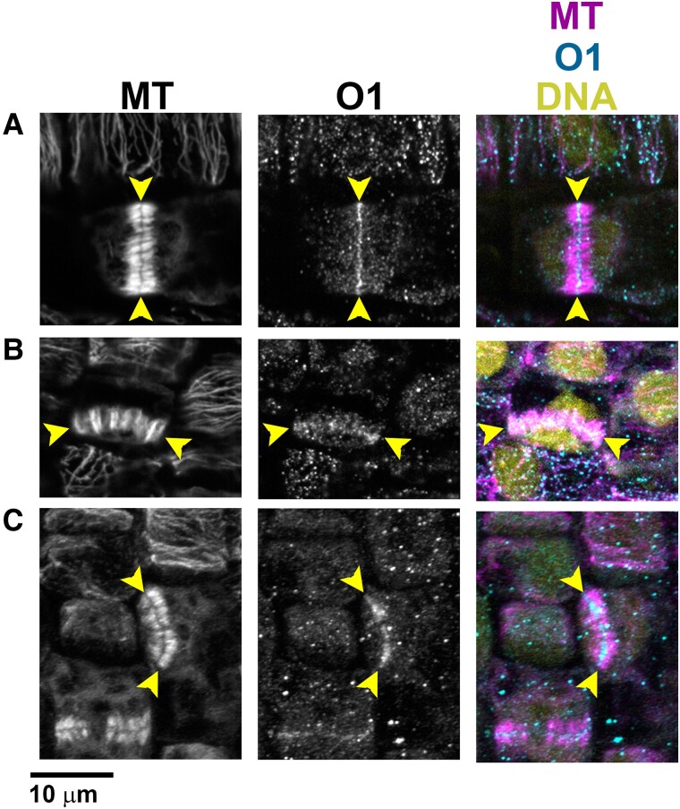
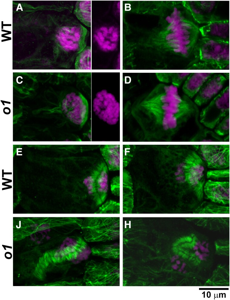
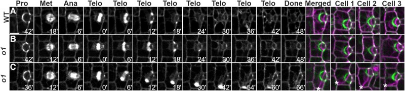
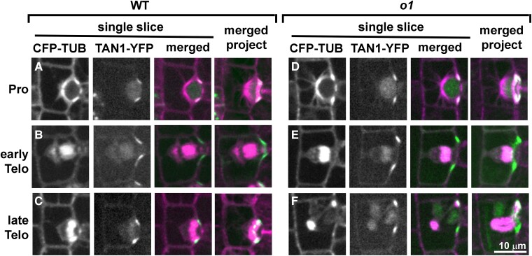
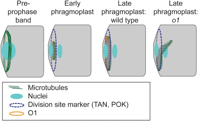
Formative asymmetric divisions produce cells with different fates and are critical for development. We show the maize (Zea mays) myosin XI protein, OPAQUE1 (O1), is necessary for asymmetric divisions during maize stomatal development. We analyzed stomatal precursor cells before and during asymmetric division to determine why o1 mutants have abnormal division planes. Cell polarization and nuclear positioning occur normally in the o1 mutant, and the future site of division is correctly specified. The defect in o1 becomes apparent during late cytokinesis, when the phragmoplast forms the nascent cell plate. Initial phragmoplast guidance in o1 is normal; however, as phragmoplast expansion continues o1 phragmoplasts become misguided. To understand how O1 contributes to phragmoplast guidance, we identified O1-interacting proteins. Maize kinesins related to the Arabidopsis thaliana division site markers PHRAGMOPLAST ORIENTING KINESINs (POKs), which are also required for correct phragmoplast guidance, physically interact with O1. We propose that different myosins are important at multiple steps of phragmoplast expansion, and the O1 actin motor and POK-like microtubule motors work together to ensure correct late-stage phragmoplast guidance.


Conflict of interest statement

Conflict of interest statement. None declared.

Figures

Figure 1.
Figure 1.
OPAQUE1 is required for normal subsidiary cell formation in maize. A) Division sequence of stomatal development in maize and other grasses. B) Percentage of abnormal subsidiary cells and C) percentage of aborted GMCs in o1 mutants (o) and their wild-type siblings (wt). In (B and C) seeds were scored based on the appearance of their endosperm (o, seeds that are phenotypically opaque; wt, translucent seeds). Each data point represents the percentage of abnormal cells from 1 plant; between 100 and 200 cells were counted per plant. Gray horizontal bars indicate means. ANOVA comparing each mutant to wild-type sibling returned P < 0.0001 for all 3 alleles. D) Methacrylate impression of the leaf epidermis of a wild-type sibling showing normal stomatal complexes. E)o1-ref mutant showing abnormal subsidiary cells (red arrowheads) and aborted GMC (blue arrowhead). Abnormal cells in (E) are highlighted in yellow.
Figure 2.
Figure 2.
Stomatal lineage cells have abnormal division planes in o1. The region of developing leaf 4 undergoing stomatal divisions was dissected, fixed, and stained with PI (red) and aniline blue (blue). Segregating wild-type (WT; A, B) and homozygous sibling o1-N1242A mutants (o1;B, D) are shown. White arrowheads mark correct divisions; yellow arrowheads mark incorrect divisions. A, B) Recently formed GMCs. C, D) Recently formed subsidiary cells, formed from SMCs. Z-projection of 3 confocal images. Scale bar, 5 µm.
Figure 3.
Figure 3.
PAN1-YFP polarizes correctly in o1. Recently formed subsidiary cells from o1-ref plants and wild-type siblings expressing PAN1-YFP were assayed for PAN1-YFP polarization. Arrows in (A–C) indicate correctly (white) or incorrectly (yellow) oriented cell walls generated from an SMC division. A) Recently divided SMC with polarized PAN1-YFP at the GMC-subsidiary cell interface. B) Correctly formed subsidiary cell from o1. PAN1-YFP is correctly polarized. C) Incorrectly oriented cell wall generated from an aberrant SMC division. PAN1-YFP is correctly polarized. D) Incorrectly oriented cell wall generated from an aberrant division of the GMC progenitor cell. Yellow asterisks in (D) indicate 4 corners of a cell formed from an aberrant GMC-generating division. Scale bar, 10 µm.
Figure 4.
Figure 4.
Actin patch formation and nuclear migration is normal in o1. A) The stomatal division zone was examined in leaf 4 of o1-N1242A mutant plants and wild-type siblings expressing the ABD2-YFP marker. GMCs in early developmental stages, found at the leaf base (lower panels), are narrow and width increases towards the leaf tip as development proceeds (upper panels). Early SMCs flanking narrow GMCs do not form an actin patch (white arrows). SMCs at later developmental stages, flanking wider GMCs, have an actin patch (yellow arrows). B) Percentage of SMCs with a polarized actin patch at progressive developmental stages in o1-N1242A mutant plants and their corresponding wild-type siblings. C) Cartoons depicting representative cell outlines at increasing GMC widths. Red arrows indicate where GMC width was measured. D) Percentage polarized nuclei in SMCs at progressive developmental stages in o1-N1242A mutant plants and their corresponding wild-type siblings. Fisher's exact tests comparing o1 mutants to their respective wild-type siblings indicate no differences between mutants and wild type at each developmental stage (P > 0.05 in all cases).
Figure 5.
Figure 5.
O1 localizes to phragmoplasts. Immunofluorescence detection of microtubules and O1 in wild-type cells (A to C). All samples are from the division zone of developing leaf 4. O1 is detected in phragmoplasts of symmetrically dividing cells (A), asymmetrically dividing stomatal lineage cells that will form GMCs (B), and asymmetrically dividing SMCs (C). Yellow arrowheads indicate phragmoplast ends. Scale bar, 10 µm.
Figure 6.
Figure 6.
O1 is required for normal phragmoplast guidance. Immunofluorescence detection of microtubules (green) and DAP-stained nuclei (magenta) in wild-type (A, B, E, F) and o1 cells (C, D, J, K). PPBs in SMCs (A, C) appear similar in wild-type siblings and o1-N1242A. Side panels in (A and C) show DAPI channel only to show condensed chromosomes. Spindles in SMCs (B, D) were similar in wild-type and o1 SMCs. In o1, SMC phragmoplasts appeared as in wild type (E, F) or misguided (J, H). Z-projections of 40 to 60 images. Scale bar, 10 µm applies to all images.
Figure 7.
Figure 7.
Time-lapse imaging confirms a phragmoplast guidance defect in o1. CFP-TUB was used to observe progression of cell division SMCs from leaf 5 or 6 in o1-N1242A and corresponding wild-type siblings. A) Wild-type cell division. B) Correctly oriented o1 cell division. C) Misoriented o1 cell division. Pro, prophase; Met, metaphase; Ana, anaphase; Telo, telophase; Done, completed division; Merged, overlay of prophase (green) and completed division. Cells 1 to 3 show 3 additional representative cells. Time (min) is listed at the bottom of each image. Misoriented cell walls are indicated by asterisks. Z-projections of 6 images. All cells displayed at the same magnification; scale bar in (A), 10 µm.
Figure 8.
Figure 8.
TAN1-YFP correctly marks the division plane during normal and abnormal o1 SMC divisions. Dividing SMCs from leaf 5 or 6 in wild-type siblings (A to C) or o1-N1242A(D to F) cells co-expressing CFP-TUB (magenta) and TAN1-YFP (green). Single planes are shown in the first 3 panels and a full projection is shown in the last panel. In wild-type cells, TAN1-YFP correctly marked the predicted division plane throughout mitosis including prophase (A; n = 33/33), metaphase (n = 30/30), anaphase (n = 12/12), early telophase (B; n = 21/21), and late telophase (C; n = 58/58). In o1-N1242A mutant cells, TAN1-YFP always correctly marked the division plane in prophase (D; n = 85/85), metaphase (n = 20/20), anaphase (n = 13/13), and early telophase (E; n = 32/32). In 2/32 cases, TAN1-YFP was also seen at an additional site during early telophase (see Supplemental Fig. S10). During late telophase, TAN1-YFP was at the cortical division site in o1 SMCs with correctly oriented phragmoplasts (n = 38/38) and incorrectly oriented phragmoplasts (F; n = 44/44). Scale bar, 10 µm applies to all images.
Figure 9.
Figure 9.
Participation of O1 in late-stage phragmoplast guidance. The cortical division site is initially marked by the PPB and later by division site markers such as TAN1 and POK proteins (which are also present in the phragmoplast). In wild-type cells, the phragmoplast is guided to the division site by actin filaments and myosin VIII (Wu and Bezanilla 2014). After meeting the cortex, the phragmoplast continues to expand and interactions are stabilized by microtubules (Bellinger et al. 2021). In wild type, the phragmoplast fuses with the existing cell wall along the established division site, which is mediated by POK, TAN, and O1. In o1 mutants, after initial contact the phragmoplast becomes misguided, resulting in abnormal division planes.

Comment in

Similar articles

Cited by

References

    1. Abu-Abied M, Belausov E, Hagay S, Peremyslov V, Dolja V, Sadot E. Myosin XI-K is involved in root organogenesis, polar auxin transport, and cell division. J Exp Bot. 2018:69(12):2869–2881. 10.1093/jxb/ery112 - DOI - PMC - PubMed
    1. Ali MF, Fatema U, Peng X, Hacker SW, Maruyama D, Sun M-X, Kawashima T. ARP2/3-independent WAVE/SCAR pathway and class XI myosin control sperm nuclear migration in flowering plants. Proc Natl Acad Sci U S A. 2020:117(51):32757–32763. 10.1073/pnas.2015550117 - DOI - PMC - PubMed
    1. Allsman LA, Dieffenbacher RN, Rasmussen CG. Glue impressions of maize leaves and their use in classifying mutants. Bio-101. 2019:e3209. 10.21769/BioProtoc.3209 - DOI
    1. Apostolakos P, Livanos P, Giannoutsou E, Panteris E, Galatis B. The intracellular and intercellular cross-talk during subsidiary cell formation in Zea mays: existing and novel components orchestrating cell polarization and asymmetric division. Ann Bot. 2018:122(5):679–696. 10.1093/aob/mcx193 - DOI - PMC - PubMed
    1. Avisar D, Abu-Abied M, Belausov E, Sadot E, Hawes C, Sparkes IA. A comparative study of the involvement of 17 Arabidopsis myosin family members on the motility of Golgi and other organelles. Plant Physiol. 2009:150(2):700–709. 10.1104/pp.109.136853 - DOI - PMC - PubMed

Publication types