. 2023 Jun 2;22(6):1682-1694.
doi: 10.1021/acs.jproteome.2c00731. Epub 2023 Apr 5.

Comparative Proteomic Analysis Revealing ActJ-Regulated Proteins in Sinorhizobium meliloti

Affiliations

Comparative Proteomic Analysis Revealing ActJ-Regulated Proteins in Sinorhizobium meliloti

Francisco Javier Albicoro et al. J Proteome Res. .

Abstract

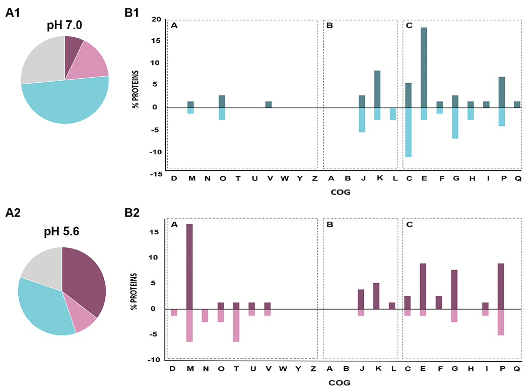
To adapt to different environmental conditions, Sinorhizobium meliloti relies on finely tuned regulatory networks, most of which are unexplored to date. We recently demonstrated that deletion of the two-component system ActJK renders an acid-vulnerable phenotype in S. meliloti and negatively impacts bacteroid development and nodule occupancy as well. To fully understand the role of ActJ in acid tolerance, S. meliloti wild-type and S. meliloti ΔactJ proteomes were compared in the presence or absence of acid stress by nanoflow ultrahigh-performance liquid chromatography coupled to mass spectrometry. The analysis demonstrated that proteins involved in the synthesis of exopolysaccharides (EPSs) were notably enriched in ΔactJ cells in acid pH. Total EPS quantification further revealed that although EPS production was augmented at pH 5.6 in both the ΔactJ and the parental strain, the lack of ActJ significantly enhanced this difference. Moreover, several efflux pumps were found to be downregulated in the ΔactJ strain. Promoter fusion assays suggested that ActJ positively modulated its own expression in an acid medium but not at under neutral conditions. The results presented here identify several ActJ-regulated genes in S. meliloti, highlighting key components associated with ActJK regulation that will contribute to a better understanding of rhizobia adaptation to acid stress.

Keywords: ActJK; Sinorhizobium meliloti; acid stress, proteomics.


Conflict of interest statement

The authors declare no competing financial interest.

Figures

Figure 1.
Figure 1.
Growth curves in GSM at (A) pH = 7.0 (neutral condition) and (B) pH = 5.6 (acid stress). In the figure, the optical density OD600nm is plotted on the ordinate as a function of time on the abscissa. The black points represent S. meliloti 2011 wild-type cells (wt) and the light blue points represent the ΔactJ mutant. The error bars in each point represent the standard deviation of the mean among three replicates.
Figure 2.
Figure 2.
Distribution of the abundance of cytoplasmic (A and C) and membrane enriched (B and D) proteins obtained from ΔactJ cells cultured in GSM at either pH 5.6 (panels A, B) or pH 7.0 (panels C, D) with respect to proteins obtained from wt cells. In each panel, the −log10(p value) is plotted on the ordinate as a function of the log2(fold change). In identifying differentially expressed proteins we included those which met the following two conditions: fold-change, FC ≥ 2 and FC ≤ −2 (corresponding to values greater than 1 or −1 on the abscissa) and p value ≤ 0.05 (corresponding to values greater than 1.3 in the ordinate). The purple circles represent overexpressed, and the pink squares represent underexpressed proteins present in ΔactJ cells relative to those in the wt at pH = 5.6. The blue circles represent overexpressed and the light-blue squares underexpressed proteins present in ΔactJ cells relative to those in the wt at pH = 7.0. The proteins located on the 0 line correspond to the on/off proteins since those proteins had no associated p-value.
Figure 3.
Figure 3.
A. Clusters of Orthologous Groups (COGs) of differentially expressed proteins obtained in GSM cultures at pH 7.0 (A1) or pH 5.6 (A2). In the panel, the two pie charts indicate the relative distribution of the cellular functions: violet represents Cellular Processes and Signaling (A), pink represents Information Storage and Processing (B), light blue represents Metabolism (C) and grey represents Poorly Characterized Proteins. B. Up- and downregulated ΔactJ vs. wt differential proteins were grouped into COG categories, B1 shows the results at pH 7.0 and B2 shows the results at pH 5.6. In each bar graph, the percent proteins is plotted on the ordinate for each of the COGs listed on the abscissa and the graph is divided in 3 zones according to the distribution of cellular functions as was mentioned before (see “A”, “B” and “C”). Percentages greater than zero correspond to upregulated proteins and those with percentages lower than zero correspond to downregulated proteins. 100% corresponds to all proteins found to be differential in each condition, excluding those belonging to the uncharacterized category of COGs. COG categories: RNA processing and modification (A), chromatin structure and dynamics (B), energy production and conversion (C), cell cycle control and mitosis (D), amino acid metabolism and transport I, nucleotide metabolism and transport (F), carbohydrate metabolism and transport (G), coenzyme metabolism (H), lipid metabolism (I), translation (J), transcription (K), replication and repair (L), membrane biogenesis (M), cell motility (N), posttranslational modification, protein turnover, chaperone functions (O), inorganic ion transport and metabolism (P), secondary structure (Q), signal transduction (T) intracellular trafficking and secretion (U), nuclear structure (Y), cytoskeleton (Z).
Figure 4.
Figure 4.
Total EPS quantification performed on cells obtained from mid-exponential cultures grown in GSM media at pH 7.0 or pH 5.6. In the figure, the EPS production in mg of glucose per mg of protein is plotted on the ordinate for each of the two S. meliloti strains indicated on the abscissa at the two respective pHs. The data are the means of three independent experiments ± standard deviations. The error bars represent standard deviations of the mean. The Tukey test was used to analyze the replicates. Common letters denote no significant difference (p > 0.05).
Figure 5.
Figure 5.
eGFP transcriptional fusions and fluorescence assays performed on cells obtained from mid-exponential cultures grown in GSM media at neutral pH (7.0, Panel A) or acid pH (5.6, Panel B). In each panel the relative fluorescence units (RFUs) per OD600 are plotted on the ordinate for each of the transcribed loci in wild-type (wt) and mutant (ΔactJ) cells indicated on the abscissa. Strains containing the promotorless pHU231eGFP plasmid are indicated as PΦ. Error bars represent the standard deviations of the mean of three replicates. ANOVA Tukey’s multiple comparison test was performed to analyze significance. Bars with same letters are not significantly different (p> 0.05).
Figure 6.
Figure 6.
Summary of most relevant pathways linked to an ActJ activity in S. meliloti. The figure constitutes an overall scheme for the current model depicting the adaptation of S. meliloti to acid stress, involving the key functional pathways operating in the different subcellular compartments at either pH 7.0 (left panel) or pH 5.6 (right panel). The intact arrows denote upregulated pathways and the blocked arrows downregulated pathways in the presence of ActJ. The putative regulations are represented by dashed arrows and the experimentally confirmed regulation by solid arrows. This bacterium exhibits a global response at low pH resulting from the cellular deterioration caused by acid stress. In S. meliloti, ActJ could trigger the various mechanisms illustrated to cope specifically with acid stress, such as an increase in the degP1 and actJK expression, a downregulation of EPS synthesis, and changes in the cell envelope, all of which modulations would contribute to the resistance to an acidic challenge.

References

    1. Evans JR; von Caemmerer S Enhancing Photosynthesis. Plant Physiol. 2011, 155 (1), 19 LP – 19. - PMC - PubMed
    1. Buckley DH; Schmidt T Exploring the Biodiversity of Soil—a Microbial Rain Forest. Biodivers. Microb. life Found. Earth’s Biosph 2001, 183–208.
    1. Kallenbach CM; Wallenstein MD; Schipanksi ME; Grandy AS Managing Agroecosystems for Soil Microbial Carbon Use Efficiency: Ecological Unknowns, Potential Outcomes, and a Path Forward. Front. Microbiol 2019, 10, 1146. - PMC - PubMed
    1. Bouton JH; Sumner ME Alfalfa Medicago Sativa L., in Highly Weathered, Acid Soils. Plant Soil 1983, 74 (3), 431–436.
    1. Fierer N; Jackson RB The Diversity and Biogeography of Soil Bacterial Communities. Proc. Natl. Acad. Sci. U. S. A 2006, 103 (3), 626 LP – 631. - PMC - PubMed

Publication types

MeSH terms

LinkOut - more resources