. 2023 Jun 19;32(13):2219-2228.
doi: 10.1093/hmg/ddad055.

Breast cancer cell-derived exosome-delivered microRNA-155 targets UBQLN1 in adipocytes and facilitates cancer cachexia-related fat loss

Affiliations

Breast cancer cell-derived exosome-delivered microRNA-155 targets UBQLN1 in adipocytes and facilitates cancer cachexia-related fat loss

S Sun et al. Hum Mol Genet. .

Abstract

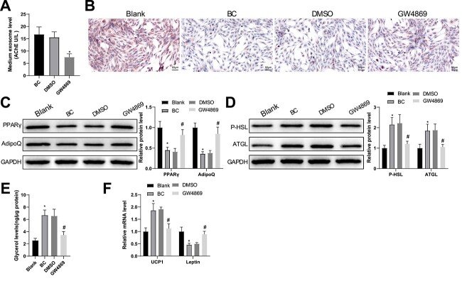
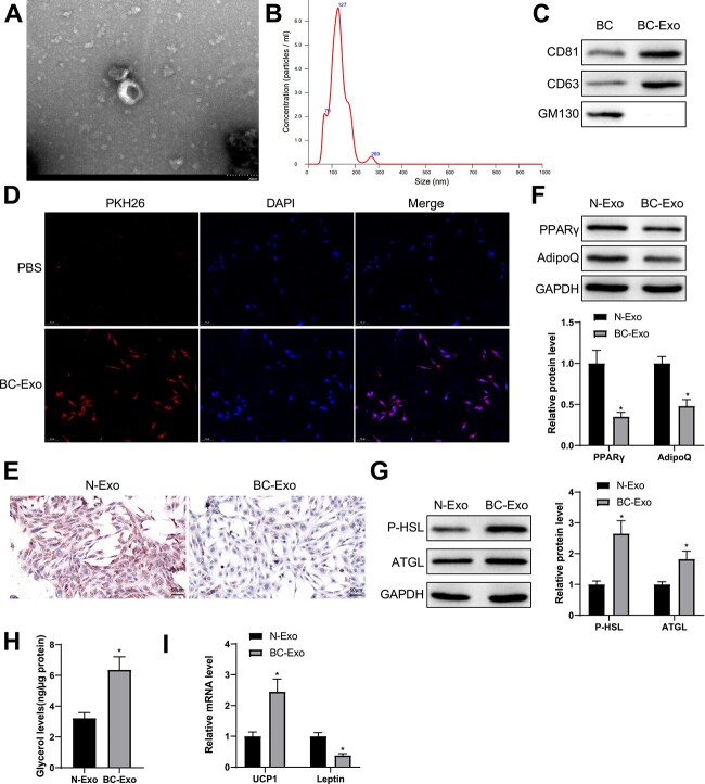
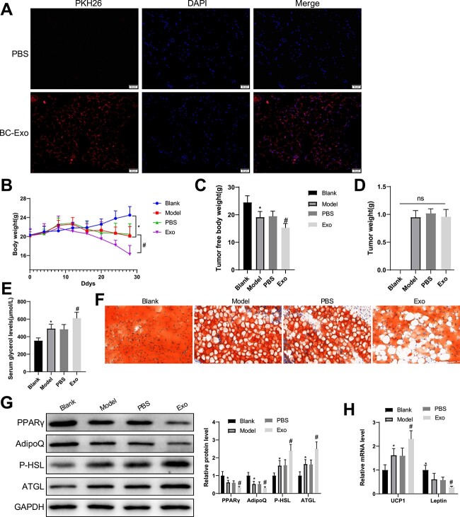
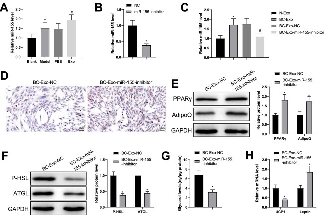
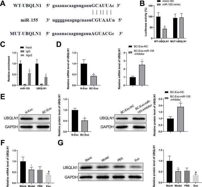
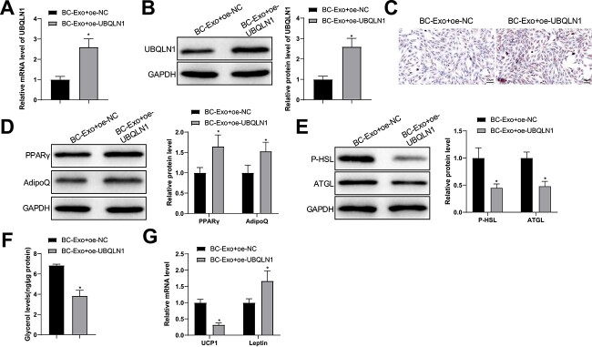
Cachexia occurrence and development are associated with loss of white adipose tissues, which may be involved with cancer-derived exosomes. This study attempted to characterize the functional mechanisms of breast cancer (BC) cell-derived exosome-loaded microRNA (miR)-155 in cancer cachexia-related fat loss. Exosomes were incubated with preadipocytes and cellular lipid droplet accumulation was observed using Oil Red O staining. Western blotting evaluated the cellular levels of lipogenesis marker peroxisome proliferator activated receptor gamma (PPARγ) and adiponectin, C1Q and collagen domain containing (AdipoQ). Differentiated adipocytes were incubated with exosomes, and phosphate hormone sensitive lipase (P-HSL), adipose triglyceride lipase (ATGL) and glycerol were detected in adipocytes, in addition to uncoupling protein 1 (UCP1) and leptin levels. A mouse model of cancer cachexia was established where cancer exosomes were injected intravenously. The changes in body weight and tumor-free body weights were recorded and serum glycerol levels and lipid accumulation in adipose tissues were determined. Also, the relationship between miR-155 and UBQLN1 was predicted and verified. BC exosome treatment reduced PPARγ and AdipoQ protein levels, promoted the levels of P-HSL and ATGL proteins, facilitated glycerol release, increased UCP1 expression and lowered leptin expression in adipocytes. Exosomal miR-155 inhibited lipogenesis in preadipocytes and boosted the browning of white adipose tissues. miR-155 downregulation alleviated cancer exosome-induced browning of white adipose tissues and fat loss. Mechanistically, miR-155 targeted UBQLN1, and UBQLN1 upregulation reversed the impacts of cancer exosomes. miR-155 loaded by BC cell-derived exosomes significantly affects white adipose browning and inhibition of cancer-derived exosomes.


Figures

Figure 1
Figure 1
BC cell-derived conditioned medium facilitates fat loss in adipocytes. (A) AchE activity in BC cell culture supernatant was tested after treatment with GW4869 for 24 h. After BC cell-derived conditioned medium was cultured with adipocytes. (B) Oil Red O staining was used to observe lipid droplet accumulation in the adipocytes; (C, D) the protein expression of PPARγ, AdipoQ (C), P-HSL and ATGL (D) was semi-quantified by western blotting; (E) free glycerin levels released by the adipocytes within 24 h were evaluated; (F) the mRNA levels of UCP1 and leptin were assessed using RT-qPCR. Measuring data were expressed as mean ± standard deviation. Comparisons among multiple groups were performed using the one-way analysis of variance test, and post hoc multiple comparisons were performed using Tukey’s multiple comparisons test. *P < 0.05.
Figure 2
Figure 2
BC cell-derived exosomes facilitate fat loss in adipocytes. (A) Exosomes isolated from BC cells were observed with a transmission electron microscope. (B) Particle size analysis of the isolated exosomes. (C) Western blotting was used to show the expression of CD81, CD63 and GM130. (D) 3T3-L1 cells was co-incubated with PKH-26-marked BC cells and observed with a fluorescence microscope. (E) Oil Red O staining was used to show lipid droplet accumulation in adipocytes. (F, G) Western blotting was used to quantify the expression of PPARγ and AdipoQ as well as P-HSL and ATGL in adipocytes. (H) The levels of free glycerol released by adipocytes within 24 h. (I) The mRNA levels of UCP1 and leptin were quantified by RT-qPCR. Measuring data were expressed as mean ± standard deviation. Comparisons between groups were performed using the t-test. *P < 0.05.
Figure 3
Figure 3
BC cell-derived exosomes promote adipose loss in mice with cancer cachexia. (A) BC cell-derived exosomes were internalized to mouse adipose tissues. (B) The changes in body weight of mice. (C) Tumor-free body weight. (D) The levels of serum glycerol. (E) Oil Red O staining was used to show lipid droplet accumulation in adipose tissues. (G) Western blotting was used to quantify the expression of serum PPARγ and AdipoQ as well as P-HSL and ATGL. (H) The mRNA levels of UCP1 and leptin were quantified by RT-qPCR. Measuring data were expressed as mean ± standard deviation. Comparisons among multiple groups were performed using the one- or two-way analysis of variance, followed by Tukey’s multiple comparisons test. *P < 0.05, #P < 0.05.
Figure 4
Figure 4
miR-155 loaded by BC cell-derived exosomes accelerates fat loss in adipocytes. (A) miR-155 levels in adipose tissues were quantified by RT-qPCR. (B) After BC cells were transduced with miR-155 inhibitor and exosomes were isolated, miR-155 levels in the exosomes were detected by RT-qPCR. (C) The isolated exosomes were cultured with adipocytes and miR-155 expression in these cells was evaluated by RT-qPCR. (D) Oil Red O staining was used to observe lipid droplet accumulation in the adipocytes; (E, F) the protein expression of PPARγ, AdipoQ (E), P-HSL and ATGL (F) was semi-quantified by western blotting; (G) free glycerin levels released by the adipocytes within 24 h were evaluated; (H) the mRNA levels of UCP1 and leptin were assessed using RT-qPCR. Measuring data were expressed as mean ± standard deviation. Comparisons among multiple groups were performed using the one-way analysis of variance test, and post hoc multiple comparisons were performed using Tukey’s multiple comparisons test. *P < 0.05.
Figure 5
Figure 5
miR-155 targets UBQLN1. (A) A binding site between miR-155 and UBQLN1 was predicted by starbase. (B, C) Dual-luciferase reporter assay (B) and RIP (C) demonstrated the interaction between miR-155 and UBQLN1. (DG) RT-qPCR (D, F) and western blotting (E, G) were used to test the levels of UBQLN mRNA and protein. Measuring data were expressed as mean ± standard deviation. Comparisons among multiple groups were performed using the one-way analysis of variance test, and post hoc multiple comparisons were performed using Tukey’s multiple comparisons test. *P < 0.05.
Figure 6
Figure 6
Exosomal miR-155 modulates fat loss in adipocytes by regulating UBQLN1 expression. (A, B) UBQLN1 levels in adipocytes were quantified by RT-qPCR (A) and western blotting (B). (C) Oil Red O staining was used to observe lipid droplet accumulation in the adipocytes; (D, E) the protein expression of PPARγ, AdipoQ (D), P-HSL and ATGL (E) was semi-quantified by western blotting; (F) free glycerin levels released by the adipocytes within 24 h were evaluated; (G) the mRNA levels of UCP1 and leptin were assessed using RT-qPCR. Measuring data were expressed as mean ± standard deviation. Comparisons among multiple groups were performed using the one-way analysis of variance test, and post hoc multiple comparisons were performed using Tukey’s multiple comparisons test. *P < 0.05.

References

    1. Nishikawa, H., Goto, M., Fukunishi, S., Asai, A., Nishiguchi, S. and Higuchi, K. (2021) Cancer cachexia: its mechanism and clinical significance. Int. J. Mol. Sci., 22(16), 22. - PMC - PubMed
    1. Zagzag, J., Hu, M.I., Fisher, S.B. and Perrier, N.D. (2018) Hypercalcemia and cancer: differential diagnosis and treatment. CA Cancer J. Clin., 68, 377–386. - PubMed
    1. Herremans, K.M., Riner, A.N., Cameron, M.E. and Trevino, J.G. (2019) The microbiota and cancer cachexia. Int. J. Mol. Sci., 20, 20. - PMC - PubMed
    1. Rybinska, I., Agresti, R., Trapani, A., Tagliabue, E. and Triulzi, T. (2020) Adipocytes in breast cancer, the thick and the thin. Cell, 9, 560–560. - PMC - PubMed
    1. Di, W., Zhang, W., Zhu, B., Li, X., Tang, Q. and Zhou, Y. (2021) Colorectal cancer prompted adipose tissue browning and cancer cachexia through transferring exosomal miR-146b-5p. J. Cell. Physiol., 236, 5399–5410. - PubMed

Publication types