Case Reports
. 2023 Apr 6;388(14):1318-1324.
doi: 10.1056/NEJMcps2210419.

An Alternate Explanation

Affiliations
Case Reports

An Alternate Explanation

Tom Alsaigh et al. N Engl J Med. .

Erratum in

  • An Alternate Explanation.
    [No authors listed] [No authors listed] N Engl J Med. 2023 Sep 21;389(12):1156. doi: 10.1056/NEJMx230005. N Engl J Med. 2023. PMID: 37733326 No abstract available.

Abstract

A 48-year-old man with long-standing type 2 diabetes mellitus (recent glycated hemoglobin level, 6.5%) and chronic kidney disease (baseline creatinine level, 3.3 mg per deciliter [292 μmol per liter]; glomerular filtration rate, 24 ml per minute per 1.73 m2 of body-surface area) presented to his primary care physician with a 3-month history of numbness, tingling, and faint violaceous discoloration of the tips of multiple fingers and toes. His physical examination showed reduced light-touch sensation in a glove-and-stocking distribution; the radial and pedal pulses were palpable. The vitamin B12 level was 260 pg per milliliter (192 pmol per liter; normal range, 190 to 950 pg per milliliter [140 to 701 pmol per liter]). He did not smoke tobacco, drink alcohol, or use illicit drugs. One month later, a nontraumatic wound developed on the left foot. The ankle–brachial index (ABI) was 1.2 on both sides (normal range, 0.91 to 1.3). Wound care was initiated for a presumed neuropathic ulcer.


Figures
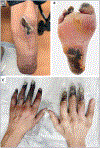
Figure 1.
Figure 1.. Photographs of Nonhealing and Necrotic Wounds.
Shown are clinical photographs of a nonhealing wound near the area of the left below-knee amputation (Panel A), necrotic wounds on the right heel and toes (Panel B), and necrotic wounds on the fingers of both hands (Panel C).
Figure 2.
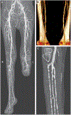
Figure 2.. CT Angiography with Runoff.
Panel A shows a two-dimensional maximum-intensity projection image of both legs (after the left below-knee amputation). Panel B shows a three-dimensional reconstruction of both superficial femoral arteries. Panel C shows a two-dimensional image of the right popliteal and below-knee arteries. L denotes left, and R right.
Figure 3.
Figure 3.. Biopsy Specimen of the Right Below-Knee Amputation.
Hematoxylin and eosin staining shows reactive capillary proliferation (arrow) and medial calcification in thick-walled vessels (arrowhead). (Image courtesy of Ryanne A. Brown, M.D.)
Figure 4.
Figure 4.. Ectopic Calcification in Arterial Calcification Due to Deficiency of CD73 (ACDC).
Shown is a depiction of the signaling cascade that leads to ectopic calcification in ACDC. Arrows show the change in CD73 and tissue-nonspecific alkaline phosphatase (TNAP) levels in ACDC. AMP denotes adenosine monophosphate.

Comment in

  • An Alternate Explanation.
    Cassini T, Montano C, Guan B. Cassini T, et al. N Engl J Med. 2023 Sep 28;389(13):1249-1250. doi: 10.1056/NEJMc2305289. N Engl J Med. 2023. PMID: 37754297 Free PMC article. No abstract available.
  • An Alternate Explanation.
    Boehm M, Brofferio A, St Hilaire C. Boehm M, et al. N Engl J Med. 2023 Sep 28;389(13):1250. doi: 10.1056/NEJMc2305289. N Engl J Med. 2023. PMID: 37754298 No abstract available.
  • An Alternate Explanation.
    O'Neill WC. O'Neill WC. N Engl J Med. 2023 Sep 28;389(13):1250-1251. doi: 10.1056/NEJMc2305289. N Engl J Med. 2023. PMID: 37754299 No abstract available.
  • An Alternate Explanation. Reply.
    Alsaigh T, Leeper NJ, Sayed N. Alsaigh T, et al. N Engl J Med. 2023 Sep 28;389(13):1251. doi: 10.1056/NEJMc2305289. N Engl J Med. 2023. PMID: 37754300 No abstract available.

References

    1. St Hilaire C. Medial arterial calcification: a significant and independent contributor of peripheral artery disease. Arterioscler Thromb Vasc Biol 2022;42:253–60. - PMC - PubMed
    1. Demer LL, Tintut Y. Vascular calcification: pathobiology of a multifaceted disease. Circulation 2008;117:2938–48. - PMC - PubMed
    1. Giachelli CM. Vascular calcification mechanisms. J Am Soc Nephrol 2004;15:2959–64. - PubMed
    1. Yerram P, Chaudhary K. Calcific uremic arteriolopathy in end stage renal disease: pathophysiology and management. Ochsner J 2014;14:380–5. - PMC - PubMed
    1. Nigwekar SU, Thadhani R, Branden-burg VM. Calciphylaxis. N Engl J Med 2018;378:1704–14. - PubMed

Publication types