Review
. 2023 Apr 27;66(8):5364-5376.
doi: 10.1021/acs.jmedchem.3c00097. Epub 2023 Apr 5.

Catalytically Active Snake Venom PLA2 Enzymes: An Overview of Its Elusive Mechanisms of Reaction

Affiliations
Review

Catalytically Active Snake Venom PLA2 Enzymes: An Overview of Its Elusive Mechanisms of Reaction

Juliana Castro-Amorim et al. J Med Chem. .

Abstract

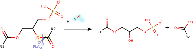
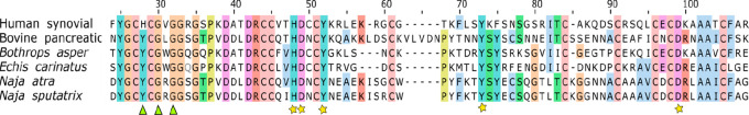
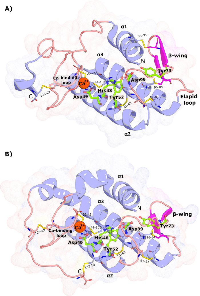
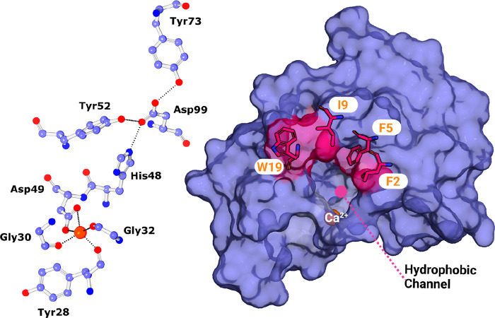
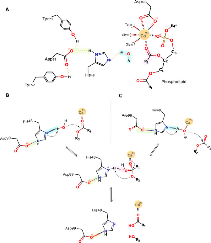
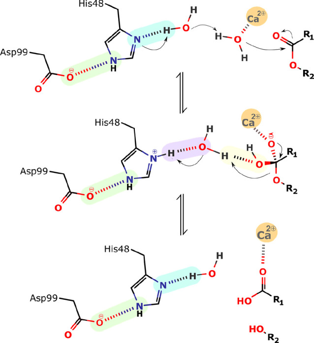
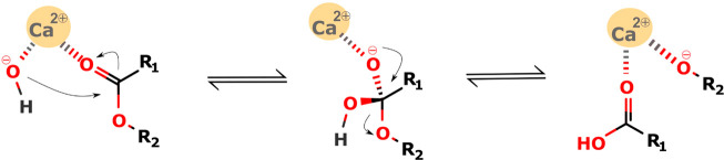
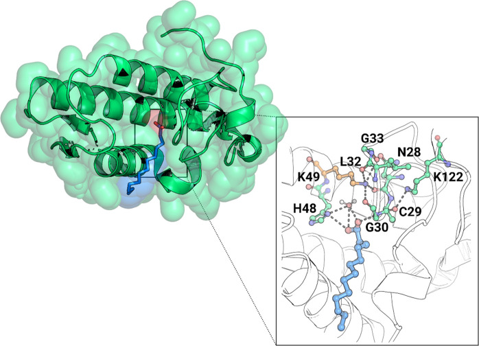
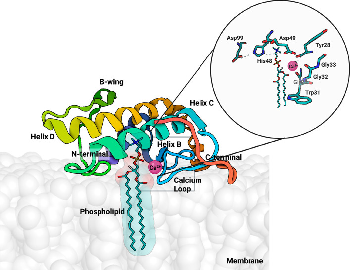
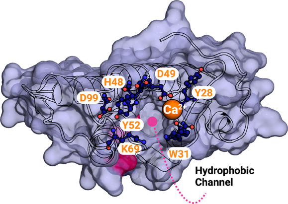
Snake venom-secreted phospholipase A2 (svPLA2) enzymes, both catalytically active and inactive, are a central component in envenoming. These are responsible for disrupting the cell membrane's integrity, inducing a wide range of pharmacological effects, such as the necrosis of the bitten limb, cardiorespiratory arrest, edema, and anticoagulation. Although extensively characterized, the reaction mechanisms of enzymatic svPLA2 are still to be thoroughly understood. This review presents and analyses the most plausible reaction mechanisms for svPLA2, such as the "single-water mechanism" or the "assisted-water mechanism" initially proposed for the homologous human PLA2. All of the mechanistic possibilities are characterized by a highly conserved Asp/His/water triad and a Ca2+ cofactor. The extraordinary increase in activity induced by binding to a lipid-water interface, known as "interfacial activation," critical for the PLA2s activity, is also discussed. Finally, a potential catalytic mechanism for the postulated noncatalytic PLA2-like proteins is anticipated.


Conflict of interest statement

The authors declare no competing financial interest.

Figures

Figure 1
Figure 1
The chemical reaction catalyzed by PLA2s. These enzymes act at the sn-2 position of glycerophospholipids and hydrolyze the ester bond releasing a lysophospholipid and a free fatty acid. R1 and R2 correspond to the fatty acid tails.
Figure 2
Figure 2
Sequence alignment of six secreted phospholipase A2 from different sources: human synovial (UniProtKB AC: P14555), bovine pancreatic (UniProtKB AC: P00593), B. asper (UniProtKB AC: P20474), E. carinatus (UniProtKB AC: Q7T3S7), N. atra (UniProtKB AC: P00598) and N. sputatrix (UniProtKB AC: Q92085). Overall, highly conserved residues can be found in the active site, Ca2+ binding loop and disulfide bonds. Amino acids residues that have an identity threshold above 40% are colored according to the ClustalX color scheme: hydrophobic (blue), positive charge (red), negative charge (magenta), polar (green), cysteines (pink), glycines (orange), prolines (yellow), aromatic (cyan), and unconserved (white). The numbering shown is that from Renetseder et al. Green triangles indicate the locations of the residues involved in the calcium-binding, while yellow stars indicate those involved in the active site.
Figure 3
Figure 3
| (A) Structure of the Chinese cobra svPLA2-IA (PDB 1POA) and (B) the Indian saw-scaled viper svPLA2-IIA (PDB 1OZ6). The active site residues (His48, Asp49, Tyr52, Tyr73, and Asp99) are shown as green sticks, the Ca2+ as an orange sphere, and the disulfide bonds as yellow lines. N- and C-terminal regions are also identified. The similarity in the folding is evident. The PyMOL molecular graphics software package was used to generate the representations.
Figure 4
Figure 4
| (left) Ball and stick representation of the residues involved in the catalytic network and respective hydrogen-bonding (dashed lines). (right) Surface representation of the GIA-PLA2 isolated from N. atra (PDB 1POA) and stick representation of the residues that constitute the hydrophobic channel. The PyMOL molecular graphics software package was used to generate the representations.
Figure 5
Figure 5
General scheme of the single-water mechanism. (A) The Ca2+ coordination shell and the catalytically relevant residues. Their representation will be simplified in the following schemes for simplicity: (B) Verheij proposal, (C) our proposal, with one calcium-bound water. Step 1: His48 abstracts a proton from the incoming water, which initiates a nucleophilic attack on the sn-2 carbonyl carbon of the substrate. Step 2: The produced tetrahedral intermediate oxyanion collapses, eliminating the alkoxyl group, which deprotonates His48. Step 3: Products release; His48 is stabilized by Asp99, which additionally forms hydrogen bonds with Tyr52 and Tyr73. The oxyanion hole that stabilizes the transition state after the nucleophilic attack is formed by the backbone HN group of Gly30 and the Ca2+ ion.
Figure 6
Figure 6
Schematic representation of the assisted-water mechanism proposed by Yu et al. Step 1: His48, which acts as a general base catalyst, abstracts a proton from the second water, which deprotonates the calcium-bound water molecule. This leads to the nucleophilic attack by the calcium-bound water on the carbonyl carbon of the substrate and the formation of the tetrahedral intermediate. Step 2: The departing alcoholate leaving group is protonated by the second water, which is itself protonated by His 48. Step 3: Collapses and the products are released.
Figure 7
Figure 7
Schematic representation of the hydroxide direct nucleophilic attack mechanism; Step 1: The hydroxide nucleophile attacks at the electrophilic C of the ester C=O leading to the formation of the tetrahedral intermediate. Step 2: The intermediate collapses, reforming the C=O. Step 3: Departure of the alkoxide leaving group, RO.
Figure 8
Figure 8
Cartoon representation of the complex between Bothrops pirajai PrTX-II and a fatty acid (tridecanoic acid) (PDB 1QLL). A close-up view of the binding site of PrTX-II, with the fatty acid molecule stabilized by a hydrogen bond with the Cys29–Gly30 peptide bond, which is hyperpolarized by the Lys122, increasing its affinity for the fatty acid. The Ca2+ binding loop interactions with the Nε atom of the Lys49 residue are also shown. PyMOL molecular graphics software package was used to generate the representations.
Figure 9
Figure 9
Representation of the svPLA (PDB 5TFV) with the phospholipid bilayer membrane, created using the CHARMM-GUI web interface. The protein orientation in the membrane was automatically set up with the PPM 2.0 server, and the substrate was manually inserted. In the active center, there is a bound phospholipid substrate in which the color red represents oxygen, phosphorus is brown, nitrogen is blue, and carbon is white; the enzyme is shown in cartoon representation, and the Ca2+ ion is shown in dark pink. Binding to the membrane makes the enzyme more active for several orders of magnitude. PyMOL molecular graphics software package was used to generate the representations.
Figure 10
Figure 10
Surface representation of the E. carinatus svPLA2-IIA (PDB 1OZ6) hydrophobic channel, with the Trp31 and Lys69 residues in evidence. Catalytic residues are also indicated. The calcium ion is represented as an orange sphere. PyMOL molecular graphics software package was used to generate the representations.

References

    1. Oliveira A. L.; Viegas M. F.; da Silva S. L.; Soares A. M.; Ramos M. J.; Fernandes P. A. The chemistry of snake venom and its medicinal potential. Nat. Rev. Chem. 2022, 6, 451.10.1038/s41570-022-00393-7. - DOI - PMC - PubMed
    1. Williams D. J.; Faiz M. A.; Abela-Ridder B.; Ainsworth S.; Bulfone T. C.; Nickerson A. D.; Habib A. G.; Junghanss T.; Fan H. W.; Turner M.; Harrison R. A.; Warrell D. A. Strategy for a globally coordinated response to a priority neglected tropical disease: Snakebite envenoming. PLoS Negl. Trop. Dis. 2019, 13, e000705910.1371/journal.pntd.0007059. - DOI - PMC - PubMed
    1. Guidelines for the Production, Control and Regulation of Snake Antivenom Immunoglobulins; World Health Organization: Geneva, 2009.
    1. Mukherjee A. K.; Mackessy S. P. Prevention and improvement of clinical management of snakebite in Southern Asian countries: a proposed road map. Toxicon 2021, 200, 140–152. 10.1016/j.toxicon.2021.07.008. - DOI - PubMed
    1. Puzari U.; Mukherjee A. K. Recent developments in diagnostic tools and bioanalytical methods for analysis of snake venom: A critical review. Anal. Chim. Acta 2020, 1137, 208–224. 10.1016/j.aca.2020.07.054. - DOI - PubMed

Publication types

LinkOut - more resources