. 2023 Jun:62:102691.
doi: 10.1016/j.redox.2023.102691. Epub 2023 Mar 29.

STING mediates hepatocyte pyroptosis in liver fibrosis by Epigenetically activating the NLRP3 inflammasome

Affiliations

STING mediates hepatocyte pyroptosis in liver fibrosis by Epigenetically activating the NLRP3 inflammasome

Yang Xiao et al. Redox Biol. 2023 Jun.

Abstract

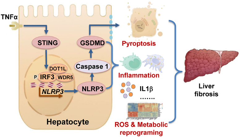
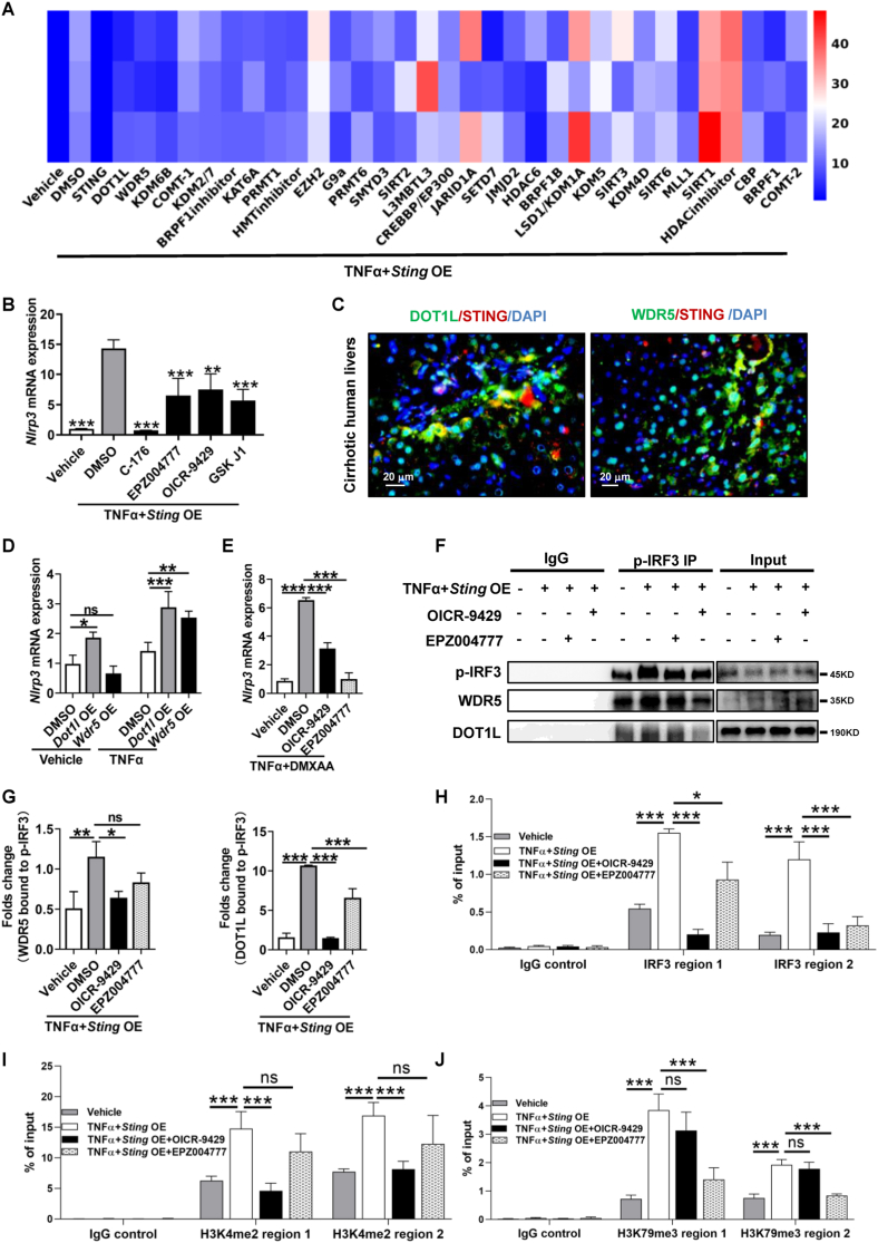
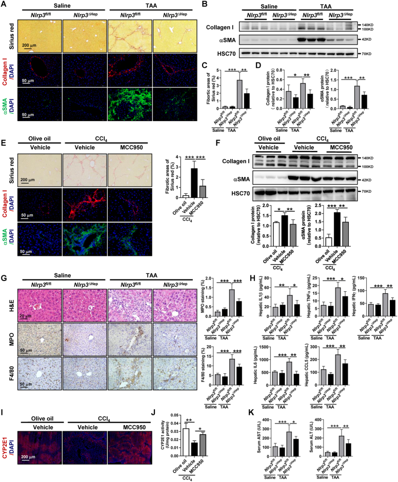
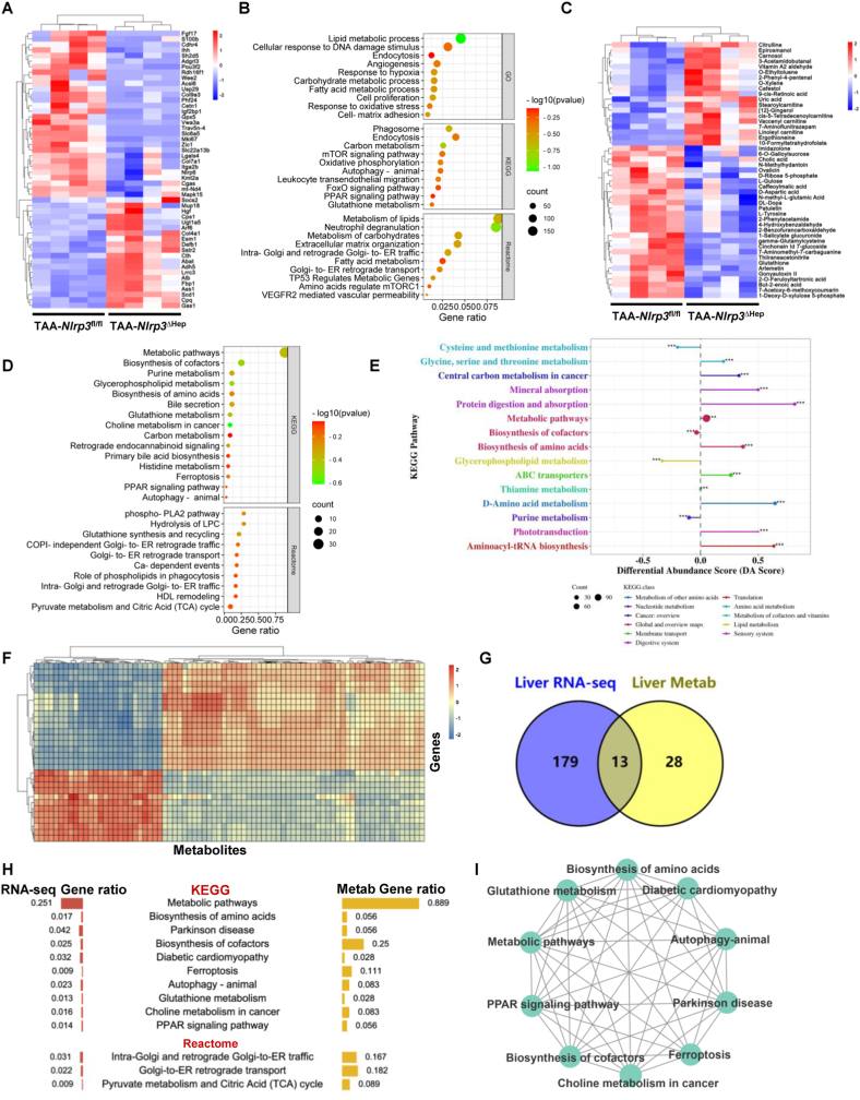
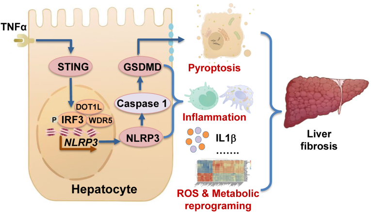
The activation of stimulator of interferon genes (STING) and NOD-like receptor protein 3 (NLRP3) inflammasome-mediated pyroptosis signaling pathways represent two distinct central mechanisms in liver disease. However, the interconnections between these two pathways and the epigenetic regulation of the STING-NLRP3 axis in hepatocyte pyroptosis during liver fibrosis remain unknown. STING and NLRP3 inflammasome signaling pathways are activated in fibrotic livers but are suppressed by Sting knockout. Sting knockout ameliorated hepatic pyroptosis, inflammation, and fibrosis. In vitro, STING induces pyroptosis in primary murine hepatocytes by activating the NLRP3 inflammasome. H3K4-specific histone methyltransferase WD repeat-containing protein 5 (WDR5) and DOT1-like histone H3K79 methyltransferase (DOT1L) are identified to regulate NLRP3 expression in STING-overexpressing AML12 hepatocytes. WDR5/DOT1L-mediated histone methylation enhances interferon regulatory transcription factor 3 (IRF3) binding to the Nlrp3 promoter and promotes STING-induced Nlrp3 transcription in hepatocytes. Moreover, hepatocyte-specific Nlrp3 deletion and downstream Gasdermin D (Gsdmd) knockout attenuate hepatic pyroptosis, inflammation, and fibrosis. RNA-sequencing and metabolomics analysis in murine livers and primary hepatocytes show that oxidative stress and metabolic reprogramming might participate in NLRP3-mediated hepatocyte pyroptosis and liver fibrosis. The STING-NLRP3-GSDMD axis inhibition suppresses hepatic ROS generation. In conclusion, this study describes a novel epigenetic mechanism by which the STING-WDR5/DOT1L/IRF3-NLRP3 signaling pathway enhances hepatocyte pyroptosis and hepatic inflammation in liver fibrosis.

Keywords: GSDMD; Histone methylation; IRF3; Liver cirrhosis; Metabolic reprogramming; Oxidative stress.


Conflict of interest statement

Declaration of competing interest All authors declare no conflict of interest.

Figures

Image 1
Graphical abstract
Fig. 1
Fig. 1
STING knockout and pharmacological inhibition attenuate liver fibrosis and hepatic inflammation (A-B) Wild-type mice were injected with olive oil or CCl4 for 6 weeks. Enriched signaling pathway analysis of upregulated (CCl4vs. olive oil) DEGs (A) and the heatmap of target DEGs (B) from RNA-sequencing were analyzed (n = 3/group). (C) Seven normal human livers and 11 cirrhotic human livers were collected. The distribution and protein levels of STING, t-IRF3, and p-IRF3 were analyzed by IHC or WB. (DG)Sting+/+ and Sting−/− mice were injected with olive oil or CCl4 for 6 weeks (n = 6/group). Liver fibrosis was analyzed by Sirius red staining, IF, and WB for collagen I and αSMA. (H) RNA-sequencing was performed in CCl4-treated Sting+/+ and Sting−/− mice. Enriched pathway analysis of downregulated DEGs (Sting−/−vs. Sting+/+) was performed (n = 3/group). (IL) Wild-type mice were treated with either olive oil or CCl4 for 6 weeks in addition to either vehicle or STING inhibitor C-176 (20 mg/kg, n = 6/group). Liver fibrosis was analyzed by Sirius red staining, IF, and WB for collagen I and αSMA. (MN) The protein levels and activity of CYP2E1 were detected by IF (M) and activity kit (N). *p < 0.05, **p < 0.01, ***p < 0.001. (For interpretation of the references to colour in this figure legend, the reader is referred to the Web version of this article.)
Fig. 2
Fig. 2
STING induces hepatocyte pyroptosis by activating the NLRP3 inflammasome (A) Wild-type mice were injected with olive oil or CCl4 for 6 weeks. Colocalization of STING and NLRP3 was assessed by IF. (B) IF was used to analyze the colocalization of STING and NLRP3 in human cirrhotic livers. (C–F) Primary hepatocytes isolated from wild-type mice were treated with DMSO or the STING inhibitor C-176 for 2 h in addition to either vehicle or TNFα+DMXAA for an additional 6 h. Colocalization of STING and NLRP3 was determined by IF (C). Morphological changes, SEM, and colocalization of Caspase 1/PI were performed (D). The protein levels of STING, p-IRF3, cl-Caspase 1, and cl-GSDMD were determined by WB (E), and the quantitative data are shown (F). (G) Primary hepatocytes isolated from Sting+/+ and Sting−/− mice were treated with vehicle or TNFα+DMXAA for 6 h. The morphological changes, SEM, and colocalization of Caspase 1/PI were analyzed. n = 3/group, *p < 0.05, **p < 0.01, ***p < 0.001.
Fig. 3
Fig. 3
STING promotes IRF3 recruitment to the Nlrp3 promoter region in hepatocytes (A) Primary hepatocytes were isolated from Sting+/+ and Sting−/− mice, and the mRNA level of Nlrp3 was then quantified by qPCR. (B) AML12 hepatocytes were transfected with empty plasmid or Sting overexpression plasmid (OE), and the mRNA level of Nlrp3 was determined by qPCR. (CD) Primary hepatocytes isolated from wild-type mice were treated with the STING agonist DMXAA (50 μg/mL) or IRF3 agonist KIN1148 (10 μg/mL) for 6 h qPCR was applied to evaluate the mRNA levels of Sting, Nlrp3, and Il18 (C), and the protein levels of NLRP3 were assessed by WB (D). (EF) Primary hepatocytes isolated from wild-type mice were treated with DMSO, the STING inhibitor C-176 (2 μM, E) or the NFκB inhibitor celastrol (50 nM, F) for 2 h in addition to either vehicle or TNFα (25 ng/mL)+DMXAA (50 μg/mL) for an additional 6 h. The mRNA levels of Nlrp3 and Il18 were analyzed by qPCR. (G) Primary hepatocytes isolated from wild-type mice were treated with the IRF3 agonist KIN1148 (10 μg/mL) for 6 h qPCR was applied to evaluate the mRNA levels of Sting, Nlrp3, and Il18. (H) Primary hepatocytes isolated from wild-type mice were treated with DMSO or TNFα (25 ng/mL)+DMXAA (50 μg/mL) for 6 h, and then colocalization of STING and p-IRF3 was determined by IF. (I) Primary hepatocytes isolated from wild-type mice were treated with DMSO, DMXAA, KIN1148, TNFα+DMXAA or TNFα+DMXAA + C-176 for 6 h. Then, p-IRF3 levels at Nlrp3 promoter regions were evaluated by ChIP for p-IRF3 followed by qPCR. n = 3/group, ns: not significant, *p < 0.05, **p < 0.01, ***p < 0.001.
Fig. 4
Fig. 4
STING increases IRF3 recruitment at Nlrp3 promoter regions by promoting histone methylation via WDR5/DOT1L in AML12 hepatocytes (A-B) A selected histone-modifying enzyme compound system with 32 targets was applied to test Nlrp3 inhibition in TNFα+Sting-overexpressing AML12 hepatocytes. The heatmap of Nlrp3 expression was analyzed by qPCR (A). Inhibitors that suppress Nlrp3 upregulation are shown (B). (C) IF was used to analyze the colocalization of STING and DOT1L or WDR5 in human cirrhotic livers. (D) AML12 hepatocytes transfected with the Dot1l or Wdr5 overexpression plasmid were treated with vehicle, TNFα (25 ng/mL) for 6 h. The mRNA level of Nlrp3 was analyzed by qPCR. (E) AML12 hepatocytes were treated with DMSO, the DOT1L inhibitor EPZ004777 (7 μM) or the WDR5 inhibitor OICR-9429 (7 μM) for 2 h in addition to either vehicle or TNFα (25 ng/mL)+DMXAA (50 μg/mL) for an additional 6 h. The mRNA level of Nlrp3 was analyzed by qPCR. (FG) AML12 hepatocytes transfected with the Sting overexpression plasmid were treated with vehicle, TNFα plus OICR-9429 or EPZ004777 for 6 h. Then, cell lysates of nuclear fractionation were subjected to co-IP against p-IRF3, and p-IRF3, WDR5, and DOT1L were then evaluated by WB. (HJ) AML12 hepatocytes transfected with the Sting overexpression plasmid were treated with vehicle, TNFα and TNFα plus OICR-9429 or EPZ004777 for 6 h. Then, p-IRF3 (H), H3K4me2 (I), and H3K79me3 (J) levels at Nlrp3 promoter regions were evaluated by ChIP‒qPCR. n = 3/group. ns: not significant, *p < 0.05, **p < 0.01, ***p < 0.001.
Fig. 5
Fig. 5
Hepatocyte NLRP3 deficiency and NLRP3 pharmacological inhibition ameliorate hepatic inflammation and liver fibrosis (A-D)Nlrp3fl/fl and Nlrp3ΔHep mice were injected with normal saline or TAA for 8 weeks. Liver fibrosis was analyzed by Sirius red staining, IF, and WB for collagen I and αSMA. (EF) Wild-type mice were treated with either olive oil or CCl4 for 6 weeks in addition to either vehicle or NLRP3 inhibitor MCC950 (20 mg/kg, n = 6/group). Liver fibrosis was analyzed by Sirius red staining, IF, and WB for collagen I and αSMA. (G) Hepatic inflammation was analyzed by H&E staining, IHC for the neutrophil marker MPO, and the macrophage marker F4/80, and the quantitative data are shown. (H) Hepatic IL1β, TNFα, IFNγ, IL6, and CCL5 levels were quantified by the Milliplex mouse multiplex assay. (IJ) The protein levels and activity of CYP2E1 were detected by IF (I) and activity kit (J). (K) Liver function was analyzed by serum AST and ALT. n = 6/group. *p < 0.05, **p < 0.01, ***p < 0.001. (For interpretation of the references to colour in this figure legend, the reader is referred to the Web version of this article.)
Fig. 6
Fig. 6
Hepatocyte-specific Nlrp3 deletion attenuates liver fibrosis via metabolic reprogramming (A-E) RNA-sequencing and metabolomics of livers from Nlrp3fl/fl and Nlrp3ΔHep mice injected with TAA for 8 weeks (n = 4/group). RNA-sequencing was performed to show the heatmap (A) and the enriched signaling pathways (B) of DEGs. Ultra HPLC-based metabolomics of livers was applied to reveal the heatmap (C), the enriched signaling pathway (D), and the KEGG metabolic pathway-based differential abundance analysis (E) of differential metabolites. (F–I) Integrative multiomics analysis of the transcriptome and metabolomics. Hierarchical clustering for canonical correlation analysis of the mRNAs and metabolomics (F). Venn diagram (G) and ontology analysis (H) displaying the shared signaling pathways between the transcriptome and metabolomics. Functional enrichment analysis was then performed to gain further insights into the KEGG of DEGs and metabolites (I).
Fig. 7
Fig. 7
STING-NLRP3-GSDMD axis inhibition suppresses hepatic ROS generation (A-B) Wild-type mice were treated with either olive oil or CCl4 for 6 weeks in addition to either vehicle STING inhibitor C-176 (20 mg/kg) or NLRP3 inhibitor MCC950 (20 mg/kg). Hepatic O was analyzed by DHE. (CD)Sting+/+ and Sting−/− mice were injected with olive oil or CCl4 for 6 weeks. Nlrp3fl/fl and Nlrp3ΔHep mice were injected with normal saline or TAA for 8 weeks. The protein level of 4HNE was analyzed by WB. (E) The levels of GSH in the liver were quantified. (F)Gsdmd+/+ and Gsdmd−/− mice were injected with normal saline or TAA for 8 weeks. The protein level of 4HNE was analyzed by WB. n = 6/group. *p < 0.05, **p < 0.01, ***p < 0.001.
Fig. 8
Fig. 8
Proposed mechanisms STING increases NLPR3 and NLRP3 inflammasome-mediated hepatocyte pyroptosis during liver injury via epigenetic regulation of the IRF3/WDR5/DOT1L transcription activator complex. STING-NLRP3 pathway leads to liver fibrosis by increasing hepatocyte pyroptosis, hepatic inflammation, and metabolic reprogramming.

References

    1. Brenner C., Galluzzi L., Kepp O., Kroemer G. Decoding cell death signals in liver inflammation. J. Hepatol. 2013;59:583–594. doi: 10.1016/j.jhep.2013.03.033. - DOI - PubMed
    1. Ibrahim S.H., Hirsova P., Malhi H., Gores G.J. Nonalcoholic steatohepatitis promoting kinases. Semin. Liver Dis. 2020;40:346–357. doi: 10.1055/s-0040-1713115. - DOI - PMC - PubMed
    1. Schuster S., Cabrera D., Arrese M., Feldstein A.E. Triggering and resolution of inflammation in NASH. Nat. Rev. Gastroenterol. Hepatol. 2018;15:349–364. doi: 10.1038/s41575-018-0009-6. - DOI - PubMed
    1. Kisseleva T., Brenner D. Molecular and cellular mechanisms of liver fibrosis and its regression. Nat. Rev. Gastroenterol. Hepatol. 2021;18:151–166. doi: 10.1038/s41575-020-00372-7. - DOI - PubMed
    1. Asrani S.K., Devarbhavi H., Eaton J., Kamath P.S. Burden of liver diseases in the world. J. Hepatol. 2019;70:151–171. doi: 10.1016/j.jhep.2018.09.014. - DOI - PubMed

Publication types