. 2023 Apr 5;13(1):5598.
doi: 10.1038/s41598-023-32679-1.

Post-reperfusion acute MR diffusion in stroke is a potential predictor for clinical outcome in rats

Affiliations

Post-reperfusion acute MR diffusion in stroke is a potential predictor for clinical outcome in rats

Szilvia Anett Nagy et al. Sci Rep. .

Abstract

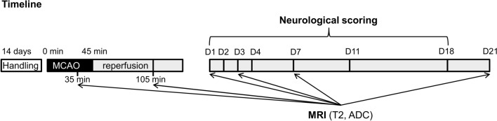
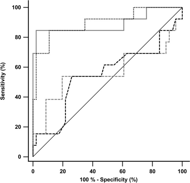
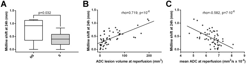
Middle cerebral artery occlusion (MCAO) models show substantial variability in outcome, introducing uncertainties in the evaluation of treatment effects. Early outcome predictors would be essential for prognostic purposes and variability control. We aimed to compare apparent diffusion coefficient (ADC) MRI data obtained during MCAO and shortly after reperfusion for their potentials in acute-phase outcome prediction. Fifty-nine male rats underwent a 45-min MCAO. Outcome was defined in three ways: 21-day survival; 24 h midline-shift and neurological scores. Animals were divided into two groups: rats surviving 21 days after MCAO (survival group, n = 46) and rats dying prematurely (non-survival/NS group, n = 13). At reperfusion, NS group showed considerably larger lesion volume and lower mean ADC of the initial lesion site (p < 0.0001), while during occlusion there were no significant group differences. At reperfusion, each survival animal showed decreased lesion volume and increased mean ADC of the initial lesion site compared to those during occlusion (p < 10-6), while NS group showed a mixed pattern. At reperfusion, lesion volume and mean ADC of the initial lesion site were significantly associated with 24 h midline-shift and neurological scores. Diffusion MRI performed soon after reperfusion has a great impact in early-phase outcome prediction, and it works better than the measurement during occlusion.


Conflict of interest statement

The authors declare no competing interests.

Figures

Figure 1
Figure 1
Schematic representation of experimental protocol. MCAO middle cerebral artery occlusion, D1-21 Day 1–21, T2 T2-weighted imaging, ADC apparent diffusion coefficient measurement; black color represents the 45-min period of filament occlusion; gray indicates the 21 day period after reperfusion.
Figure 2
Figure 2
Apparent diffusion coefficient (ADC) maps show larger ischemic stroke volume at reperfusion in rats not surviving ischemia–reperfusion injury. (A) Representative ADC maps measured from a survival (S) rat (surviving ischemia–reperfusion injury) and from a non-survival (NS) rat (died at 48 h after reperfusion) during left middle cerebral artery occlusion (MCAO) and at reperfusion. (B) Summary data of differences between survival (S) and non-survival (NS) rats (n = 46, n = 13 respectively) in ADC lesion volume (mm3) during occlusion of left middle cerebral arteries and at reperfusion. (C) Differences between survival (S) and non-survival (NS) rats in mean ADC of the initial lesion site (mm2/s × 10–4) during occlusion of left middle cerebral arteries and at reperfusion. Whiskers are set at minimum and maximum, the horizontal line marks the median, whereas box indicates the interquartile range (25–75%). 2-sided exact p-values are based on Mann–Whitney U-test.
Figure 3
Figure 3
Receiver operating characteristic (ROC) curves of the acute stage diffusion measures for identification of animals dying prematurely. ROC curves of lesion volume during occlusion (dashed gray line), lesion volume at reperfusion (solid gray line), mean ADC of the initial lesion site during occlusion (dashed black line) and mean ADC of the initial lesion site at reperfusion (dotted black line) are shown.
Figure 4
Figure 4
Cerebral midline-shift (assessed 24 h post injury) is increased in rats not surviving ischemia–reperfusion injury, which correlates with lesion volumes calculated by apparent diffusion coefficient (ADC) maps at reperfusion. (A) Summary data of differences between survival (S) and non-survival (NS) rats (n = 46, n = 5 respectively) in midline-shift (mm) measured 24 h post-reperfusion. Whiskers are set at minimum and maximum, the horizontal line marks the median, whereas box indicates the interquartile range (25–75%). 2-sided exact p-value is based on Mann–Whitney U-test. (B) Correlation between ADC lesion volume (mm3) at reperfusion and midline-shift (mm) measured 24 h post-reperfusion in the studied rats (n = 51). rho = Spearman’s rank correlation coefficient. (C) Correlation between mean ADC of the initial lesion site at reperfusion (mm2/s × 10–4) and midline-shift (mm) measured 24 h post-perfusion in the studied animals (n = 51). rho = Spearman’s rank correlation coefficient.
Figure 5
Figure 5
Neurological deficit (assessed 24 h post injury) is increased in rats not surviving ischemia–reperfusion injury, which correlates with lesion volumes calculated by apparent diffusion coefficient (ADC) maps at reperfusion. (A) Differences between survival (S) and non-survival (NS) rats (n = 46, n = 8 respectively) in general, focal and total neurological deficit scores assessed at 24 h after reperfusion. Whiskers are set at minimum and maximum, the horizontal line marks the median, whereas box indicates the interquartile range (25–75%). 2-sided exact p-values are based on Mann–Whitney U-test. (B) Correlation between ADC lesion volume (mm3) at reperfusion and total deficit measured 24 h post-reperfusion in the studied rats (n = 54). rho = Spearman’s rank correlation coefficient. (C) Correlation between mean ADC of the initial lesion site at reperfusion (mm2/s × 10–4) and total deficit measured 24 h post-reperfusion in the studied rats (n = 54). rho = Spearman’s rank correlation coefficient.

References

    1. Go AS, et al. Heart disease and stroke statistics—2014 update: A report from the American Heart Association. Circulation. 2014;129:e28–e292. doi: 10.1161/01.cir.0000441139.02102.80. - DOI - PMC - PubMed
    1. Luengo-Fernandez R, Violato M, Candio P, Leal J. Economic burden of stroke across Europe: A population-based cost analysis. Eur. Stroke J. 2020;5:17–25. doi: 10.1177/2396987319883160. - DOI - PMC - PubMed
    1. Hankey GJ. Secondary stroke prevention. Lancet Neurol. 2014;13:178–194. doi: 10.1016/S1474-4422(13)70255-2. - DOI - PubMed
    1. Wafa HA, et al. Burden of stroke in Europe: Thirty-year projections of incidence, prevalence, deaths, and disability-adjusted life years. Stroke. 2020;51:2418–2427. doi: 10.1161/STROKEAHA.120.029606. - DOI - PMC - PubMed
    1. Ng YS, Stein J, Ning M, Black-Schaffer RM. Comparison of clinical characteristics and functional outcomes of ischemic stroke in different vascular territories. Stroke. 2007;38:2309–2314. doi: 10.1161/STROKEAHA.106.475483. - DOI - PubMed

Publication types