. 2023 May;37(5):988-1005.
doi: 10.1038/s41375-023-01877-1. Epub 2023 Apr 5.

The KMT2A recombinome of acute leukemias in 2023

Affiliations

The KMT2A recombinome of acute leukemias in 2023

C Meyer et al. Leukemia. 2023 May.

Abstract

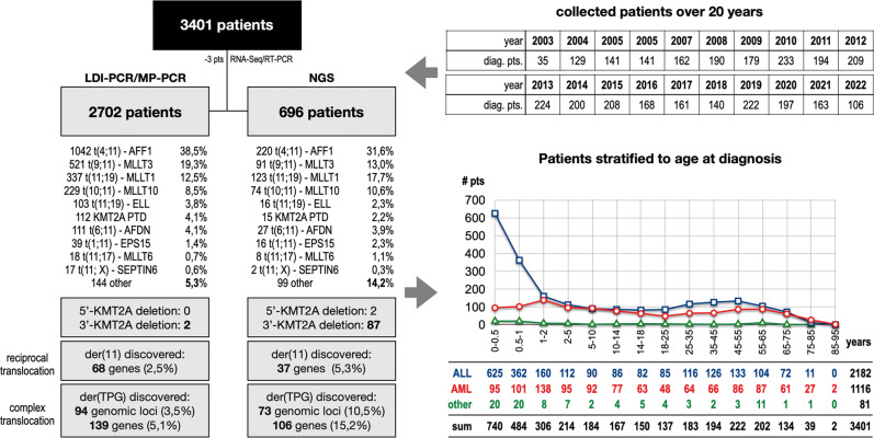
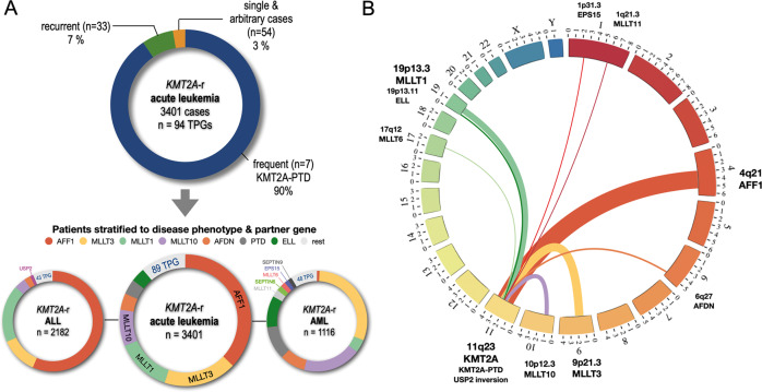
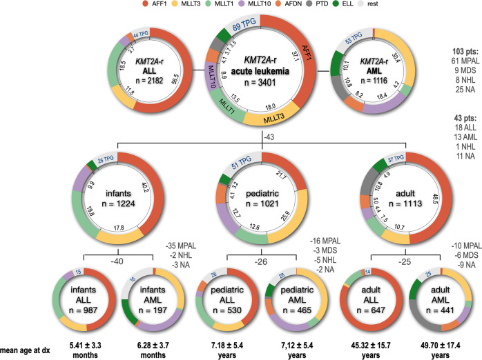
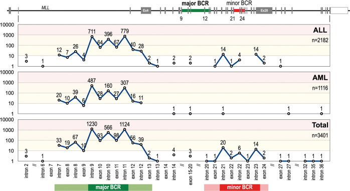
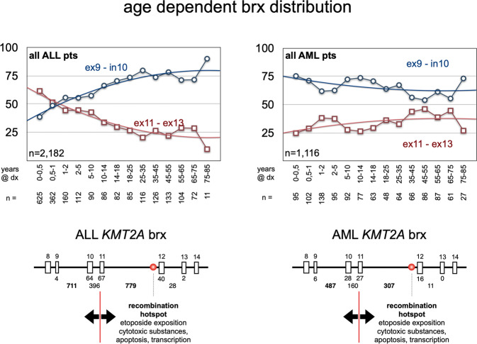
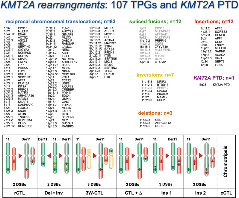
Chromosomal rearrangements of the human KMT2A/MLL gene are associated with de novo as well as therapy-induced infant, pediatric, and adult acute leukemias. Here, we present the data obtained from 3401 acute leukemia patients that have been analyzed between 2003 and 2022. Genomic breakpoints within the KMT2A gene and the involved translocation partner genes (TPGs) and KMT2A-partial tandem duplications (PTDs) were determined. Including the published data from the literature, a total of 107 in-frame KMT2A gene fusions have been identified so far. Further 16 rearrangements were out-of-frame fusions, 18 patients had no partner gene fused to 5'-KMT2A, two patients had a 5'-KMT2A deletion, and one ETV6::RUNX1 patient had an KMT2A insertion at the breakpoint. The seven most frequent TPGs and PTDs account for more than 90% of all recombinations of the KMT2A, 37 occur recurrently and 63 were identified so far only once. This study provides a comprehensive analysis of the KMT2A recombinome in acute leukemia patients. Besides the scientific gain of information, genomic breakpoint sequences of these patients were used to monitor minimal residual disease (MRD). Thus, this work may be directly translated from the bench to the bedside of patients and meet the clinical needs to improve patient survival.


Conflict of interest statement

The authors declare no competing interests.

Figures

Fig. 1
Fig. 1. Overview of all analyzed patients.
In the last 20 years a total of 3401 acute leukemia patients have been analyzed at the DCAL. Most of these patients were analyzed by the described LDI-PCR technology (2702 patients), a technique that has been successfully substituted by targeted NGS over the last years (696 pts). The benefit of using NGS over LDI-PCR (see left part) is clearly shown by the differences in novel target gene identification (15.2% vs. 5.1%), as well as the identification of 5’- and 3’-KMT2A deletions, which is per se impossible by using PCR-based techniques. The age stratification of all investigated patients is shown on the right bottom, clearly indicating that patients with ALL below 1 year of age at diagnosis represent a unique patient population with a high incidence for KMT2A rearrangements.
Fig. 2
Fig. 2. Classification of patients according to TPG and disease phenotype.
A The 7 most frequent KMT2A fusion partners (AFF1, MLLT3, MLLT1, MLLT10, AFDN, KMT2A -PTDs and ELL) represented more than 90% of the investigated patients. The next 33 recurrently diagnosed fusion partners represented 7% of cases, while all 54 unique fusion partners represent only 3% of the cohort (n = 3401). This patient cohort was divided into 2182 ALL (left) and 1116 AML patients (right). The 7 most frequently diagnosed fusion partners are color coded as indicated on top of the circular plots. Noteworthy, AFF1, MLLT1 and MLLT3 represented 87% of all diagnosed ALL patients. KMT2A::USP2 fusion were solely diagnosed in the ALL patient group and are indicated separately. Within the AML group, the fusion partners MLLT3, MLLT1, MLLT10, AFDN, KMT2A-PTDs and ELL cases accounted for 82% of diagnosed patients. Genes like MLLT11, SEPTIN6, MLLT6, EPS15 and SEPTIN 9 account for additional 8.3% of cases and are indicated separately. B Circos plot for the 11 most frequent KMT2A fusion partner genes: EPS15, MLLT11, AFF1, AFDN, MLLT3, MLLT10, KMT2A-PTDs, USP2, MLLT6, ELL and MLLT1 (sorted according to their chromosomal order).
Fig. 3
Fig. 3. Classification of all fusion partner genes by disease phenotype and age classification.
All 3401 diagnosed patients were grouped by their diagnosed disease type (ALL: 2182; AML: 1116; 103 pts had other diseases listed on the right). Since we had for 43 pts no age information at diagnosis, they were excluded from being further subdivided into the age groups infant (n = 1224), pediatric (n = 1021) and adult patients (n = 1113). All 3 age groups were again subdivided in ALL or AML subgroups (infant ALL = 987 pts; infant AML = 197 pts; pediatric ALL = 530 pts; pediatric AML = 465 pts; adult ALL = 647 pts; adult AML = 441 pts). Number of patients with missing information or different disease subtypes are indicated (grey letters). The mean age for all 6 subgroups is given below, either in months or years ± SD. The distribution of the 7 most frequent fusion partners is given by different colors (color code on top) and their frequency in percent. The additional number of identified fusion partner genes are given by blue numbers for each subgroup.
Fig. 4
Fig. 4. Breakpoint distribution within the KMT2A gene.
Top: The KMT2A gene with its 37 exons gene structure (NM_001412597.1) The major and minor BCR are indicated by green and red areas. Below: the number of breakpoint starting from intron 2 until intron 36 is displayed in a logarithmic scale for the disease subgroups ALL (n = 2182), AML (n = 1116) and the total analyzed patients (n = 3401). From this analysis it became clear that breakpoints in the minor BCR of KMT2A is a ALL-specific feature, which is nearly absent (only 1 patient) in AML patients. Noteworthy, the 4 breakpoints upstream of the major BCR were associated with ALL, the interim breakpoints (between intron 12 and exon 20) with ALL, AML and MPAL, while the breakpoints downstream of the minor BCR were associated with ALL, AML, MPAL and NHL. the most prominent areas for major and minor BCR are indicated by darker colors (major BCR is intron 9 - intron 11; minor BCR is intron 21 - intron 23).
Fig. 5
Fig. 5. Breakpoint distribution A/B vs. C in the disease subgroups ALL and AML.
The age and breakpoint distribution within the KMT2A gene. As indicated in Suppl. Table 3, the breakpoints in KMT2A distributed differently in infant, pediatric and adult patients. Here, patients were categorized by disease subtype (ALL or AML) and age at diagnoses in years (indicated under the plots). The amount of breakpoints in the regions KMT2A ex9-in10 (region A/B; blue lines) was compared to the breakpoints in region KMT2A ex11-ex13 (region C; red lines). From this analysis it became clear that ALL patients below 6 months at diagnosis have much more breakpoints in the region C than in region A/B. After 6 months, this changes into the opposite distribution with having at the end 90% of breakpoints within region A/B and only 10% of breakpoints in region C. This is completely different in AML patients, where breakpoints start already in the first months of life at 75% within regions A/B and slowly decreasing with age. Vice versa, breakpoints in region C slightly increase with age in AML patients, starting from 25% and ending in elderly patients at much higher rates. This again demonstrates that infant ALL patients up to 6 months at diagnosis are probably different from all other patients.
Fig. 6
Fig. 6. The KMT2A recombinome 2023.
All known KMT2A gene rearrangements are subclassified either into reciprocal (balanced) chromosomal translocations (n = 91), spliced fusions (n = 3 + 9), inversion on chromosome 11 (n = 6 + 1), deletions on chromosome 11 (n = 3) and TPG chromatin fragment insertions into the KMT2A gene, or vice versa, KMT2A gene fragment insertions into the TPG’s (n = 12). A few of the possible gene rearrangements are depicted at the bottom where different genetic scenarios are indicated. Since we had analyzed more than 400 complex KMT2A rearrangements, most of these scenarios have been identified, apart from chromotripsis which is a known mechanism to generate a multitude of gene fusions in solid cancer, but not in hemato-malignant tumors.

References

    1. Pui CH, Gaynon PS, Boyett JM, Chessells JM, Baruchel A, Kamps W, et al. Outcome of treatment in childhood acute lymphoblastic leukaemia with rearrangements of the 11q23 chromosomal region. The Lancet. 2002;359:1909–15. doi: 10.1016/S0140-6736(02)08782-2. - DOI - PubMed
    1. Pui CH, Chessells JM, Camitta B, Baruchel A, Biondi A, Boyett JM, et al. Clinical heterogeneity in childhood acute lymphoblastic leukemia with 11q23 rearrangements. Leukemia. 2003;17:700–6. doi: 10.1038/sj.leu.2402883. - DOI - PubMed
    1. Balgobind BV, Raimondi SC, Harbott J, Zimmermann M, Alonzo TA, Auvrignon A, et al. Novel prognostic subgroups in childhood 11q23/MLL-rearranged acute myeloid leukemia: results of an international retrospective study. Blood. 2009;114:2489–96. doi: 10.1182/blood-2009-04-215152. - DOI - PMC - PubMed
    1. Szczepański T, Harrison CJ, van Dongen JJ. Genetic aberrations in paediatric acute leukaemias and implications for management of patients. Lancet Oncol. 2010;11:880–9. doi: 10.1016/S1470-2045(09)70369-9. - DOI - PubMed
    1. Burmeister T, Marschalek R, Schneider B, Meyer C, Gökbuget N, Schwartz S, et al. Monitoring minimal residual disease by quantification of genomic chromosomal breakpoint sequences in acute leukemias with MLL aberrations. Leukemia. 2006;20:451–7. doi: 10.1038/sj.leu.2404082. - DOI - PubMed

Publication types

Substances