. 2023 Aug;36(4):1712-1722.
doi: 10.1007/s10278-023-00819-8. Epub 2023 Apr 5.

Skin Lesion Segmentation in Dermoscopic Images with Noisy Data

Affiliations

Skin Lesion Segmentation in Dermoscopic Images with Noisy Data

Norsang Lama et al. J Digit Imaging. 2023 Aug.

Abstract

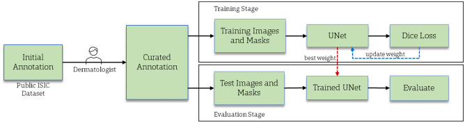
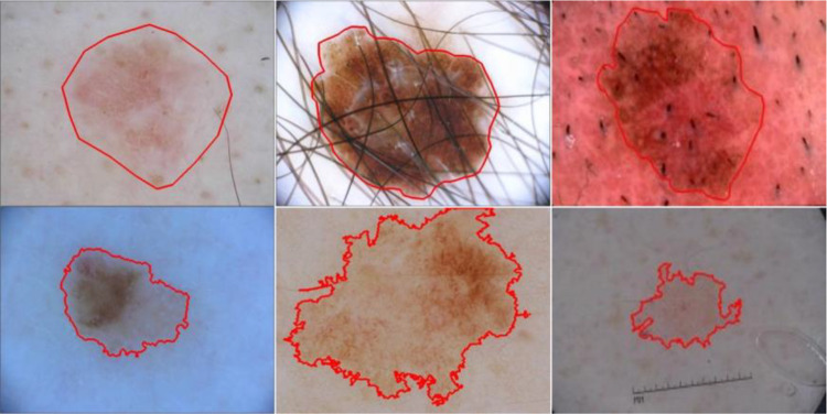
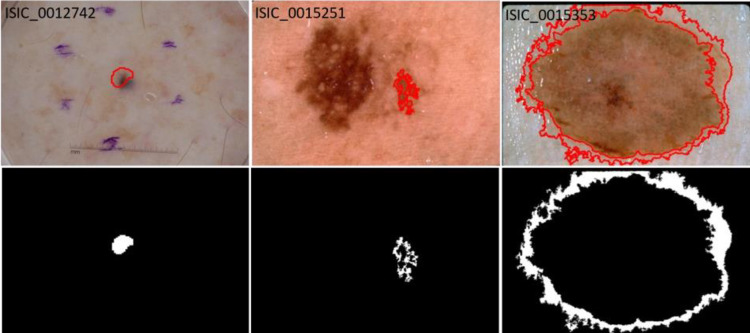
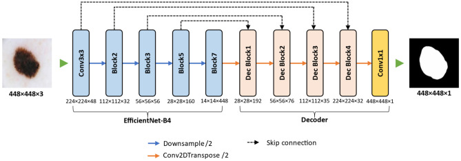
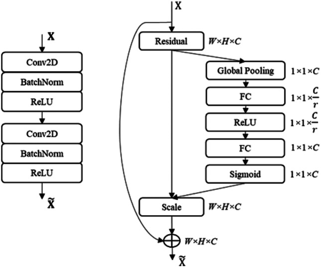
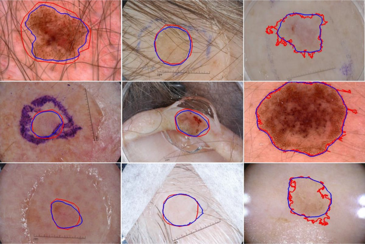
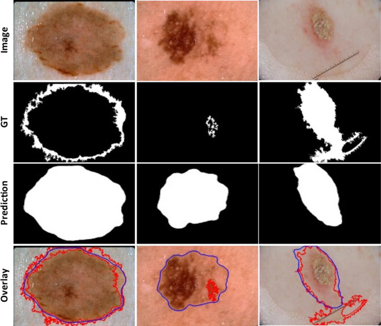
We propose a deep learning approach to segment the skin lesion in dermoscopic images. The proposed network architecture uses a pretrained EfficientNet model in the encoder and squeeze-and-excitation residual structures in the decoder. We applied this approach on the publicly available International Skin Imaging Collaboration (ISIC) 2017 Challenge skin lesion segmentation dataset. This benchmark dataset has been widely used in previous studies. We observed many inaccurate or noisy ground truth labels. To reduce noisy data, we manually sorted all ground truth labels into three categories - good, mildly noisy, and noisy labels. Furthermore, we investigated the effect of such noisy labels in training and test sets. Our test results show that the proposed method achieved Jaccard scores of 0.807 on the official ISIC 2017 test set and 0.832 on the curated ISIC 2017 test set, exhibiting better performance than previously reported methods. Furthermore, the experimental results showed that the noisy labels in the training set did not lower the segmentation performance. However, the noisy labels in the test set adversely affected the evaluation scores. We recommend that the noisy labels should be avoided in the test set in future studies for accurate evaluation of the segmentation algorithms.

Keywords: Deep learning; Dermoscopy; Image segmentation; Melanoma; Noisy data.


Conflict of interest statement

The authors declare no competing interests.

Figures

Fig. 1
Fig. 1
The overall flow diagram of a proposed skin lesion segmentation method
Fig. 2
Fig. 2
Skin lesion dermoscopy images with ground truth lesion boundary (red) from publicly available ISIC skin lesion datasets. The masks are manually drawn (first row) or generated using a semi-automated process (second row)
Fig. 3
Fig. 3
Examples of inaccurate or noisy ground truths on ISIC lesion segmentation dataset. Overlays show GT lesion boundaries on lesion images (top row) and ground truth lesion segmentation mask (bottom row). The lesion boundary (red) fails to cover the whole lesion in all examples
Fig. 4
Fig. 4
Proposed architecture for skin lesion segmentation. An encoder-decoder architecture with pretrained EfficientNet model as the encoder network, and the decoder network comprised four squeeze-and-excitation residual blocks
Fig. 5
Fig. 5
Structures of convolution blocks in the decoder network. Double convolution block (left) and squeeze-and-excitation residual block (right)
Fig. 6
Fig. 6
Segmentation results of the proposed method on ISIC 2017 test set. Overlays of ground truth lesion boundary (red) and predicted lesion boundary (blue) on skin lesion images. Lesion border predictions are accurate even in the presence of artifacts like hair, ruler marks, and ink markers
Fig. 7
Fig. 7
Segmentation results of the proposed method on examples having noisy (or inaccurate) ground truth (GT) on an official ISIC 2017 test set. The predicted lesion borders (blue) cover the lesion area more accurately than the GT lesion border (red)

Similar articles

Cited by

References

    1. Siegel RL, Miller KD, Fuchs HE, Jemal A. Cancer statistics, 2022. CA Cancer J Clin. 2022;72(1):7–33. doi: 10.3322/caac.21708. - DOI - PubMed
    1. Pehamberger H, Binder M, Steiner A, Wolff K. In vivo epiluminescence microscopy: improvement of early diagnosis of melanoma. Journal of Investigative Dermatology. 1993;100(3 SUPPL.):S356–S362. doi: 10.1038/jid.1993.63. - DOI - PubMed
    1. Soyer HP, Argenziano G, Talamini R, Chimenti S. Is dermoscopy useful for the diagnosis of melanoma? Arch Dermatol. 2001;137(10):1361–1363. doi: 10.1001/archderm.137.10.1361. - DOI - PubMed
    1. Braun RP, Rabinovitz HS, Oliviero M, Kopf AW, Saurat JH. Pattern analysis: a two-step procedure for the dermoscopic diagnosis of melanoma. Clin Dermatol. 2002;20(3):236–239. doi: 10.1016/S0738-081X(02)00216-X. - DOI - PubMed
    1. A. Krizhevsky, I. Sutskever, and G. Hinton, ImageNet classification with deep convolutional neural networks, in Advances in Neural Information and Processing Systems (NIPS), vol. 25, 2012, pp. 1097–1105.

LinkOut - more resources