. 2023 Apr 5;18(1):279.
doi: 10.1186/s13018-023-03762-0.

Practical considerations for determination of scapular internal rotation and its relevance in reverse total shoulder arthroplasty planning

Affiliations

Practical considerations for determination of scapular internal rotation and its relevance in reverse total shoulder arthroplasty planning

Paul Siegert et al. J Orthop Surg Res. .

Abstract

Background: Scapulothoracic orientation, especially scapular internal rotation (SIR) may influence range of motion in reverse total shoulder arthroplasty (RTSA) and is subjected to body posture. Clinical measurements of SIR rely on apical bony landmarks, which depend on changes in scapulothoracic orientation, while radiographic measurements are often limited by the restricted field of view (FOV) in CT scans. Therefore, the goal of this study was (1) to determine whether the use of CT scans with a limited FOV to measure SIR is reliable and (2) if a clinical measurement could be a valuable alternative.

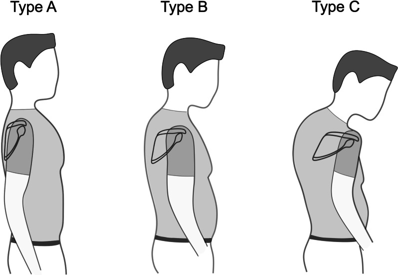
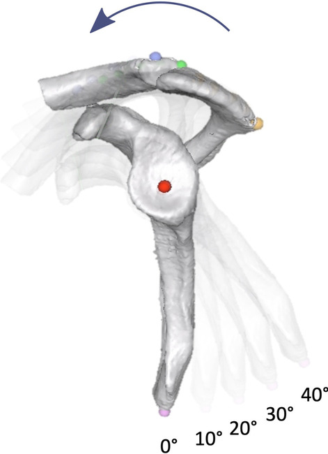
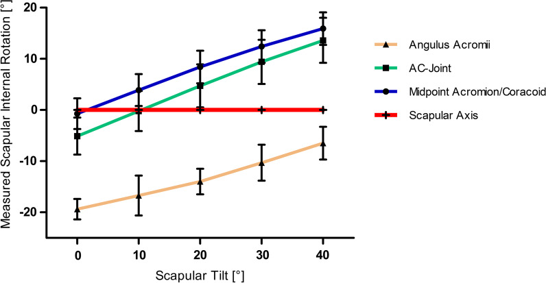
Methods: This anatomical study analyzed the whole-body CT scans of 100 shoulders in 50 patients (32 male and 18 female) with a mean age of 61.2 ± 20.1 years (range 18; 91). (1) CT scans were rendered into 3D models and SIR was determined as previously described. Results were compared to measurements taken in 2D CT scans with a limited FOV. (2) Three apical bony landmarks were defined: (the angulus acromii (AA), the midpoint between the AA and the coracoid process tip (C) and the acromioclavicular (AC) joint. The scapular axis was determined connecting the trigonum scapulae with these landmarks and referenced to the glenoid center. The measurements were repeated with 0°, 10°, 20°, 30° and 40° anterior scapular tilt.

Results: Mean SIR was 44.8° ± 5.9° and 45.6° ± 6.6° in the 3D and 2D model, respectively (p < 0.371). Mean difference between the measurements was 0.8° ± 2.5° with a maximum of 10.5°. Midpoint AA/C showed no significant difference to the scapular axis at 0° (p = 0.203) as did the AC-joint at 10° anterior scapular tilt (p = 0.949). All other points showed a significant difference from the scapular axis at all degrees of tilt.

Conclusion: 2D CT scans are reliable to determine SIR, even if the spine is not depicted. Clinical measurements using apical superficial scapula landmarks are a possible alternative; however, anterior tilt influenced by posture alters measured SIR.

Keywords: Posture types; Reverse total shoulder arthroplasty; Scapular internal rotation; Scapulothoracic orientation.


Conflict of interest statement

PM is a consultant for Arthrex. All other authors declare that they have no competing interests.

Figures

Fig. 1
Fig. 1
Illustration of three different posture types. From Type A over B to C patients show increasing scapular internal rotation, anterior tilt, protraction, and drooping as well as kyphosis and a barrel-shaped chest according to Moroder et al. [7]
Fig. 2
Fig. 2
A Measurement of scapular internal rotation (SIR) in a 3D model in respect to the sagittal body axis. B Anterior scapular tilt was measured on an “en face” view of the glenoid in respect to the examination table as a reference of the coronar body axis. C 2D measurement of SIR in respect to the standardized transversal reference line with a limited field of view
Fig. 3
Fig. 3
Various landmarks on the A parasagittal, B posterior-lateral C axial and D posterior view. 1: center of the glenoid; 2: most apical point located on a perpendicular line from the midpoint between the coracoid process and the angulus acromii; 3: posterior AC-joint; 4: angulus acromii, 5: medial root of scapular spine (trigonum scapulae); 6: inferior angulus; C: coracoid process tip
Fig. 4
Fig. 4
Parasagittal view with 0°, 10°, 20°, 30° and 40° anterior scapular tilt
Fig. 5
Fig. 5
Examples of axial view of the scapula with A 0° and B 30° anterior tilt. Scapular axis is marked with a red line. An angle was measured from the glenoid center to the trigonum scapulae and to the respective landmark (blue line). 1: midpoint of the glenoid; 2: most apical point located on a perpendicular line from the midpoint between the coracoid process and the angulus acromii; 3: posterior AC-joint; 4: angulus acromii, 5: medial root of scapular spine; 6: inferior angulus
Fig. 6
Fig. 6
Bland–Altman-Plot showing differences between 3 and 2D measurements for scapular internal rotation (SIR). Mean difference is marked by blue line and upper and lower bound (± 1.96 SD) marked by dashed red lines
Fig. 7
Fig. 7
Results of measurement of scapular internal rotation (SIR) with SD based on different landmarks (Angulus acromii, AC- joint and midpoint AA/C) dependent on progressive anterior scapular tilt in 0°, 10°, 20°, 30° and 40°. Scapular axis is shown as a red line
Fig. 8
Fig. 8
3D-CT reconstruction of a patient with advanced kyphoscoliosis. 3D measurement of scapular internal rotation (SIR) is referenced as a perpendicular line to the sagittal axis through vertebra Th1 and the sternum (blue line). With a limited field of view (black rectangle) the standardized CT reference (black dashed line) is used as the transversal axis

References

    1. Karelse AT, Bhatia DN, De Wilde LF. Prosthetic component relationship of the reverse Delta III total shoulder prosthesis in the transverse plane of the body. J Shoulder Elbow Surg. 2008;17(4):602–607. doi: 10.1016/j.jse.2008.02.005. - DOI - PubMed
    1. Moroder P, Urvoy M, Raiss P, Werthel JD, Akgun D, Chaoui J, et al. Patient posture affects simulated ROM in reverse total shoulder arthroplasty: a modeling study using preoperative planning software. Clin Orthop Relat Res. 2022;480(3):619–631. doi: 10.1097/CORR.0000000000002003. - DOI - PMC - PubMed
    1. Stephenson DR, Oh JH, McGarry MH, Rick Hatch GF, Lee TQ. Effect of humeral component version on impingement in reverse total shoulder arthroplasty. J Shoulder Elbow Surg. 2011;20(4):652–658. doi: 10.1016/j.jse.2010.08.020. - DOI - PubMed
    1. Khan WS, Longo UG, Ahrens PM, Denaro V, Maffulli N. A systematic review of the reverse shoulder replacement in rotator cuff arthropathy, rotator cuff tears, and rheumatoid arthritis. Sports Med Arthrosc Rev. 2011;19(4):366–379. doi: 10.1097/JSA.0b013e318224e44e. - DOI - PubMed
    1. Sulkar HJ, Aliaj K, Tashjian RZ, Chalmers PN, Foreman KB, Henninger HB. High and low performers in internal rotation after reverse total shoulder arthroplasty: a biplane fluoroscopic study. J Shoulder Elbow Surg;2022. - PMC - PubMed

LinkOut - more resources