. 2023 Mar 20:11:1101513.
doi: 10.3389/fbioe.2023.1101513. eCollection 2023.

Synthesis by solid route and physicochemical characterizations of blends of calcium orthophosphate powders and mesoporous silicon particles

Affiliations

Synthesis by solid route and physicochemical characterizations of blends of calcium orthophosphate powders and mesoporous silicon particles

Caroline Richard et al. Front Bioeng Biotechnol. .

Abstract

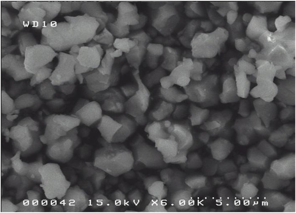
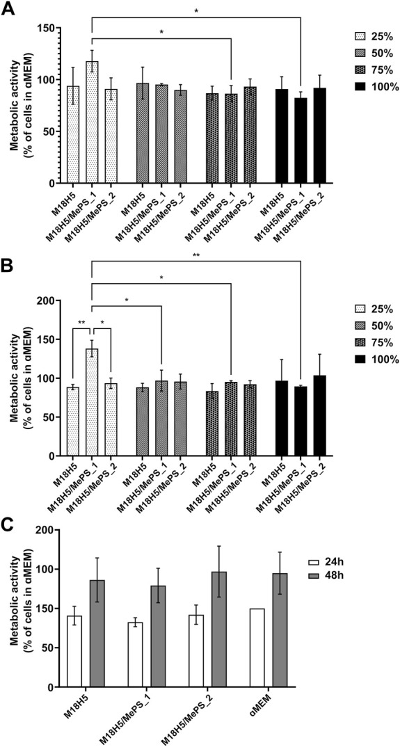
The purpose of the study was to investigate the synthesis of economic calcium phosphate powders from recycled oyster shells, using a ball milling method. The oyster shell powder and a calcium pyrophosphate powder were used as starting materials and ball milled, then heat treated at 1,050°C for 5 h to produce calcium phosphate powders through a solid-state reaction. Electrochemically synthesized mesoporous silicon microparticles were then added to the prepared phosphate powders by mechanical mixer. The final powders were characterized using X-ray diffraction, Fourier transform infrared spectroscopy, and scanning electron microscopy to analyze their chemical composition and determine the most suitable process conditions. The biocompatibility of the produced powders was also tested in vitro using murine cells and the results showed good biocompatibility.

Keywords: ball milling method; biocompatibility; hydroxyapatite (HAP); mesoporous silicon; tricalcium phosphate (TCP).


Conflict of interest statement

The authors declare that the research was conducted in the absence of any commercial or financial relationships that could be construed as a potential conflict of interest.

Figures

FIGURE 1
FIGURE 1
SEM images (SE mode) of selected raw oyster shell powder for the synthesis. (A) Detail of the surface of a particle (bar: 1 µm). (B) Global view and morphology of the particles (bar: 200 µm).
FIGURE 2
FIGURE 2
Physicochemical characterization of oyster shell powder. (A) X ray diffractogram compared with JCPDS standard with the diffracting crystal planes and (B) FTIR pattern with the characteristic carbonate vibrational bands. (C) TGA/DTA of raw oyster shell powder.
FIGURE 3
FIGURE 3
SEM images of crushed OSP sample (Milling duration: 18 h).
FIGURE 4
FIGURE 4
SEM Images of M18H5 sample.
FIGURE 5
FIGURE 5
XRD pattern of M18H5 powder.
FIGURE 6
FIGURE 6
Rietveld analysis pattern of obtained powder. The black solid line concerns the calculated intensities and the red asterisk line, the observed intensities. The short vertical lines show the position of possible Bragg reflections. The difference between the observed and calculated intensities is plotted below the profile.
FIGURE 7
FIGURE 7
(A) SEM images of the mixture M18H5 and MePS. (B) Detail of particles of MePS and M18H5 particles.
FIGURE 8
FIGURE 8
Evaluation of the metabolic activity of MC3T3-E1 cells cultured in presence of M18H5, M18H5/MePS_1 and M18H5/MePS_2 powder extracts, pure (100%) or diluted at d = 1/4 (75%), d = 1/2 (50%) and d = 3/4 (25%) for (A): 24 h and (B): 48 h. The metabolic activity is expressed as a percentage of the control, i.e. the value obtained for the cells cultured in complete culture medium (ɑMEM). (C): Metabolic activity of the cell measured after 24 h and 48 h of cultivation and expressed as a percentage of the value obtained for the cell cultured in complete culture medium (ɑMEM) after 24 h as a control. Statistical analysis: ANOVA one-way followed by a Tukey post hoc test. *: p ≤ 0.05, **: p < 0.01. n = 3 independent experiments.
FIGURE 9
FIGURE 9
Density and proliferation rate of MC3T3-E1 cells cultured in presence of M18H5, M18H5/MePS_1 and M18H5/MePS_2 powder extracts. (A) Micrographs of cells cultured in presence of M18H5 (A–C), M18H5/MePS _1 (D–F) and M18H5/MePS_2 (G–I) powder extracts stained after EdU incorporation assay. A, D and (G) EdU after conjugation with AlexaFluor488; B, E and (F) nuclei stained by Hoechst 33,342; C, F and (I) merge. Scale bar: 250 µm (insets: 100 µm). Upper right: insets showing the magnification of areas in the main image (B) Cell density, determined by image analysis and expressed as cells/cm2. Statistical analysis: ANOVA one way followed by a Tukey post hoc test; ns: p > 0.05. (C) Cell proliferation, expressed in % of cells positive for EdU in total cell population (Hoechst 3,342 positive nuclei) determined by image analysis. Statistical analysis: Kruskall-Wallis followed by a Dunn’s post hoc test; ns: p > 0.05. n = 3 independent experiments.
FIGURE 10
FIGURE 10
Morphology of MC3T3-E1 cells cultured in presence of M18H5, M18H5/MePS_1 and M18H5/MePS_2 powder extracts after staining of actin cytoskeleton and ɑ-tubulin. M18H5 (A–D), M18H5/MePN_1 (E–H), M18H5/MePN_2 (I–L) and complete culture medium (M–P). A, E, I and M: actin cytoskeleton; B, F, J and N: ɑ-tubulin; C, G, K, and O: nuclei; D, H, L and P: merge. Scale bar: 50 μm. n = 3 independent experiments.

References

    1. Astrova E., Tolmachev V. (2000). Effective refractive index and composition of oxidized porous silicon films. Mat. Sci. Eng. B 69–70, 142–148. 10.1016/S0921-5107(99)00236-6 - DOI
    1. Bobbio A. (1972). The first endosseous alloplastic implant in the history of man. Bull. Hist. Dent. 20, 1–6. PMID: 4505221. - PubMed
    1. Bose S., Fielding G., Tarafder S., Bandyopadhyay A. (2013). Understanding of dopant-induced osteogenesis and angiogenesis in calcium phosphate ceramics. Trends Biotechnol. 31, 594–605. 10.1016/j.tibtech.2013.06.005 - DOI - PMC - PubMed
    1. Bowditch A., Waters K., Gale H., Rice P., Scott E., Canham L., et al. (1998). In-vivo assessment of tissue compatibility and calcification of bulk and porous silicon. MRS Proc. 536, 149. 10.1557/PROC-536-149 - DOI
    1. Brundavanam R-K., Fawcett D., Jai Poinern G-E. (2017). Synthesis of a bone like composite material derived from waste pearl oyster shells for potential bone tissue bioengineering applications. Int. J. Res. Med. Sci. 5 (6), 2454–2461. 10.18203/2320-6012.ijrms20172428 - DOI

LinkOut - more resources