. 2023 Mar 20:11:1147943.
doi: 10.3389/fbioe.2023.1147943. eCollection 2023.

Optimization of guanosine-based hydrogels with boric acid derivatives for enhanced long-term stability and cell survival

Affiliations

Optimization of guanosine-based hydrogels with boric acid derivatives for enhanced long-term stability and cell survival

Maria Merino-Gómez et al. Front Bioeng Biotechnol. .

Abstract

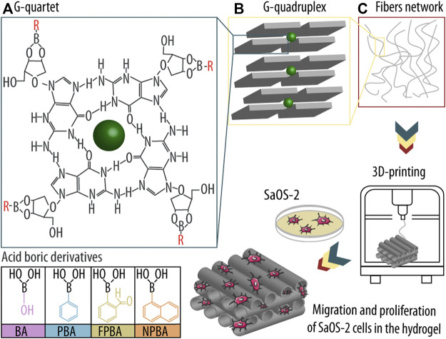
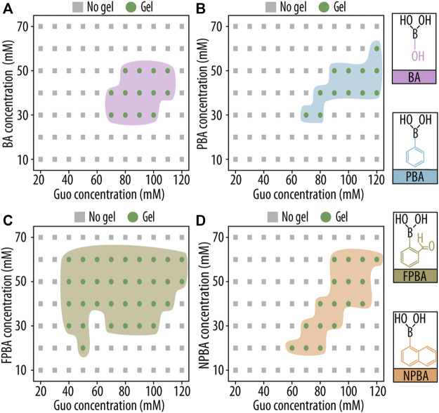
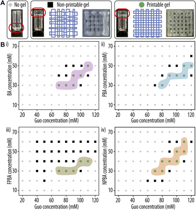
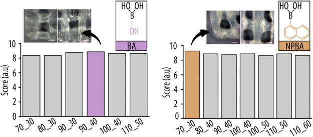
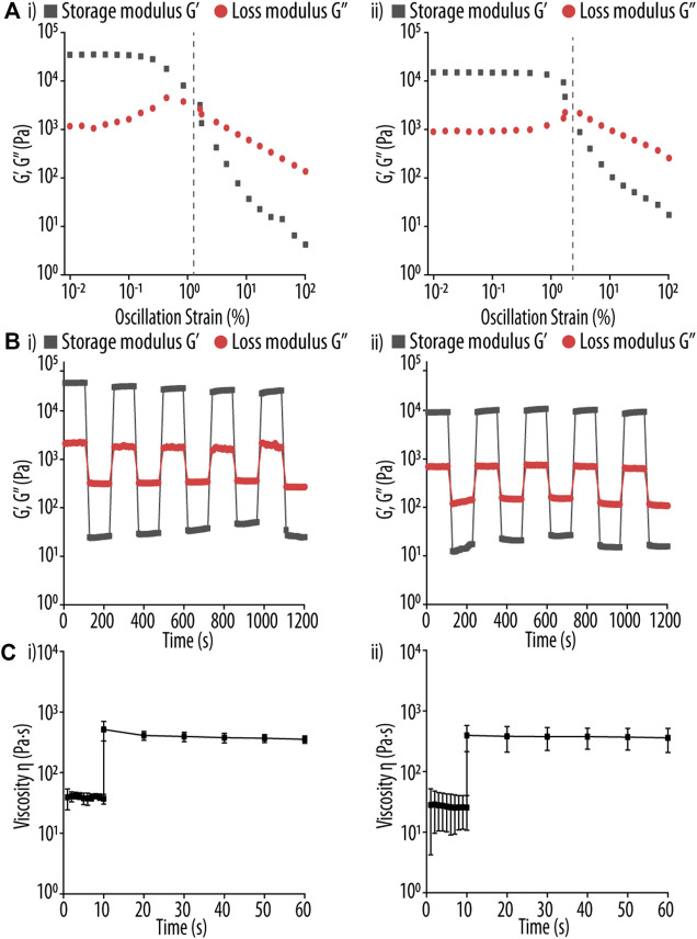
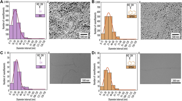
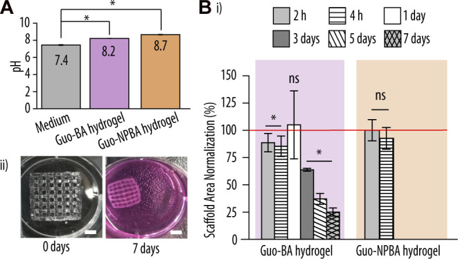
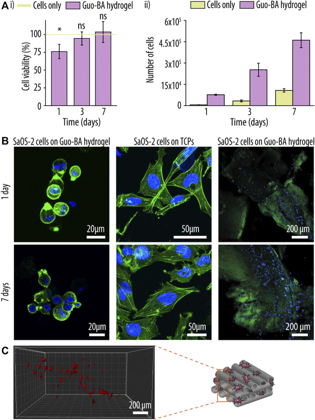
Tissue defects can lead to serious health problems and often require grafts or transplants to repair damaged soft tissues. However, these procedures can be complex and may not always be feasible due to a lack of available tissue. Hydrogels have shown potential as a replacement for tissue grafts due to their ability to support cell survival and encapsulate biomolecules such as growth factors. In particular, guanosine-based hydrogels have been explored as a potential solution, but they often exhibit limited stability which hampers their use in the biofabrication of complex grafts. To address this issue, we explored the use of borate ester chemistry and more complex boric acid derivatives to improve the stability and properties of guanosine-based hydrogels. We hypothesized that the aromatic rings in these derivatives would enhance the stability and printability of the hydrogels through added π-π stack interactions. After optimization, 13 compositions containing either 2-naphthylboronic acid or boric acid were selected. Morphology studies shows a well-defined nanofibrilar structure with good printable properties (thixotropic behaviour, print fidelity and printability). Moreover, the pH of all tested hydrogels was within the range suitable for cell viability (7.4-8.3). Nevertheless, only the boric acid-based formulations were stable for at least 7 days. Thus, our results clearly demonstrated that the presence of additional aromatic rings did actually impair the hydrogel properties. We speculate that this is due to steric hindrance caused by adjacent groups, which disrupt the correct orientation of the aromatic groups required for effective π-π stack interactions of the guanosine building block. Despite this drawback, the developed guanosine-boric acid hydrogel exhibited good thixotropic properties and was able to support cell survival, proliferation, and migration. For instance, SaOS-2 cells planted on these printed structures readily migrated into the hydrogel and showed nearly 100% cell viability after 7 days. In conclusion, our findings highlight the potential of guanosine-boric acid hydrogels as tissue engineering scaffolds that can be readily enhanced with living cells and bioactive molecules. Thus, our work represents a significant advancement towards the development of functionalized guanosine-based hydrogels.

Keywords: 3D printing; boric acid derivatives; guanosine-based hydrogels; nucleoside; printable hydrogels.


Conflict of interest statement

The authors declare that the research was conducted in the absence of any commercial or financial relationships that could be construed as a potential conflict of interest.

Figures

SCHEME 1
SCHEME 1
Guanosine (Guo)-based hydrogels formation using boric acid derivatives (dBAs) and subsequent three-dimensional (3D) printing and seeding of osteogenic sarcoma cells (SaOS-2). (A) First, Guo, dBAs, and potassium ions (K+) form an ordered G-quartet structure. (B) Four G-quartets interact and form a higher structure known as G-quadruplex, and ultimately (C) nanofibers, which build the hydrogel network. The assembly is then printed in a 3D printer, and SaOS-2 cells are seeded onto the scaffold, allowing them to migrate into the matrix and proliferate in the hydrogel system.
FIGURE 1
FIGURE 1
Printability assessment of hydrogel-based inks. (A) Three-dimensional (3D) printed platform used for the evaluation of the filament collapse area. While Ae represents the experimentally determined area, At corresponds to the theoretical area under the printed filament. (B) (i) Printing pattern used for the filament fusion test. Two filament layers were deposited along 0° and 90°, and the resulting areas between filaments (Ae) were measured and compared to their theoretical counterparts (At). Adapted with permission. [37] Copyright 2018, MDPI.
FIGURE 2
FIGURE 2
Gelation study of guanosine (Guo)-based hydrogels with boric acid derivatives (GB hydrogels) using a vial inversion test. Phase diagrams of gelation after 1 h at room temperature (RT) are shown as functions of Guo concentration and (A) boric acid (BA), (B) phenyl boronic acid (PBA), (C) 2-formilphenylboronic acid (FPBA), and (D) 2-naphtylboronic acid (NPBA) concentration.
FIGURE 3
FIGURE 3
Printability assessment of guanosine (Guo)-based hydrogels with boric acid derivatives (GB hydrogels). (A) Representative images of hydrogel formulations yielding either no gel (left), a non-printable gel (middle), or a printable hydrogel (right) are shown. (B) Phase diagram of GB hydrogel gelation after 1 h at room temperature (RT) as a functions of Guo concentration and (i) boric acid (BA), (ii) phenyl boronic acid (PBA), (iii) 2-formilphenylboronic acid (FPBA) and (iv) 2-naphtylboronic acid (NPBA) concentration. Compositions resulting in three-dimensional (3D)-printable hydrogels are indicated as green circles, non-printable solutions as black squares.
FIGURE 4
FIGURE 4
In-depth evaluation of two different ink parameters after printing to enable a comparative assessment of the tested guanosine (Guo)-acid boric derivatives (dBAs) (GB) formulation. Two dBAs were tested (A) boric acid (BA) and (B) 2-naphtylboronic acid (NPBA), and four different parameters evaluated: (i) collapse area factor, corresponding to the area of the hanging filament, (ii) diffusion rate, representing the material spreading on the printing surface, (iii) grid printability, corresponding to the perimeter of the printed grid squares, and (iv) angle deviation rate, reflecting the shape and rectangularity of the printed square holes, respectively. Samples are coded as X_Y, where X and Y represent the millimolar concentration of Guo and dBAs/KOH, respectively.
FIGURE 5
FIGURE 5
Summed and normalized printability scores of the individual hydrogel formulations obtained from filament fusion and collapse test. Inserts show representative images of the three dimensional (3D)-printed grid structures, highlighting corners (left) and filament cross-sections (right) for the best hydrogel compositions of boric acid (BA) and 2-naphtylboronic acid (NPBA), respectively. Scale bar 0.5 mm.
FIGURE 6
FIGURE 6
(A) Strain sweep test of guanosine (Guo)-based hydrogels using (I) boric acid (BA) and (ii) 2-naphtylboronic acid (NPBA). Storage modulus (G′, black square) and loss modulus (G″, red circle) were determined using a strain range of 0.01%–100% and a fixed oscillation frequency of 1 Hz. The respective turning point of both Guo-BA and Guo-NPBA hydrogels formulations in indicated by a vertical gray dashed line. (B) Rheological test of Guo-based hydrogels using (i) BA, and (ii) NPBA at 37°C. The G′ and G″ of the hydrogels are shown using a strain amplitude of 0.1% and 100% for the recovery and shear step, respectively, and a fixed angular frequency of 10 rad s−1. Each interval was constant at 100 s. (C) Rheological test of Guo-based hydrogels using (i) BA and (ii) NPBA at 37°C. A shear stress of 3.45 s−1 was applied for 10 s, mimicking the conditions during three dimensional (3D)-printing, and then the hydrogels were allowed to recover for up to 50 s at 0.1 s−1.
FIGURE 7
FIGURE 7
Scanning electron microscopy (SEM) analysis of printed hydrogels for guanosine (Guo)-based hydrogels using (A) boric acid (BA) and (B) 2-naphtylboronic acid (NPBA). (i) Diameter distribution histograms of the nanofibrillar network and (ii) SEM images. Transmission electron microscopy (TEM) analysis for (C) Guo-BA hydrogels and (D) Guo-NPBA hydrogels. (i) Diameter distribution histograms of the nanofibrillar network and (ii) TEM image.
FIGURE 8
FIGURE 8
Biocompatibility evaluation of the printed hydrogel scaffolds. (A) pH values of the printed guanosine (Guo)-based hydrogels using boric acid (BA) and 2-naphtylboronic acid (NPBA) hydrogels vs. complete McCoy 5A medium. (B) (i) Normalized scaffold areas of printed Guo-BA and Guo-NPBA hydrogels after incubation in McCoy 5A medium at 37°C for up to 7 days. The red line represents 100% viability of the cells at 0 h (control). Difference testing was done versus control (ii) Representative image of guanosine-boric acid derivatives (GB) hydrogels after 0 and 7 days of immersion in complete McCoy 5A medium at 37°C and 5% CO2. Scale bar, 5 mm.
FIGURE 9
FIGURE 9
Post-printing cell functionalization of a guanosine (Guo)-based hydrogels with boric acid (BA) hydrogel. (A) Cell viability (i) and proliferation (ii) experiment of sarcoma osteogenic (SaOS-2) cells seeded onto the Guo-BA hydrogels up to 7 days. The green line represents 100% viability of the cells seeded on tissue culture polystyrene (TCPs) (control). Difference testing was done versus control. (B) Merged confocal laser scanning images of SaOS-2 cells morphology seeded on top of Guo-BA hydrogels, SaOS-2 cells morphology seeded on TCP and SaOS-2 cell distribution on Guo-BA hydrogels after 1 and 7 days of incubation. Actin filaments were stained with phalloidin-Atto 488 (green fluorescence signal) and nuclei with DAPI (blue fluorescence signal). (C) three dimensional (3D) representation of SaOS-2 cells migration through the Guo-BA hydrogel after 7 days. Cells were stained with CellTracker™ Deep Red reagent.

Similar articles

Cited by

References

    1. Adhikari B., Shah A., Kraatz H. B. (2014). Self-assembly of guanosine and deoxy-guanosine into hydrogels: Monovalent cation guided modulation of gelation, morphology and self-healing properties. J. Mater. Chem. B 2, 4802–4810. 10.1039/c4tb00702f - DOI - PubMed
    1. Bhattacharyya T., Kumar Y. P., Dash J. (2017). Supramolecular hydrogel inspired from DNA structures mimics peroxidase activity. ACS Biomater. Sci. Eng. 3, 2358–2365. 10.1021/acsbiomaterials.7b00563 - DOI - PubMed
    1. Bhattacharyya T., Saha P., Dash J. (2018). Guanosine-derived supramolecular hydrogels: Recent developments and future opportunities. ACS Omega 3, 2230–2241. 10.1021/acsomega.7b02039 - DOI - PMC - PubMed
    1. Biswas A., Maiti S., Kalaskar D. M., Das A. K. (2018a). Redox-active dynamic self-supporting thixotropic 3D-printable G-quadruplex hydrogels. Chem. - An Asian J. 13, 3928–3934. 10.1002/asia.201801409 - DOI - PubMed
    1. Biswas A., Malferrari S., Kalaskar D. M., Das A. K. (2018b). Arylboronate esters mediated self-healable and biocompatible dynamic G-quadruplex hydrogels as promising 3D-bioinks. Chem. Commun. 54, 1778–1781. 10.1039/c7cc09051j - DOI - PubMed