Editorial
. 2023 Oct 1;78(4):1106-1117.
doi: 10.1097/HEP.0000000000000367. Epub 2023 Apr 7.

Role of HNF4alpha-cMyc interaction in liver regeneration and recovery after acetaminophen-induced acute liver injury

Affiliations
Editorial

Role of HNF4alpha-cMyc interaction in liver regeneration and recovery after acetaminophen-induced acute liver injury

Manasi Kotulkar et al. Hepatology. .

Abstract

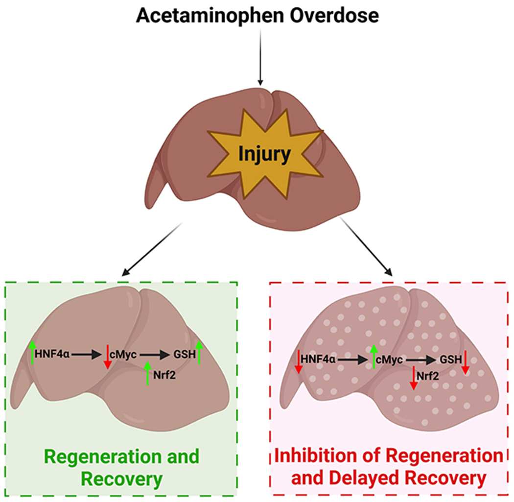
Background and aims: Overdose of acetaminophen (APAP) is the major cause of acute liver failure in the western world. We report a novel signaling interaction between hepatocyte nuclear factor 4 alpha (HNF4α) cMyc and nuclear factor erythroid 2-related factor 2 (Nrf2) during liver injury and regeneration after APAP overdose.

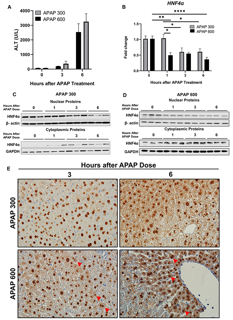
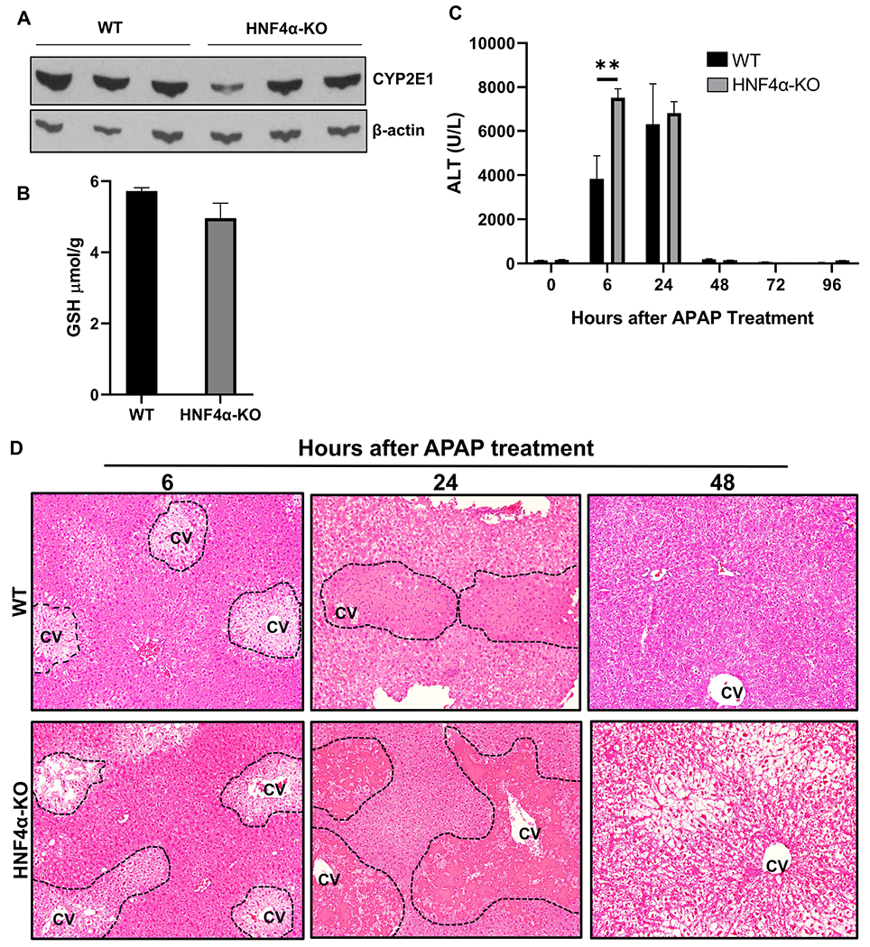
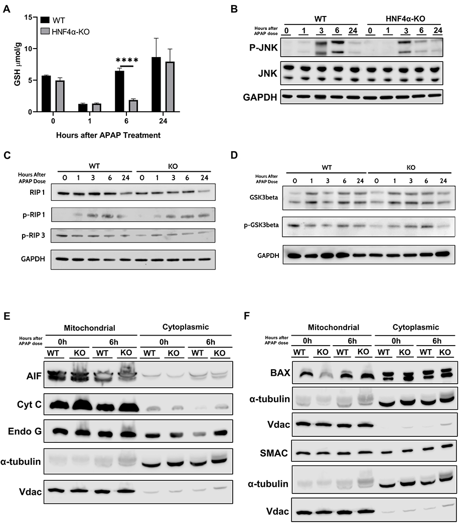
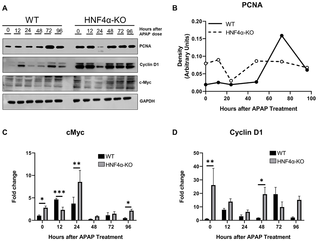
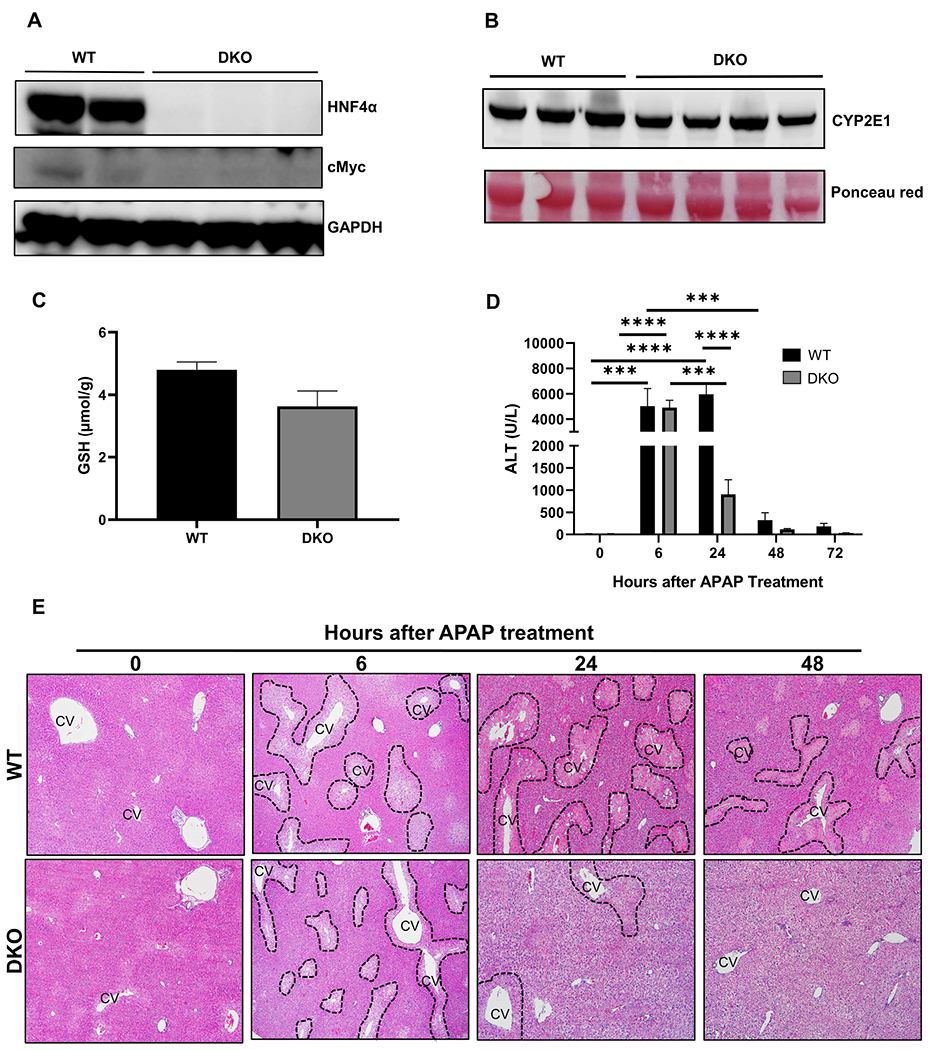
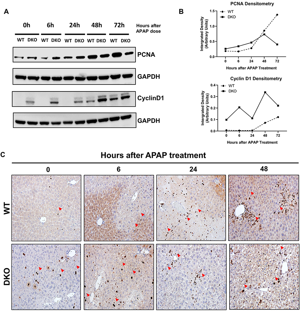
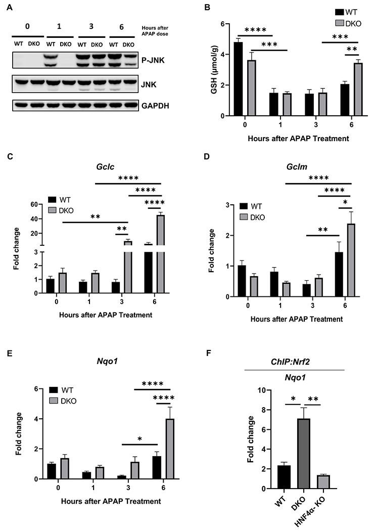
Approach and results: APAP-induced liver injury and regeneration were studied in male C57BL/6J (WT) mice, hepatocyte-specific HNF4α knockout mice (HNF4α-KO), and HNF4α-cMyc double knockout mice (DKO). C57BL/6J mice treated with 300 mg/kg maintained nuclear HNF4α expression and exhibited liver regeneration, resulting in recovery. However, treatment with 600-mg/kg APAP, where liver regeneration was inhibited and recovery was delayed, showed a rapid decline in HNF4α expression. HNF4α-KO mice developed significantly higher liver injury due to delayed glutathione recovery after APAP overdose. HNF4α-KO mice also exhibited significant induction of cMyc, and the deletion of cMyc in HNF4α-KO mice (DKO mice) reduced the APAP-induced liver injury. The DKO mice had significantly faster glutathione replenishment due to rapid induction in Gclc and Gclm genes. Coimmunoprecipitation and ChIP analyses revealed that HNF4α interacts with Nrf2 and affects its DNA binding. Furthermore, DKO mice showed significantly faster initiation of cell proliferation resulting in rapid liver regeneration and recovery.

Conclusions: These data show that HNF4α interacts with Nrf2 and promotes glutathione replenishment aiding in recovery from APAP-induced liver injury, a process inhibited by cMyc. These studies indicate that maintaining the HNF4α function is critical for regeneration and recovery after APAP overdose.


Conflict of interest statement

Conflict of interest:

The authors declared no potential conflicts of interest with respect to the research, authorship, and/or publication of this article.

Figures

Figure 1:
Figure 1:. Rapid HNF4α nuclear to cytoplasmic translocation after non-regenerating dose of APAP
(A) Serum ALT levels (B) qPCR analysis of HNF4α mRNA at various time points after administration of 300 mg/kg and 600 mg/kg APAP in C57BL/6J mice. (C) Western blot analysis of HNF4α using nuclear and cytoplasmic extracts showing nuclear to cytoplasmic translocation of HNF4α after 300 mg/kg dose of APAP and (D) 600 mg/kg dose of APAP. (E) Representative photomicrographs of HNF4α immunohistochemistry staining at 3 and 6 h after 300 mg/kg and 600 mg/kg dose of APAP. Arrows pointing to cytoplasmic HNF4α staining. Original magnification, 600X; * Indicates significant difference at, *=P<0.05, **=P<0.01, ****=P<0.0001
Figure 2:
Figure 2:. Kinetics of APAP-induced liver injury in HNF4α-KO mice
(A) Western blot analysis of hepatic CYP2E1 and (B) Hepatic GSH levels in WT and HNF4α KO mice at 0 h time point (C) Serum ALT levels and (D) representative photomicrographs of H&E-stained liver sections of WT and HNF4α-KO mice treated with 300 mg/kg APAP. Dotted lines demark area of necrosis. CV, central vein; Original magnification, 200X; *=P<0.05
Figure 3:
Figure 3:. Mechanisms of liver regeneration in HNF4α-KO mice
(A) Total glutathione levels in liver lysates of WT and HNF4α-KO mice at various time points after APAP 300 mg/kg dose. Western blot analysis of (B) total and phospho-JNK, (C) total and phospho-RIP1 and phospho-RIP3, and (D) total and phospho-GSK3β performed using pooled (n= 3-5) liver lysates and (E) AIF, Cyt C, Endo G (F) BAX and SMAC performed using pooled (n=3-5) liver mitochondrial and cytoplasmic fractions of WT and HNF4α-KO mice treated with APAP 300 mg/kg. *=P<0.05
Figure 4:
Figure 4:. Sustained cell proliferation in HNF4α-KO mice after APAP overdose accompanied by cMyc and CylinD1 induction
(A) Western blot analysis of proliferating cell nuclear antigen (PCNA), cMyc and Cyclin D1 performed using pooled (n=3-5) liver lysates of WT and HNF4α-KO mice treated with APAP 300 mg/kg. (B) Densitometric analysis of protein levels of PCNA in WT and HNF4α-KO mice. (C) qPCR analysis of cMyc and (D) Cyclin D1. *=P<0.05, **=P<0.01, ***=P<0.001
Figure 5:
Figure 5:. cMyc deletion in HNF4α-KO mice reduced APAP induced liver injury
(A) Western blot analysis of HNF4α and cMyc showing deletion of HNF4α and cMyc in DKO mice (B) Western blot analysis of hepatic CYP2E1 and (C) Hepatic GSH levels in WT and DKO mice at 0 h time point. (D) Serum ALT levels of WT and DKO mice treated with APAP 300 mg/kg dose at various time points. (E) Representative photomicrographs of H&E-stained liver sections. Dotted lines demark area of necrosis. CV, central vein; Original magnification, 200X; * Indicates significant difference at, **=P<0.01, ***=P<0.001, ****=P<0.0001.
Figure 6:
Figure 6:. Faster initiation of cell proliferation in HNF4α-cMyc DKO mice after APAP overdose
(A) Western blot analysis of PCNA and CyclinD1 performed using pooled (n=3-5) liver lysates from WT and DKO mice at various time points.(B) Densitometric analysis of PCNA and CyclinD1 Western blots. (C) Ki67 immunohistochemistry of WT and DKO mice at 0, 6, 24, 48 h after APAP overdose. Red arrows pointing to Ki67 positive cells. Original magnification, 400X
Figure 7:
Figure 7:. Rapid GSH replenishment in DKO mice after APAP treatment
(A) Western blot analysis of JNK and phospho- JNK performed using pooled (n=3-5) liver lysates from WT and DKO mice after 300 mg/kg dose of APAP. (B) Total glutathione levels in liver lysates of WT and DKO mice. qPCR analysis of (C) Gclc, (D) Gclm and, (E) Nqo1 genes. (F) ChIP of Nrf2 in control WT, DKO and HNF4α-KO mice using the frozen liver tissues. Nqo1 qPCR was performed to confirm the pulldown of Nrf2. * Indicates significant difference at, *=P<0.05, **=P<0.01, ***=P<0.001, ****=P<0.0001.
Figure 8:
Figure 8:. Graphical Abstract
Schematic representation of final conclusions.

Comment on

Similar articles

Cited by

References

    1. Bernal W and Wendon J, Acute liver failure. N Engl J Med, 2013. 369(26): p. 2525–34. - PubMed
    1. Larson AM, et al., Acetaminophen-induced acute liver failure: results of a United States multicenter, prospective study. Hepatology, 2005. 42(6): p. 1364–72. - PubMed
    1. Shehab N, et al., US Emergency Department Visits for Outpatient Adverse Drug Events, 2013-2014. JAMA, 2016. 316(20): p. 2115–2125. - PMC - PubMed
    1. Jaeschke H and Bajt ML, Intracellular signaling mechanisms of acetaminophen-induced liver cell death. Toxicol Sci, 2006. 89(1): p. 31–41. - PubMed
    1. Bhushan B and Apte U, Acetaminophen Test Battery (ATB): A Comprehensive Method to Study Acetaminophen-Induced Acute Liver Injury. Gene Expr, 2020. 20(2): p. 125–138. - PMC - PubMed

MeSH terms