Review
. 2023 Oct 1;103(4):2349-2422.
doi: 10.1152/physrev.00058.2021. Epub 2023 Apr 6.

Mitochondria in health, disease, and aging

Affiliations
Review

Mitochondria in health, disease, and aging

John S Harrington et al. Physiol Rev. .

Abstract

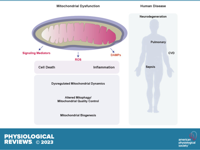
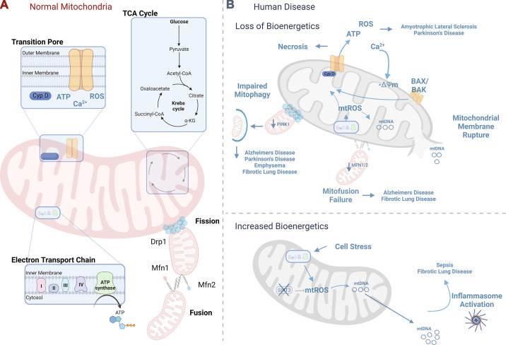
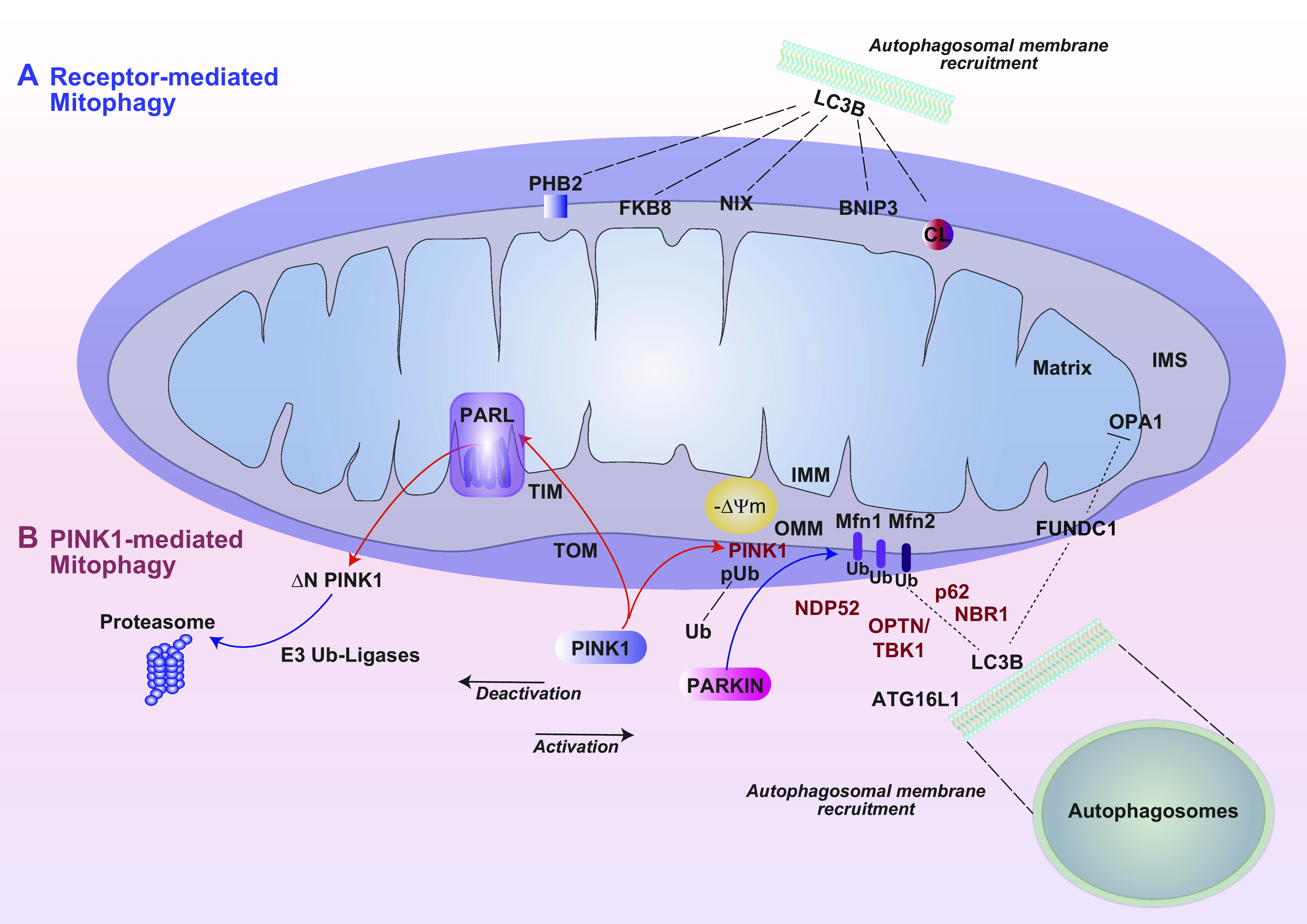
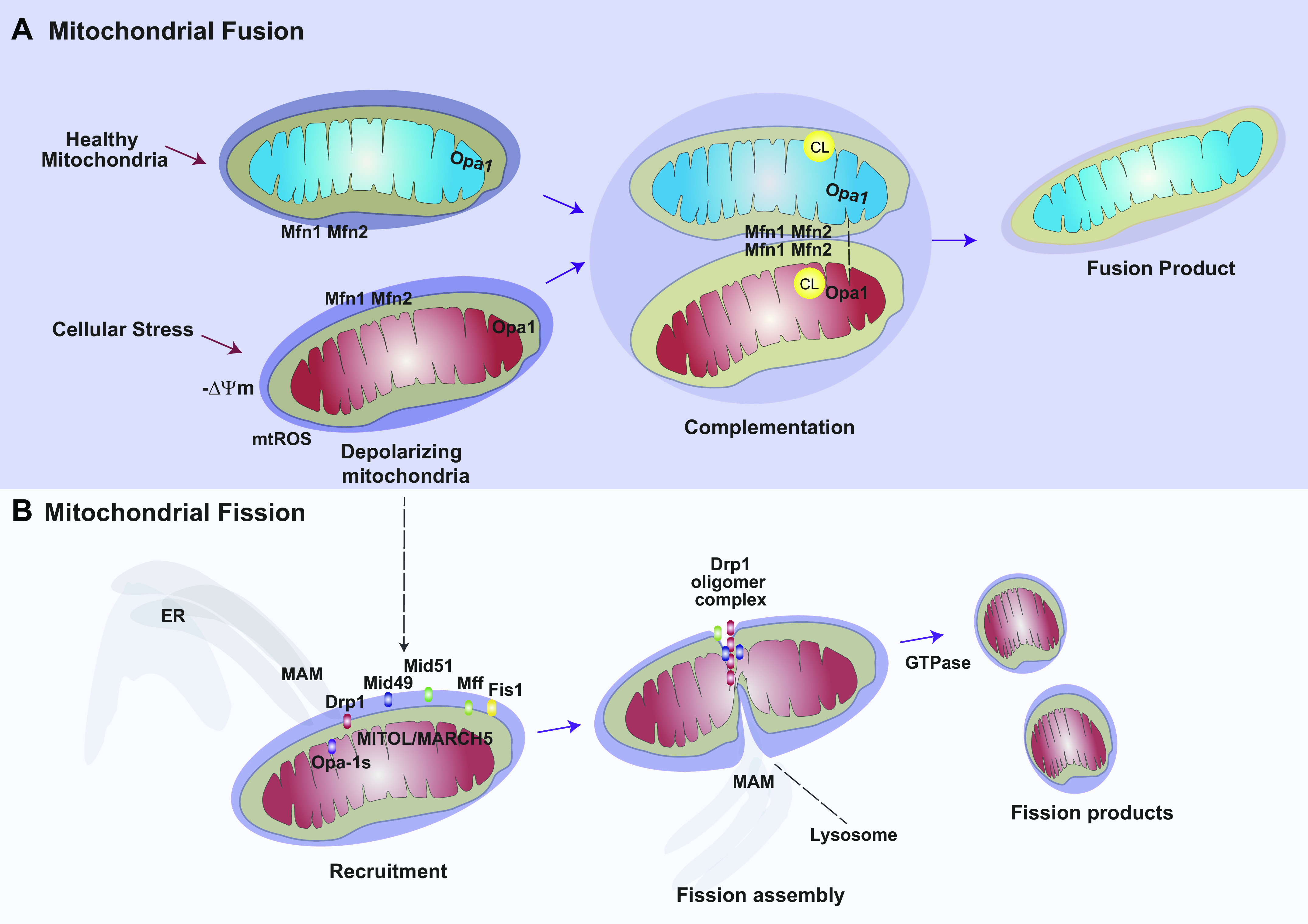
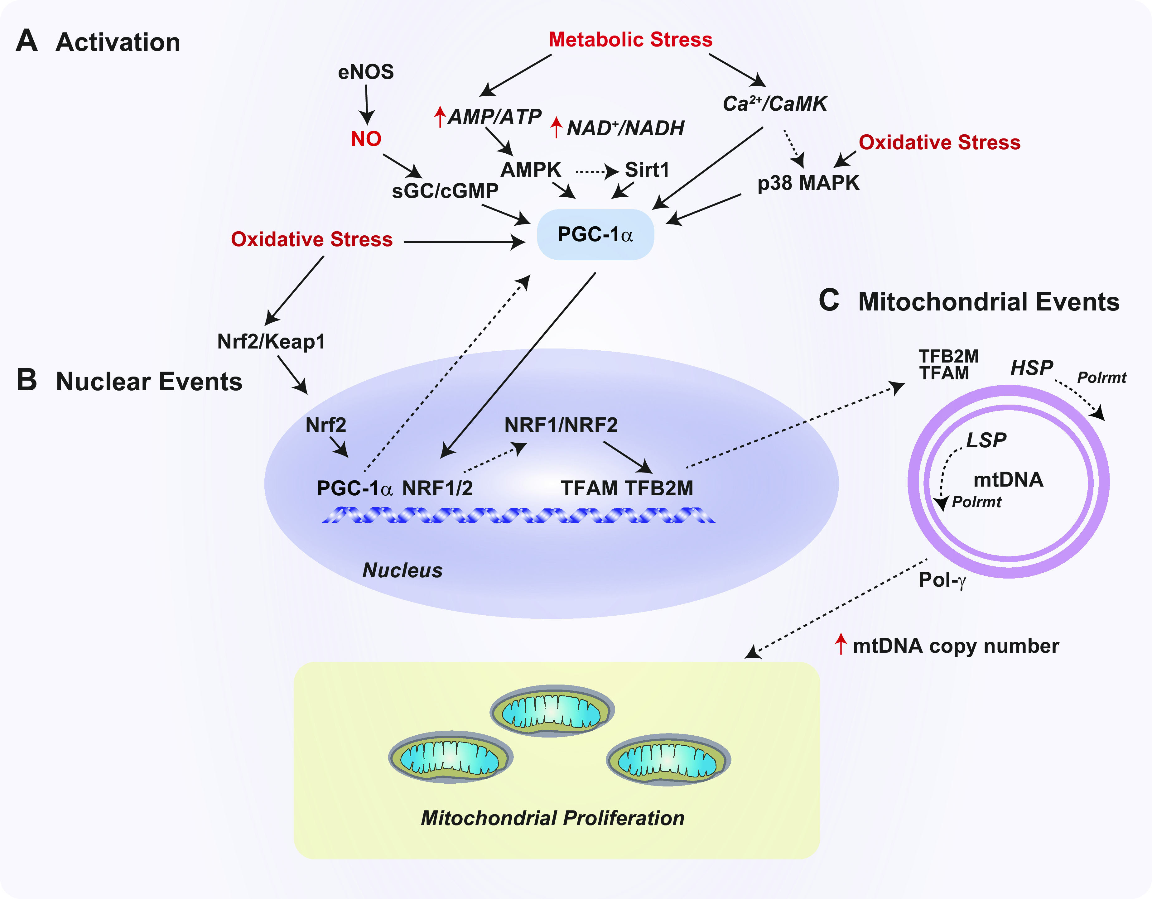
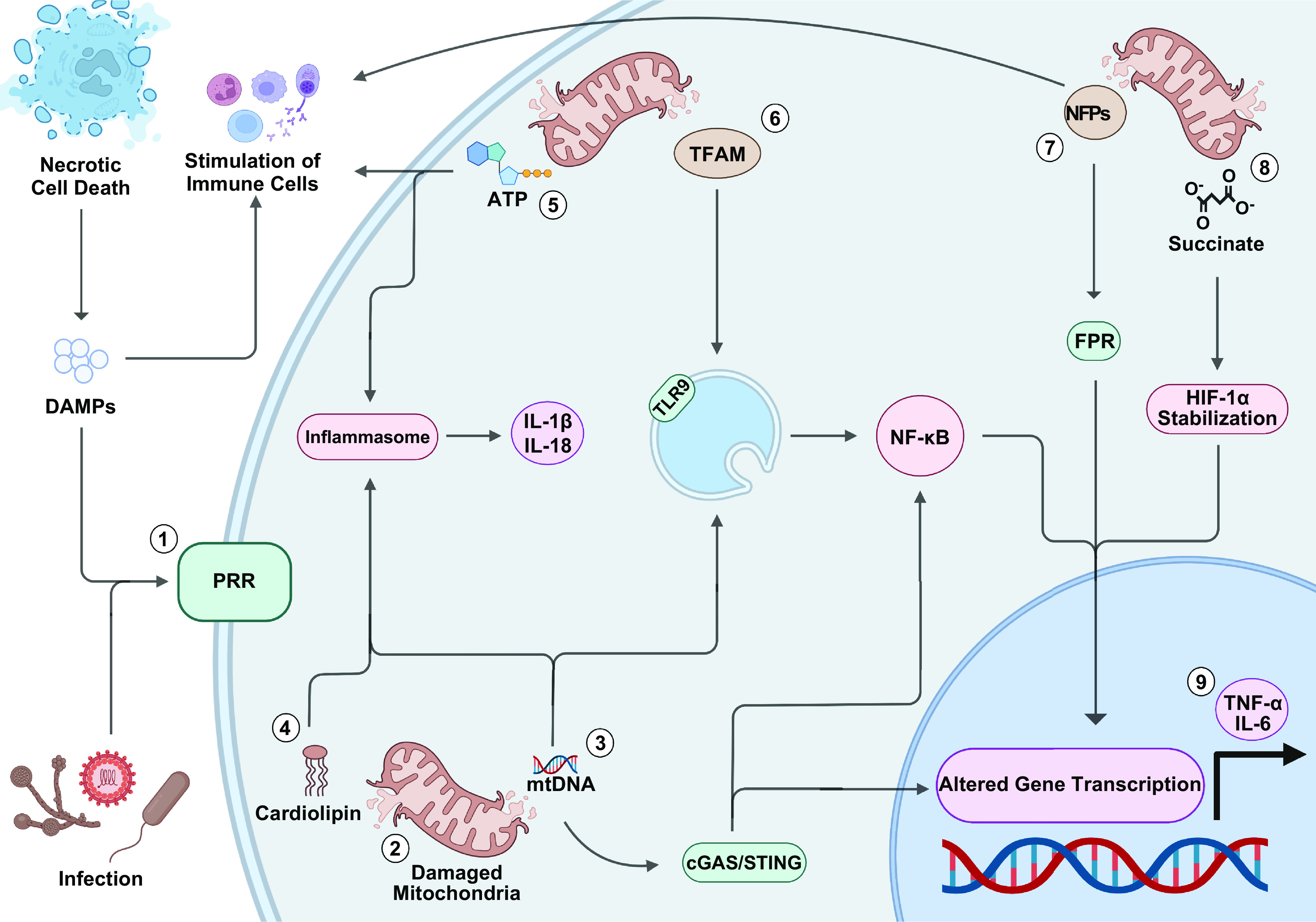
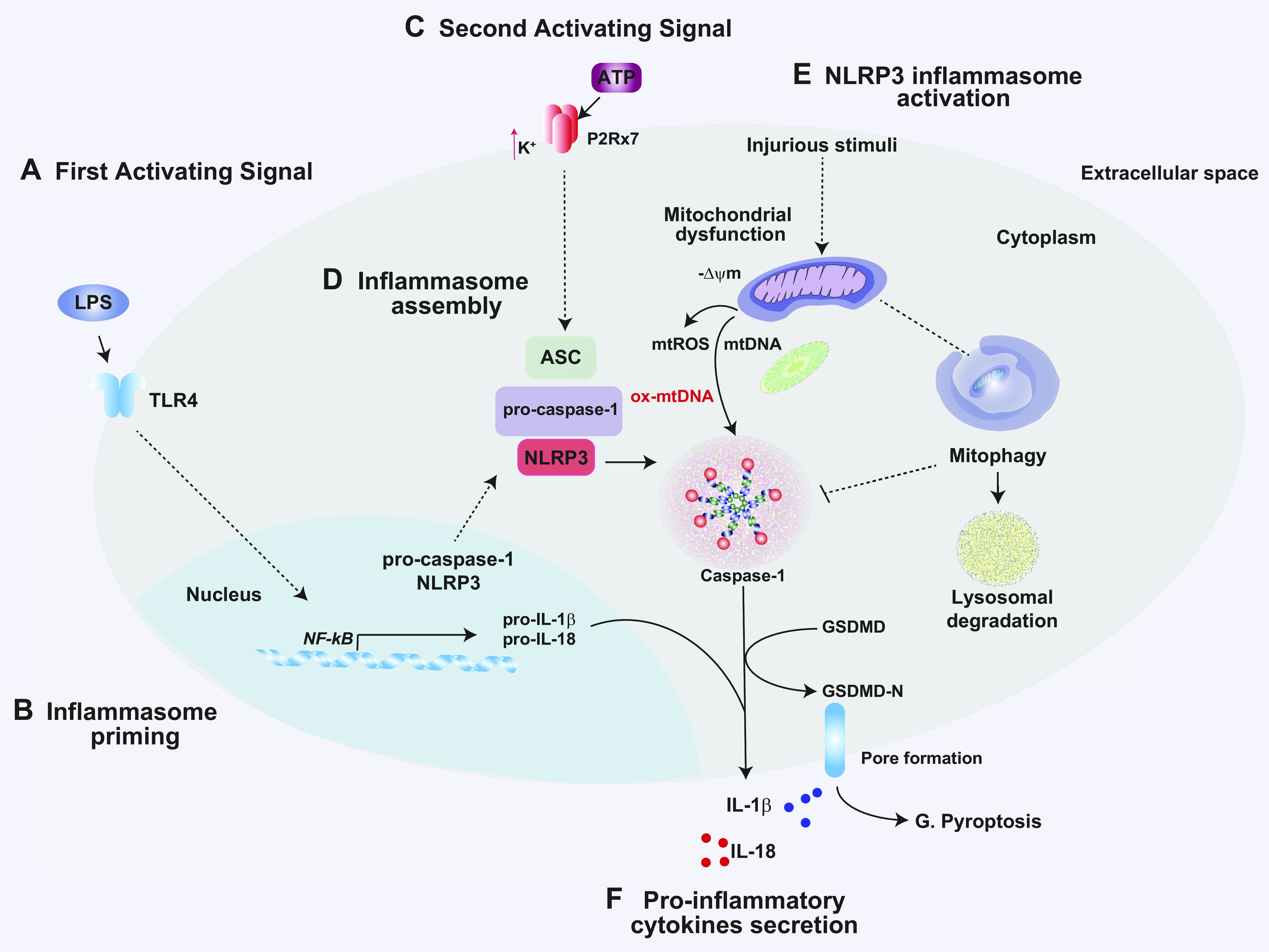
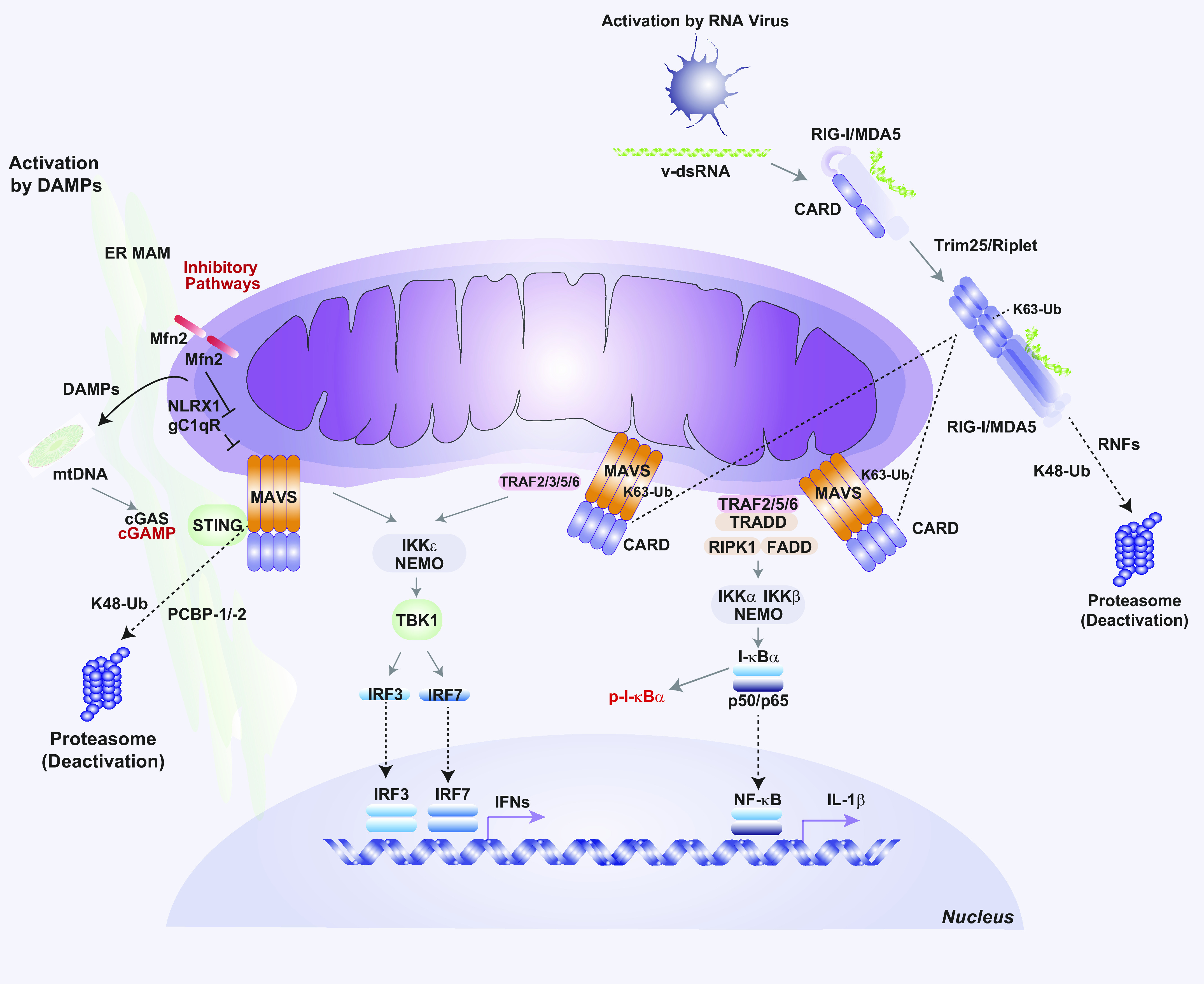
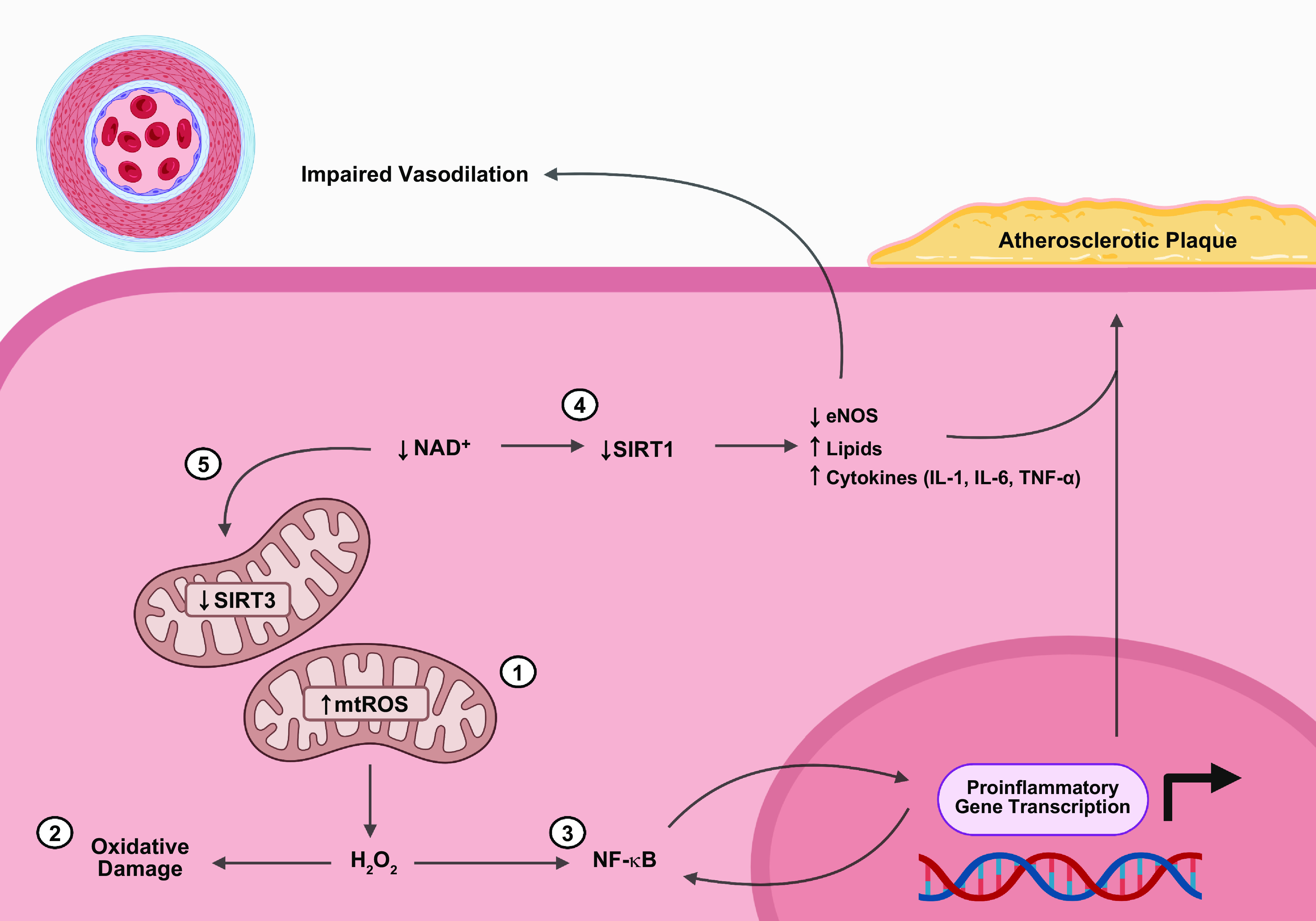
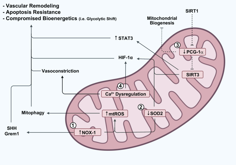
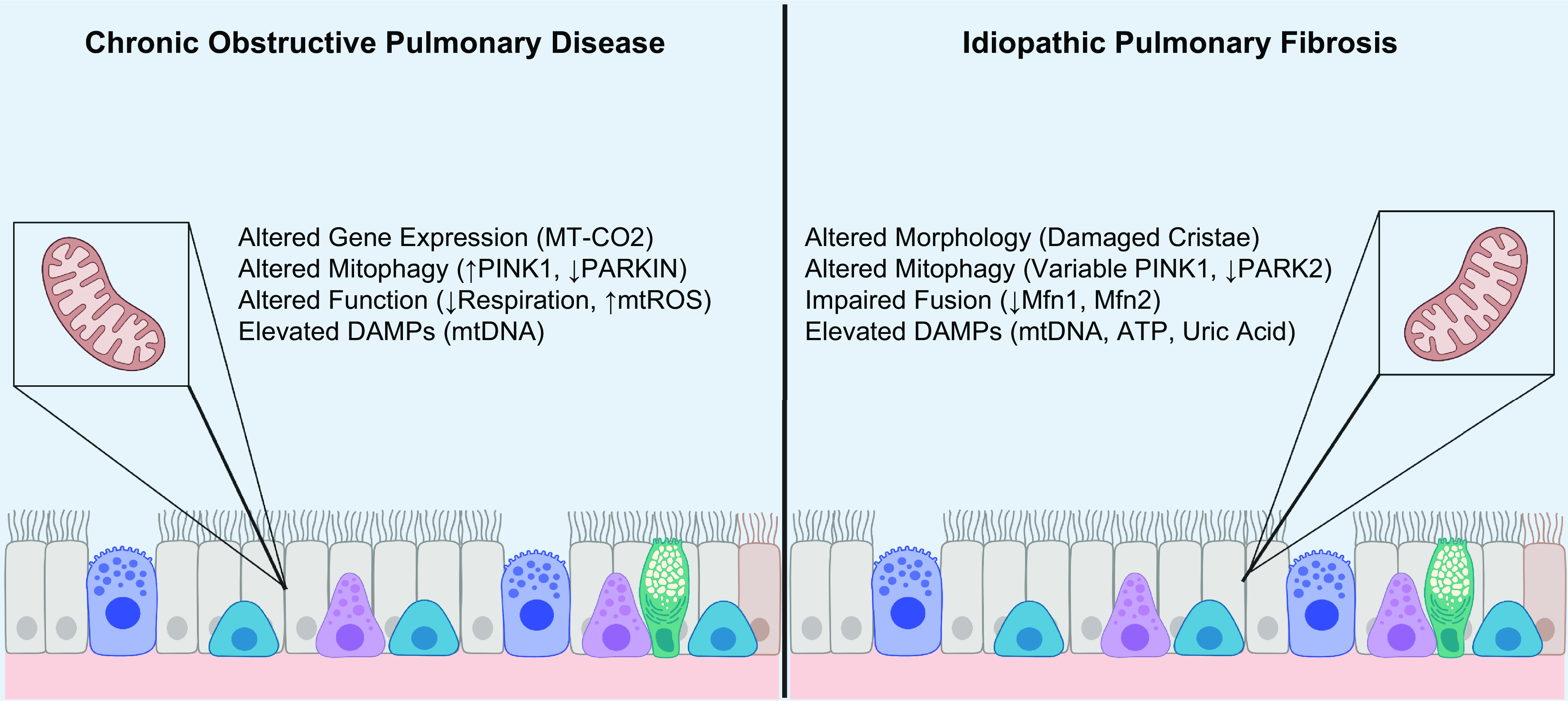
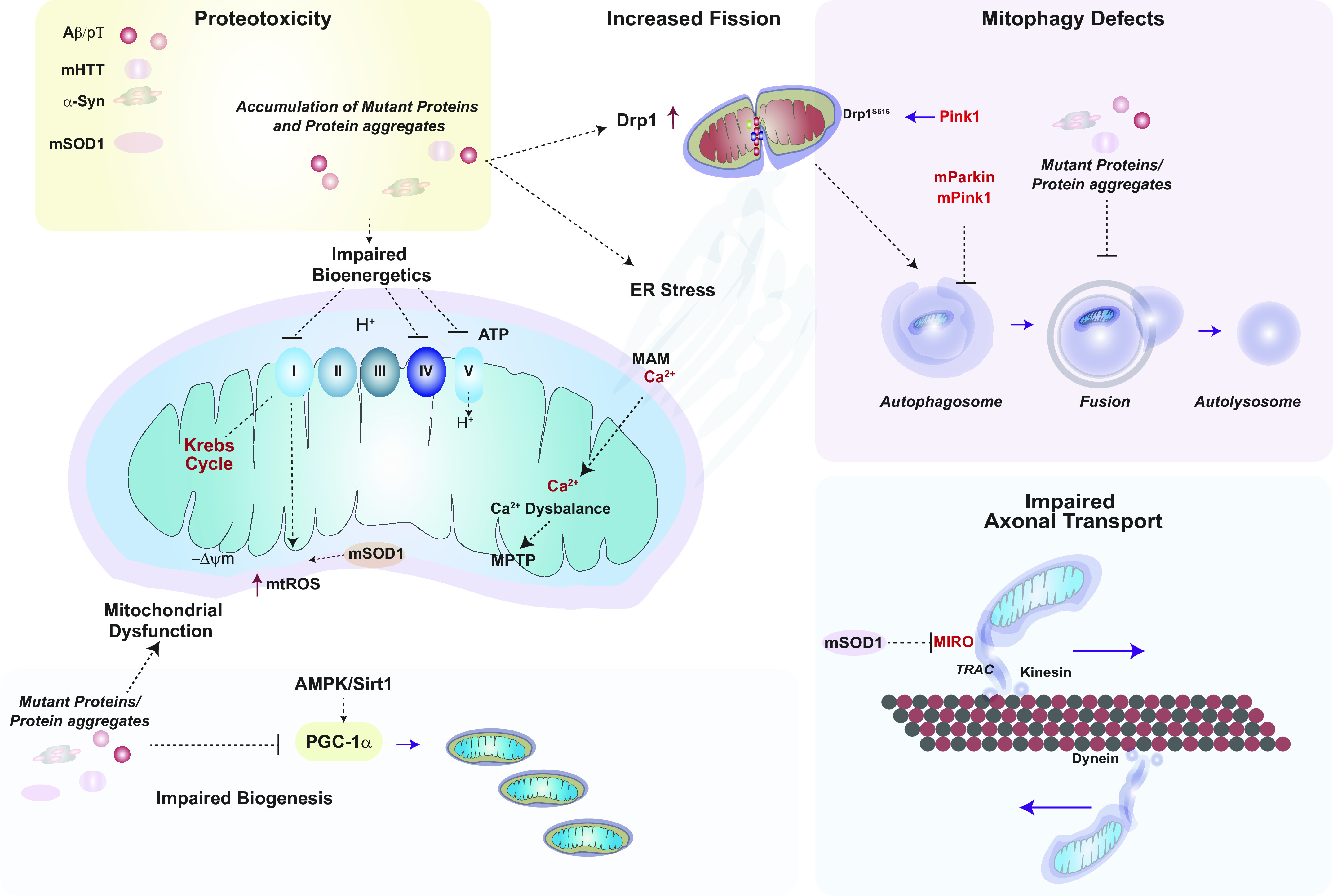
Mitochondria are well known as organelles responsible for the maintenance of cellular bioenergetics through the production of ATP. Although oxidative phosphorylation may be their most important function, mitochondria are also integral for the synthesis of metabolic precursors, calcium regulation, the production of reactive oxygen species, immune signaling, and apoptosis. Considering the breadth of their responsibilities, mitochondria are fundamental for cellular metabolism and homeostasis. Appreciating this significance, translational medicine has begun to investigate how mitochondrial dysfunction can represent a harbinger of disease. In this review, we provide a detailed overview of mitochondrial metabolism, cellular bioenergetics, mitochondrial dynamics, autophagy, mitochondrial damage-associated molecular patterns, mitochondria-mediated cell death pathways, and how mitochondrial dysfunction at any of these levels is associated with disease pathogenesis. Mitochondria-dependent pathways may thereby represent an attractive therapeutic target for ameliorating human disease.

Keywords: inflammation; mitochondria; mitochondrial dynamics; mitochondrial dysfunction; mitophagy.


Conflict of interest statement

S.W.R. is a current employee and stockholder of Proterris Inc. and a former Weill Cornell employee. A.M.K.C. is a cofounder and equity stockholder of Proterris, which develops therapeutic uses for carbon monoxide. A.M.K.C. has a use patent on CO. Additionally, A.M.K.C. has a patent in COPD. None of the other authors has any conflicts of interest, financial or otherwise, to disclose.

Figures

None
Graphical abstract
FIGURE 1.
FIGURE 1.
Mitochondrial bioenergetics. A: normal mitochondria serve essential functions for the cell including ATP generation as a result of a functioning electron transport chain, calcium handling and metabolism regulated through the mitochondrial transition pore, energy generation through the oxidation of acetyl-CoA in the Krebs cycle, and maintenance of mitochondrial integrity through mitochondrial dynamics, including mitochondrial fission and fusion. B: loss of mitochondrial bioenergetics has been linked to human disease pathogenesis. Mitochondrial reactive oxygen species (mtROS) can induce cyclophilin D, leading to mitochondrial permeability transition pore-driven necrosis and loss of mitochondrial bioenergetics, a process previously linked to amyotrophic lateral sclerosis and Parkinson’s disease onset. Mitochondrial membrane depolarization or loss of membrane potential (−ΔΨm) can ultimately lead to mitochondrial membrane rupture with the release of mitochondrial DAMPs, with implications for sepsis and fibrotic lung disease. Failure of mitofission-related mitochondrial maintenance has been linked to Alzheimer’s disease and fibrotic lung disease, whereas failure to properly dispose of abnormal mitochondria through mitophagy has been linked to neurodegenerative diseases as well as chronic lung disease. Cell death processes, including intrinsic and extrinsic apoptosis and necroptosis, cross talk to mitochondrial energetics, with implications for human disease. Cell stress secondary to inflammatory signaling can increase mitochondrial bioenergetics, leading to mtROS-driven mitochondrial DNA release (mtDNA) that mediates inflammatory diseases including sepsis and fibrotic lung disease. See glossary for abbreviations. Image created with BioRender.com, with permission.
FIGURE 2.
FIGURE 2.
Mitophagy. Mitophagy is a selective process for the turnover of mitochondria by the lysosome-dependent autophagy pathway. Distinct modes of mitophagy include receptor-dependent mitophagy (A) and PINK1/Parkin-dependent mitophagy (B), the primary pathway by which dysfunctional or depolarizing mitochondria are cleared. Under resting conditions PINK1 is imported into the mitochondria via TIM and TOM and processed by the IMM rhomboid protease PARL. A truncated form of PINK1 (ΔPINK1) is exported from the mitochondria and processed for proteasomal degradation via E3 ubiquitin ligases. In response to mitochondrial depolarization, PINK1 is stabilized on the OMM, phosphorylates ubiquitin (UB), and recruits Parkin to the mitochondria. Parkin, an E3 ubiquitin ligase, ubiquitinates OMM proteins, including Mfn1/Mfn2 and others. Ubiquitinated OMM proteins interact with mitophagy cargo adaptors, which, through LIR domains, interact with LC3 on nascent autophagosome membranes. This process facilitates recruitment of additional autophagy initiating factors, including Atg16L, and others. Mitophagy cargo adaptors include NDP52, p62 (SQSTM1), NBR1, and OPTN activated by TBK1. Receptor-dependent mitophagy (A) is important for physiological regulation of mitochondrial populations and utilizes OMM receptor proteins that interact directly with autophagosomal membrane protein LC3. These include BNIP3, Nix1, the membrane lipid cardiolipin (CL), and others (i.e., PHB2, FKB8). FUNDC1 is a mitophagy receptor that is specifically activated by hypoxia and that can also inhibit fusion via interaction with Opa1. Targeting of mitochondria to autophagosome membranes ultimately results in their assimilation in mature autophagosomes and degradation in the lysosomal compartment. See glossary for abbreviations.
FIGURE 3.
FIGURE 3.
Mitochondrial dynamics. The mitochondrial population is subject to genetic regulation of its morphology and distribution, in processes referred to as mitochondrial dynamics. Mitochondrial biogenesis ensures the proliferation and maintenance of a healthy mitochondrial population via its principal regulatory components, PGC-1α, NRF1, and TFAM. Stress conditions lead to mitochondrial dysfunction including enhanced mtROS production and mitochondrial depolarization. A: the process of fusion creates larger mitochondria from 2 or more mitochondria. This process may serve to repair dysfunctional mitochondria via complementation with healthy mitochondria. B: the process of fission generates smaller mitochondria by dividing injured mitochondria. Drp1 is the principal regulator of fission. Drp1 is activated by phosphorylation at Ser637. Initiation of fission involves recruitment and oligomerization of Drp1 by accessory molecules including Mff, Fis1, and Mid59/Mid51. Opa-1s (short form) promotes fission. Fission is enabled by interactions with the ER MAM and requires GTPase activity for execution. See glossary for abbreviations.
FIGURE 4.
FIGURE 4.
Regulation of mitochondrial biogenesis. The mitochondrial biogenesis pathway governs mitochondrial DNA (mtDNA) replication and mitochondrial proliferation. This process is regulated by peroxisome proliferator-activated receptor gamma (PPARγ) coactivator 1-α (PGC-1α) and related factors. A: several signaling pathways can trigger PGC-1α activation and downstream processes. These include the sirtuin-1 (Sirt1)-5′ adenosine monophosphate-activated protein kinase (AMPK) axis and endothelial nitric oxide synthase (eNOS)-derived nitric oxide (NO), production which activates the soluble guanylate cyclase (sGC)/guanosine 3′,5′ cyclic monophosphate (cGMP) pathway. Metabolic signals can also activate PGC-1α via the Ca2+/calmodulin-dependent kinase (CaMK) pathway and/or increased oxidative stress and mtROS production, which culminate in p38 mitogen-activated protein kinase (p38 MAPK) activation. Redox imbalance can activate PGC-1α transcription via the nuclear factor erythroid 2-related factor-2 (Nrf2)/Kelch-like ECH-associated protein 1 (Keap1) pathway. B: in the nucleus, PGC-1α drives the transcription of nuclear respiratory factors (NRF1 and NRF2). NRF1 regulates the expression of mitochondrial transcription factor-A (mitochondrial) (TFAM) and transcription factor-B2 (mitochondrial) (TFB2M). C: these nuclear-encoded factors regulate mtDNA transcription in the mitochondria via activation of mitochondrial RNA polymerase (Polrmt) at 2 promoters, the light strain promoter (LSP) and the heavy strand promoter (HSP). TFAM can also promote mtDNA replication via activation of mtDNA polymerase-gamma (Pol-γ). Increased mtDNA replication supports mitochondrial proliferation.
FIGURE 5.
FIGURE 5.
Mitochondrial damage-associated molecular patterns (DAMPs). 1: DAMPs released by necrotic cells or pathogen-associated molecular patterns (PAMPs) released during infection bind pattern recognition receptors (PRRs). Once activated, PRRs initiate a signal transduction cascade causing increased production of mitochondrial reactive oxygen species (mtROS). 2: The increased oxidative stress can fragment the mitochondrial genome and damage mitochondria, allowing mitochondrial DAMPs to enter the cytosol. 3: Cytosolic mitochondrial DNA (mtDNA) can bind toll-like receptor 9 (TLR-9), leading to activation of NF-κB, inflammasomes leading to caspase-mediated production of IL-1β and IL-18 from their zymogens, or cGAS/STING-mediated activation of NF-κB and interferon-stimulated genes (ISGs). 4: Cardiolipin can facilitate organization of the inflammasome. 5: ATP can activate inflammasome or facilitate recruitment and activation of immune cells if released to the extracellular space. 6: Mitochondrial transcription factor A (TFAM) facilitates internalization of mtDNA by endosomes where it can activate TLR-9. 7: N-formyl peptides (NFPs) can stimulate immune cells or alter gene expression through binding formyl peptide receptors (FPRs). 8: Succinate can alter gene expression by stabilizing Hypoxia-inducible factor-1α (HIF-1α). 9: Mitochondrial DAMPs can alter gene transcription, resulting in the production of tumor necrosis factor-α (TNF-α), IL-6, and other proinflammatory cytokines. Image created with BioRender.com, with permission.
FIGURE 6.
FIGURE 6.
Regulation of the NOD-, LRR- and pyrin domain-containing protein 3 (NLRP3) inflammasome by mitochondrial DAMPs. The NLRP3 inflammasome is activated by a 2-signal activation mechanism. A: first, a Toll-like receptor (TLR) ligand such as lipopolysaccharide (LPS) binds to TLR4 and induces inflammasome priming via nuclear factor-κB (NF-κB) activation. B: NF-κB drives NLRP3 transcription and the expression of pro-forms of caspase-1, IL-1β, and IL-18. C: a second activating signal, typically ATP, activates P2X purinoceptor 7 (P2Rx7) and induces K+ efflux. D: NLRP3 forms a complex with the adaptor molecule apoptosis-associated speck-like protein containing a COOH-terminal caspase recruitment domain (ASC) and pro-caspase-1. E: mitochondrial dysfunction by injurious stimuli will trigger the release of mitochondrial reactive oxygen species (mtROS) and mitochondrial DNA (mtDNA); the oxidized form of mtDNA (ox-mtDNA) has been implicated in NLRP3 inflammasome regulation. Pink1/Parkin-dependent mitophagy removes depolarizing mitochondria via the lysosomal degradation pathway and thereby acts as an inhibitor of NLRP3 inflammasome activation. F: inflammasome activation triggers the cleavage of caspase-1, which in turn promotes the maturation and secretion of inflammasome cytokines IL-1β and IL-18. G: activated caspase-1 also triggers membrane disruption and pyroptotic cell death via activation of gasdermin-D (GSDMD) via its active NH2 terminal (GSDMD-N). −ΔΨm, mitochondrial membrane potential (loss).
FIGURE 7.
FIGURE 7.
Mitochondria-dependent anti-viral responses. Mitochondrial antiviral-signaling protein (MAVS) is an OMM protein that serves as a platform for the regulation of antiviral responses. During viral infections, RIG-I (retinoic acid-inducible gene I) and MDA5 serve as cytosolic pattern recognition receptors (PRR) responsible for initiating the type-1 interferon (IFN1) response. Viral double-stranded RNA (v-dsRNA) binds to RIG1 (or MDA5) to activate the protein. RIG-I is covalently modified with K63-Ub chains by the E3 ligase tripartite motif 25 (TRIM25). The NH2-terminal caspase activation and recruitment domains (CARDs) of activated RIG-I target and bind the CARDs of MAVS. Activated MAVS forms filamentous structures that are prerequisite for downstream signaling. One arm of the MAVs pathway activates a canonical nuclear factor kappa-b (NF-kB)-dependent pathway that regulates pro-inflammatory cytokines (e.g., IL-1b) production. This pathway depends on complexes with TNF receptor-associated factors (TRAF)-2/-5/-6, TNFR1-associated death domain protein (TRADD), tripartite motif 14 (TRIM14) and other proteins such as FADD and RIPK1. A second arm of the MAVS pathway involves complexes of TRAFs-2/-3/-5/-6 to activate the IFN1 response. This pathway also requires NEMO/IKKe and TBK1, which phosphorylates and activates IRF3 and IRF7. Homodimerization of IRF3 and IRF7 triggers the transcription of IFN genes. MAVS can be stimulated via release of DAMPs from mitochondria, such as mtDNA, which activates MAVS via a cGAS/cGAMP/STING axis. MAVS activation can be inhibited by fusion protein MFN2 and interactions with the ER MAM, and by other factors including NLRX1 and gC1qR. MAVS and RIG-I are marked for proteasomal degradation via K48-Ub modification by PCB1/PCBP2 or RNFs, respectively. cGAS, cyclic GMP-AMP synthase; cGAMP, cyclic GMP-AMP; gC1qR, globular C1q receptor; DAMP, damage associated molecular pattern; ER, endoplasmic reticulum; FADD, Fas-associated protein with death domain; IFN1, interferon type 1; IKK, I-kB kinase (a, b, e); IRF-3/-7, interferon regulatory factor-3/-7; I-kBa, inhibitor of NF-kB (alpha); MAM, mitochondria-associated membrane; MDA5: melanoma differentiation-associated protein-5; MFN2: mitofusin-2; mtDNA, mitochondrial DNA; NEMO, NF-kB essential modulator; NLRX1, NLR family member X1; PCBP-1/-2, poly(RC)-binding protein (PCBP)-1,-2; RIPK1, receptor-interacting serine/threonine-protein kinase 1; RNF, ring finger proteins; STING, stimulator of interferon genes; TBK1, TANK-binding kinase-1; TRADD, TNFR1-associated death domain protein.
FIGURE 8.
FIGURE 8.
Mitochondrial dysfunction in age-related cardiovascular disease (CVD). 1: The proximity of the mitochondrial genome to the ETC is associated with the age-related accrual of mutations, which ultimately impair mitochondrial bioenergetics and increase mtROS. 2: To manage the rise in mtROS, superoxide dismutase 2 (SOD2) converts O2•− to H2O2, an uncharged lipophilic molecule that transverses mitochondrial membranes to enter the cytosol. Once in the cytosol, H2O2 can impair cell function by causing oxidative damage to cellular components (DNA, proteins, and lipids) 3: Alternatively, H2O2 can stimulate NF-κB nuclear translocation to promote the expression of proinflammatory genes such as TNF-α, IL-6, VCAM-1, ICAM-1, and MMP-9 that are important for atherosclerotic plaque formation. By upregulating proinflammatory cytokines, the NF-κB signal transduction cascade can become self-sustaining, resulting in a chronic inflammatory state. Of note, several other pathways, such as the renin-angiotensin system and TNF-α, can contribute to the development of age-related CVD by converging on NF-κB (not shown). 4: During aging, NAD+ levels decline, impairing sirtuin function. Decreased sirtuin 1 (SIRT1) activity is associated with downregulation of endothelial nitric oxide synthase (eNOS) and increased production of lipids, and inflammatory cytokines. 5: In contrast, decreased sirtuin 3 (SIRT3) activity is associated with downregulation of SOD2 and PGC-1α, which further compromises mitochondrial function, bioenergetics, and biogenesis. See glossary for abbreviations. Image created with BioRender.com, with permission.
FIGURE 9.
FIGURE 9.
Mitochondrial dysfunction in pulmonary hypertension. PAH is associated with a series of mitochondria-mediated metabolic derangements that contribute to vascular remodeling, apoptosis resistance, and bioenergetic compromise. 1: Increased NOX-1 encourages vascular remodeling via ROS-mediated stimulation of sonic hedgehog (SHH) and gremlin-1 (Grem1). Furthermore, the increased mtROS stimulates mitophagy. As mitochondria are removed, endothelial cells rely on glycolysis to meet their energetic demands. This glycolytic shift, also known as the Warburg effect, is associated with altered mitochondrial membrane polarization, which promotes apoptosis resistance by inhibiting the release of proapoptotic factors. 2: Decreased superoxide dismutase 2 (SOD2) is not only associated with increased mtROS but also with HIF-1α stabilization. 3: Additionally, decreased PGC-1α activity inhibits SIRT3 expression through impaired coactivation of ERR-α. This promotes HIF-1α stabilization and STAT3 activation, which promotes hyperproliferation and apoptosis resistance. 4: Mitochondrial calcium dysregulation, which can occur through multiple mechanisms in PAH, promotes pulmonary vasoconstriction and HIF-1α stabilization. See glossary for other abbreviations. Image created with BioRender.com, with permission.
FIGURE 10.
FIGURE 10.
Mitochondrial dysfunction in chronic lung disease. Mitochondrial dysfunction plays a central role in the pathogenesis of chronic lung diseases, such as COPD and IPF. Genomic studies in COPD have identified differential gene expression among patients, with MT-CO2 being upregulated in emphysema. Additionally, mitophagy and cell death pathways appear to be dysregulated in COPD, with some studies noting increased PINK1 associated with upregulation of RIPK3-mediated necroptosis and others observing decreased PARKIN expression leading to mitophagy impairment and cellular senescence. COPD is also associated with impaired cellular respiration and increased generation of mtROS. In contrast, IPF is associated with morphological changes to mitochondria, such as the swelling of cristae. Mitophagy also appears dysregulated with variable expression of PINK1 and reduced expression of PARK2. Mitochondrial fusion is also involved in IPF, with Mfn1 and Mfn2 deficiency associated with lung fibrosis. Disease severity in both COPD and IPF has also been associated with elevated levels of circulating mitochondrial DAMPs. See glossary for abbreviations. Image created with BioRender.com, with permission.
FIGURE 11.
FIGURE 11.
Mitochondrial dysfunction in neurodegenerative disease. Mitochondrial dysfunction is a hallmark of neurodegenerative diseases. Mutant proteins and their aggregates have been implicated in the pathogenesis of various neurodegenerative diseases, including AD, HD, PD, and ALS, by disrupting proteostasis and mitochondrial function. Mutant Aβ and hyperphosphorylated Tau (p-T), mutant Huntingtin (mHTT), and α-synuclein (α-Syn) aggregates can block electron transport chain (ETC) activities at various complexes. α-Syn and mHTT may also trigger mitochondrial dysfunction and mitochondrial reactive oxygen species (mtROS) production. mHTT, p-T, and mutant Aβ can interfere with the Sirt1-PGC-1α axis and its initiation of mitochondrial biogenesis. mHTT, p-T, and mutant Aβ can promote mitochondrial fragmentation via interactions with dynamin-related protein-1 (Drp1). Mutant forms of PTEN-induced kinase 1 (PINK1) can also interfere with PINK1-dependent Drp1 S616 phosphorylation. Autophagy and mitophagy processes may be disrupted by mutant proteins: mHTT can interfere with autophagosome formation, whereas p-T and mutant Aβ block autophagosome-lysosome fusion. Mutant PINK1 (mPINK1) and Parkin (mParkin) may cause mitophagy dysfunction. Superoxide dismutase 1 (SOD1) catalyzes the dismutation of superoxide generated by mitochondria, whereas in ALS the mutant form (mSOD1) is defective at removing superoxide, which favors peroxynitrite formation. mSOD1 can also interfere with mitochondrial axonal transport via inhibiting the transport regulator protein mitochondrial Rho GTPase (MIRO) and by promoting the autophagosome-dependent degradation of MIRO. −ΔΨm, mitochondrial membrane potential (loss); ER, endoplasmic reticulum; MAM, mitochondria-associated membrane; p62, SQSTM1 autophagy cargo adaptor protein. See glossary for other abbreviations.

Similar articles

Cited by

References

    1. Vakifahmetoglu-Norberg H, Ouchida AT, Norberg E. The role of mitochondria in metabolism and cell death. Biochem Biophys Res Commun 482: 426–431, 2017. doi:10.1016/j.bbrc.2016.11.088. - DOI - PubMed
    1. Rizzuto R, De Stefani D, Raffaello A, Mammucari C. Mitochondria as sensors and regulators of calcium signalling. Nat Rev Mol Cell Biol 13: 566–578, 2012. doi:10.1038/nrm3412. - DOI - PubMed
    1. Orrenius S, Zhivotovsky B, Nicotera P. Regulation of cell death: the calcium-apoptosis link. Nat Rev Mol Cell Biol 4: 552–565, 2003. doi:10.1038/nrm1150. - DOI - PubMed
    1. Jin HS, Suh HW, Kim SJ, Jo EK. Mitochondrial control of innate immunity and inflammation. Immune Netw 17: 77–88, 2017. doi:10.4110/in.2017.17.2.77. - DOI - PMC - PubMed
    1. Balaban RS, Nemoto S, Finkel T. Mitochondria, oxidants, and aging. Cell 120: 483–495, 2005. doi:10.1016/j.cell.2005.02.001. - DOI - PubMed

Publication types

Substances

LinkOut - more resources