

The effect of weight loss following 18 months of lifestyle intervention on brain age assessed with resting-state functional connectivity
- PMID: 37022140
- PMCID: PMC10174688
- DOI: 10.7554/eLife.83604
The effect of weight loss following 18 months of lifestyle intervention on brain age assessed with resting-state functional connectivity
Abstract
Background: Obesity negatively impacts multiple bodily systems, including the central nervous system. Retrospective studies that estimated chronological age from neuroimaging have found accelerated brain aging in obesity, but it is unclear how this estimation would be affected by weight loss following a lifestyle intervention.
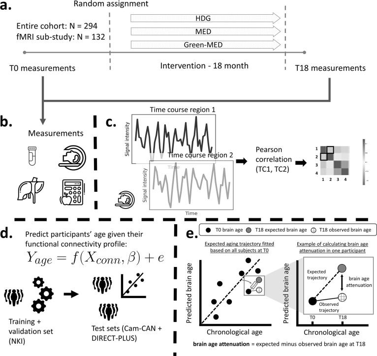
Methods: In a sub-study of 102 participants of the Dietary Intervention Randomized Controlled Trial Polyphenols Unprocessed Study (DIRECT-PLUS) trial, we tested the effect of weight loss following 18 months of lifestyle intervention on predicted brain age based on magnetic resonance imaging (MRI)-assessed resting-state functional connectivity (RSFC). We further examined how dynamics in multiple health factors, including anthropometric measurements, blood biomarkers, and fat deposition, can account for changes in brain age.
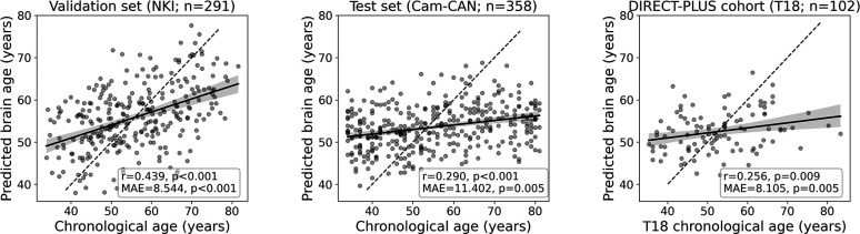
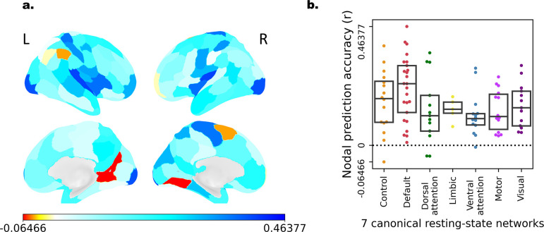
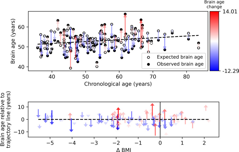
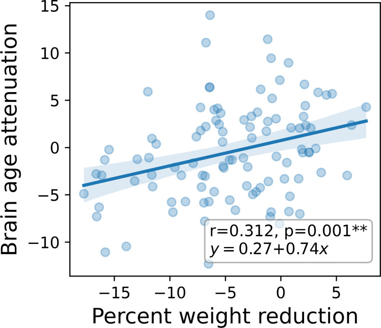
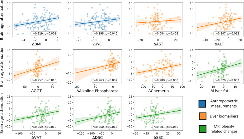
Results: To establish our method, we first demonstrated that our model could successfully predict chronological age from RSFC in three cohorts (n=291;358;102). We then found that among the DIRECT-PLUS participants, 1% of body weight loss resulted in an 8.9 months' attenuation of brain age. Attenuation of brain age was significantly associated with improved liver biomarkers, decreased liver fat, and visceral and deep subcutaneous adipose tissues after 18 months of intervention. Finally, we showed that lower consumption of processed food, sweets and beverages were associated with attenuated brain age.
Conclusions: Successful weight loss following lifestyle intervention might have a beneficial effect on the trajectory of brain aging.
Funding: The German Research Foundation (DFG), German Research Foundation - project number 209933838 - SFB 1052; B11, Israel Ministry of Health grant 87472511 (to I Shai); Israel Ministry of Science and Technology grant 3-13604 (to I Shai); and the California Walnuts Commission 09933838 SFB 105 (to I Shai).
Keywords: MRI; Mediterranean diet; brain age; epidemiology; functional connectivity; global health; human; lifestyle intervention; obesity.
Plain language summary
Obesity is linked with the brain aging faster than would normally be expected. Researchers are able to capture this process by calculating a person’s ‘brain age’ – how old their brain appears on detailed scans, regardless of chronological age. This approach also helps to monitor how certain factors, such as lifestyle, can influence brain aging over relatively short time scales. It is not clear whether lifestyle interventions that promote weight loss can help to slow obesity-driven brain aging. To answer this question, Levakov et al. studied 102 individuals who met the criteria for obesity and took part in a lifestyle intervention aimed to improve diet and physical activity levels over 18 months. The participants received a brain scan at the beginning and the end of the program; additional tests and measurements were also conducted at these times to capture other biological processes impacted by obesity, such as liver health. Levakov et al. used the brain scans taken at the start and end of the study to examine the impact of the lifestyle intervention on the aging trajectory. The results revealed that a reduction in body weight of 1% led to the participants’ brain age being nearly 9 months younger than the expected brain age after 18 months. This attenuated aging was associated with changes in other biological measures, such as decreased liver fat and liver enzymes. Increases in liver fat and production of specific liver enzymes were previously shown to negatively impact brain health in Alzheimer’s disease. Finally, examining more closely the food consumption reports completed by participants showed that reduced consumption of processed food, sweets and beverages were linked to attenuated brain aging. The findings show that lifestyle interventions which promote weight loss can have a beneficial impact on the aging trajectory of the brain observed with obesity. The next steps will include determining whether slowing down obesity-driven brain aging results in better clinical outcomes for patients. In addition, the work by Levakov et al. demonstrates a potential strategy to evaluate the success of lifestyle changes on brain health. With global rates of obesity rising, identifying interventions that have a positive impact on brain health could have important clinical, educational and social impacts.
© 2023, Levakov, Kaplan et al.
Conflict of interest statement
GL, AK, AY, ER, GT, HZ, UC, MS, IS, GA, IS No competing interests declared, MB has received consulting fees from Amgen, Astra Zeneca, Boehringer-Ingelheim, Bayer, Lilly, Novo Nordisk, Novartis, Sanofi and Pfizer; and fees for lectures/ presentations from Amgen, Astra Zeneca, Boehringer-Ingelheim, Bayer, Daiichi-Sankyo, Lilly, Novo Nordisk, Novartis, Sanofi and Pfizer. The author is also on the advisory board for Boehringer-Ingelheim. The author has no other competing interests to declare
Figures
Update of
- doi: 10.1101/2022.09.21.22280182
Similar articles
-
The effect of weight management interventions that include a diet component on weight-related outcomes in pregnant and postpartum women: a systematic review protocol.JBI Database System Rev Implement Rep. 2015 Jan;13(1):88-98. doi: 10.11124/jbisrir-2015-1812. JBI Database System Rev Implement Rep. 2015. PMID: 26447010
-
Lifestyle weight-loss intervention may attenuate methylation aging: the CENTRAL MRI randomized controlled trial.Clin Epigenetics. 2021 Mar 4;13(1):48. doi: 10.1186/s13148-021-01038-0. Clin Epigenetics. 2021. PMID: 33663610 Free PMC article. Clinical Trial.
-
The effectiveness of web-based programs on the reduction of childhood obesity in school-aged children: A systematic review.JBI Libr Syst Rev. 2012;10(42 Suppl):1-14. doi: 10.11124/jbisrir-2012-248. JBI Libr Syst Rev. 2012. PMID: 27820152
-
The Minderoo-Monaco Commission on Plastics and Human Health.Ann Glob Health. 2023 Mar 21;89(1):23. doi: 10.5334/aogh.4056. eCollection 2023. Ann Glob Health. 2023. PMID: 36969097 Free PMC article. Review.
-
Offspring body size and metabolic profile - effects of lifestyle intervention in obese pregnant women.Dan Med J. 2014 Jul;61(7):B4893. Dan Med J. 2014. PMID: 25123127 Review.
Cited by
-
Deep learning-based BMI inference from structural brain MRI reflects brain alterations following lifestyle intervention.Hum Brain Mapp. 2024 Feb 15;45(3):e26595. doi: 10.1002/hbm.26595. Hum Brain Mapp. 2024. PMID: 38375968 Free PMC article. Clinical Trial.
-
Dilemmas in Elderly Diabetes and Clinical Practice Involving Traditional Chinese Medicine.Pharmaceuticals (Basel). 2024 Jul 16;17(7):953. doi: 10.3390/ph17070953. Pharmaceuticals (Basel). 2024. PMID: 39065801 Free PMC article. Review.
-
Mitigating Age-Related Cognitive Decline and Oxidative Status in Rats Treated with Catechin and Polyphenon-60.Nutrients. 2024 Jan 26;16(3):368. doi: 10.3390/nu16030368. Nutrients. 2024. PMID: 38337652 Free PMC article.
-
The Role of Gut Microbiota in the Development and Treatment of Obesity and Overweight: A Literature Review.J Clin Med. 2025 Jul 11;14(14):4933. doi: 10.3390/jcm14144933. J Clin Med. 2025. PMID: 40725626 Free PMC article. Review.
-
A narrative review about cognitive impairment in Metabolic Dysfunction-Associated Steatotic Liver Disease (MASLD): Another matter to face through a holistic approach.J Adv Res. 2025 Feb;68:231-240. doi: 10.1016/j.jare.2024.02.007. Epub 2024 Feb 17. J Adv Res. 2025. PMID: 38369241 Free PMC article. Review.
References
-
- Abarca-Gómez L, Abdeen ZA, Hamid ZA, Abu-Rmeileh NM, Acosta-Cazares B, Acuin C, Adams RJ, Aekplakorn W, Afsana K, Aguilar-Salinas CA, Agyemang C, Ahmadvand A, Ahrens W, Ajlouni K, Akhtaeva N, Al-Hazzaa HM, Al-Othman AR, Al-Raddadi R, Al Buhairan F, Al Dhukair S, Ali MM, Ali O, Alkerwi A, Alvarez-Pedrerol M, Aly E, Amarapurkar DN, Amouyel P, Amuzu A, Andersen LB, Anderssen SA, Andrade DS, Ängquist LH, Anjana RM, Aounallah-Skhiri H, Araújo J, Ariansen I, Aris T, Arlappa N, Arveiler D, Aryal KK, Aspelund T, Assah FK, Assunção MCF, Aung MS, Avdicová M, Azevedo A, Azizi F, Babu BV, Bahijri S, Baker JL, Balakrishna N, Bamoshmoosh M, Banach M, Bandosz P, Banegas JR, Barbagallo CM, Barceló A, Barkat A, Barros AJ, Barros MV, Bata I, Batieha AM, Batista RL, Batyrbek A, Baur LA, Beaglehole R, Romdhane HB, Benedics J, Benet M, Bennett JE, Bernabe-Ortiz A, Bernotiene G, Bettiol H, Bhagyalaxmi A, Bharadwaj S, Bhargava SK, Bhatti Z, Bhutta ZA, Bi H, Bi Y, Biehl A, Bikbov M, Bista B, Bjelica DJ, Bjerregaard P, Bjertness E, Bjertness MB, Björkelund C, Blokstra A, Bo S, Bobak M, Boddy LM, Boehm BO, Boeing H, Boggia JG, Boissonnet CP, Bonaccio M, Bongard V, Bovet P, Braeckevelt L, Braeckman L, Bragt MC, Brajkovich I, Branca F, Breckenkamp J, Breda J, Brenner H, Brewster LM, Brian GR, Brinduse L, Bruno G, Bueno-de-Mesquita HB, Bugge A, Buoncristiano M, Burazeri G, Burns C, de León AC, Cacciottolo J, Cai H, Cama T, Cameron C, Camolas J, Can G, Cândido APC, Capanzana M, Capuano V, Cardoso VC, Carlsson AC, Carvalho MJ, Casanueva FF, Casas JP, Caserta CA, Chamukuttan S, Chan AW, Chan Q, Chaturvedi HK, Chaturvedi N, Chen CJ, Chen F, Chen H, Chen S, Chen Z, Cheng CY, Chetrit A, Chikova-Iscener E, Chiolero A, Chiou ST, Chirita-Emandi A, Chirlaque MD, Cho B, Cho Y, Christensen K, Christofaro DG, Chudek J, Cifkova R, Cinteza E, Claessens F, Clays E, Concin H, Confortin SC, Cooper C, Cooper R, Coppinger TC, Costanzo S, Cottel D, Cowell C, Craig CL, Crujeiras AB, Cucu A, D’Arrigo G, d’Orsi E, Dallongeville J, Damasceno A, Damsgaard CT, Danaei G, Dankner R, Dantoft TM, Dastgiri S, Dauchet L, Davletov K, De Backer G, De Bacquer D, De Curtis A, de Gaetano G, De Henauw S, de Oliveira PD, De Ridder K, De Smedt D, Deepa M, Deev AD, Dehghan A, Delisle H, Delpeuch F, Deschamps V, Dhana K, Di Castelnuovo AF, Dias-da-Costa JS, Diaz A, Dika Z, Djalalinia S, Do HT, Dobson AJ, Donati MB, Donfrancesco C, Donoso SP, Döring A, Dorobantu M, Dorosty AR, Doua K, Drygas W, Duan JL, Duante C, Duleva V, Dulskiene V, Dzerve V, Dziankowska-Zaborszczyk E, Egbagbe EE, Eggertsen R, Eiben G, Ekelund U, El Ati J, Elliott P, Engle-Stone R, Erasmus RT, Erem C, Eriksen L, Eriksson JG, la Peña JE, Evans A, Faeh D, Fall CH, Sant’Angelo VF, Farzadfar F, Felix-Redondo FJ, Ferguson TS, Fernandes RA, Fernández-Bergés D, Ferrante D, Ferrari M, Ferreccio C, Ferrieres J, Finn JD, Fischer K, Flores EM, Föger B, Foo LH, Forslund AS, Forsner M, Fouad HM, Francis DK, Franco M, Franco OH, Frontera G, Fuchs FD, Fuchs SC, Fujita Y, Furusawa T, Gaciong Z, Gafencu M, Galeone D, Galvano F, Garcia-de-la-Hera M, Gareta D, Garnett SP, Gaspoz JM, Gasull M, Gates L, Geiger H, Geleijnse JM, Ghasemian A, Giampaoli S, Gianfagna F, Gill TK, Giovannelli J, Giwercman A, Godos J, Gogen S, Goldsmith RA, Goltzman D, Gonçalves H, González-Leon M, González-Rivas JP, Gonzalez-Gross M, Gottrand F, Graça AP, Graff-Iversen S, Grafnetter D, Grajda A, Grammatikopoulou MG, Gregor RD, Grodzicki T, Grøntved A, Grosso G, Gruden G, Grujic V, Gu D, Gualdi-Russo E, Guallar-Castillón P, Guan OP, Gudmundsson EF, Gudnason V, Guerrero R, Guessous I, Guimaraes AL, Gulliford MC, Gunnlaugsdottir J, Gunter M, Guo X, Guo Y, Gupta PC, Gupta R, Gureje O, Gurzkowska B, Gutierrez L, Gutzwiller F, Hadaegh F, Hadjigeorgiou CA, Si-Ramlee K, Halkjær J, Hambleton IR, Hardy R, Kumar RH, Hassapidou M, Hata J, Hayes AJ, He J, Heidinger-Felso R, Heinen M, Hendriks ME, Henriques A, Cadena LH, Herrala S, Herrera VM, Herter-Aeberli I, Heshmat R, Hihtaniemi IT, Ho SY, Ho SC, Hobbs M, Hofman A, Hopman WM, Horimoto AR, Hormiga CM, Horta BL, Houti L, Howitt C, Htay TT, Htet AS, Htike MMT, Hu Y, Huerta JM, Petrescu CH, Huisman M, Husseini A, Huu CN, Huybrechts I, Hwalla N, Hyska J, Iacoviello L, Iannone AG, Ibarluzea JM, Ibrahim MM, Ikeda N, Ikram MA, Irazola VE, Islam M, Ismail A, Ivkovic V, Iwasaki M, Jackson RT, Jacobs JM, Jaddou H, Jafar T, Jamil KM, Jamrozik K, Janszky I, Jarani J, Jasienska G, Jelakovic A, Jelakovic B, Jennings G, Jeong SL, Jiang CQ, Jiménez-Acosta SM, Joffres M, Johansson M, Jonas JB, Jørgensen T, Joshi P, Jovic DP, Józwiak J, Juolevi A, Jurak G, Jureša V, Kaaks R, Kafatos A, Kajantie EO, Kalter-Leibovici O, Kamaruddin NA, Kapantais E, Karki KB, Kasaeian A, Katz J, Kauhanen J, Kaur P, Kavousi M, Kazakbaeva G, Keil U, Boker LK, Keinänen-Kiukaanniemi S, Kelishadi R, Kelleher C, Kemper HC, Kengne AP, Kerimkulova A, Kersting M, Key T, Khader YS, Khalili D, Khang YH, Khateeb M, Khaw KT, Khouw IM, Kiechl-Kohlendorfer U, Kiechl S, Killewo J, Kim J, Kim YY, Klimont J, Klumbiene J, Knoflach M, Koirala B, Kolle E, Kolsteren P, Korrovits P, Kos J, Koskinen S, Kouda K, Kovacs VA, Kowlessur S, Koziel S, Kratzer W, Kriemler S, Kristensen PL, Krokstad S, Kromhout D, Kruger HS, Kubinova R, Kuciene R, Kuh D, Kujala UM, Kulaga Z, Kumar RK, Kunešová M, Kurjata P, Kusuma YS, Kuulasmaa K, Kyobutungi C, La QN, Laamiri FZ, Laatikainen T, Lachat C, Laid Y, Lam TH, Landrove O, Lanska V, Lappas G, Larijani B, Laugsand LE, Lauria L, Laxmaiah A, Bao KLN, Le TD, Lebanan MAO, Leclercq C, Lee J, Lee J, Lehtimäki T, León-Muñoz LM, Levitt NS, Li Y, Lilly CL, Lim WY, Lima-Costa MF, Lin HH, Lin X, Lind L, Linneberg A, Lissner L, Litwin M, Liu J, Loit HM, Lopes L, Lorbeer R, Lotufo PA, Lozano JE, Luksiene D, Lundqvist A, Lunet N, Lytsy P, Ma G, Ma J, Machado-Coelho GL, Machado-Rodrigues AM, Machi S, Maggi S, Magliano DJ, Magriplis E, Mahaletchumy A, Maire B, Majer M, Makdisse M, Malekzadeh R, Malhotra R, Rao KM, Malyutina S, Manios Y, Mann JI, Manzato E, Margozzini P, Markaki A, Markey O, Marques LP, Marques-Vidal P, Marrugat J, Martin-Prevel Y, Martin R, Martorell R, Martos E, Marventano S, Masoodi SR, Mathiesen EB, Matijasevich A, Matsha TE, Mazur A, Mbanya JCN, McFarlane SR, McGarvey ST, McKee M, McLachlan S, McLean RM, McLean SB, McNulty BA, Yusof SM, Mediene-Benchekor S, Medzioniene J, Meirhaeghe A, Meisfjord J, Meisinger C, Menezes AMB, Menon GR, Mensink GB, Meshram II, Metspalu A, Meyer HE, Mi J, Michaelsen KF, Michels N, Mikkel K, Miller JC, Minderico CS, Miquel JF, Miranda JJ, Mirkopoulou D, Mirrakhimov E, Mišigoj-Durakovic M, Mistretta A, Mocanu V, Modesti PA, Mohamed MK, Mohammad K, Mohammadifard N, Mohan V, Mohanna S, Yusoff MFM, Molbo D, Møllehave LT, Møller NC, Molnár D, Momenan A, Mondo CK, Monterrubio EA, Monyeki KDK, Moon JS, Moreira LB, Morejon A, Moreno LA, Morgan K, Mortensen EL, Moschonis G, Mossakowska M, Mostafa A, Mota J, Mota-Pinto A, Motlagh ME, Motta J, Mu TT, Muc M, Muiesan ML, Müller-Nurasyid M, Murphy N, Mursu J, Murtagh EM, Musil V, Nabipour I, Nagel G, Naidu BM, Nakamura H, Námešná J, Nang EEK, Nangia VB, Nankap M, Narake S, Nardone P, Navarrete-Muñoz EM, Neal WA, Nenko I, Neovius M, Nervi F, Nguyen CT, Nguyen ND, Nguyen QN, Nieto-Martínez RE, Ning G, Ninomiya T, Nishtar S, Noale M, Noboa OA, Norat T, Norie S, Noto D, Nsour MA, O’Reilly D, Obreja G, Oda E, Oehlers G, Oh K, Ohara K, Olafsson Ö, Olinto MTA, Oliveira IO, Oltarzewski M, Omar MA, Onat A, Ong SK, Ono LM, Ordunez P, Ornelas R, Ortiz AP, Osler M, Osmond C, Ostojic SM, Ostovar A, Otero JA, Overvad K, Owusu-Dabo E, Paccaud FM, Padez C, Pahomova E, Pajak A, Palli D, Palloni A, Palmieri L, Pan WH, Panda-Jonas S, Pandey A, Panza F, Papandreou D, Park SW, Parnell WR, Parsaeian M, Pascanu IM, Patel ND, Pecin I, Pednekar MS, Peer N, Peeters PH, Peixoto SV, Peltonen M, Pereira AC, Perez-Farinos N, Pérez CM, Peters A, Petkeviciene J, Petrauskiene A, Peykari N, Pham ST, Pierannunzio D, Pigeot I, Pikhart H, Pilav A, Pilotto L, Pistelli F, Pitakaka F, Piwonska A, Plans-Rubió P, Poh BK, Pohlabeln H, Pop RM, Popovic SR, Porta M, Portegies ML, Posch G, Poulimeneas D, Pouraram H, Pourshams A, Poustchi H, Pradeepa R, Prashant M, Price JF, Puder JJ, Pudule I, Puiu M, Punab M, Qasrawi RF, Qorbani M, Bao TQ, Radic I, Radisauskas R, Rahman M, Rahman M, Raitakari O, Raj M, Rao SR, Ramachandran A, Ramke J, Ramos E, Ramos R, Rampal L, Rampal S, Rascon-Pacheco RA, Redon J, Reganit PFM, Ribas-Barba L, Ribeiro R, Riboli E, Rigo F, de Wit TFR, Rito A, Ritti-Dias RM, Rivera JA, Robinson SM, Robitaille C, Rodrigues D, Rodríguez-Artalejo F, del Cristo Rodriguez-Perez M, Rodríguez-Villamizar LA, Rojas-Martinez R, Rojroongwasinkul N, Romaguera D, Ronkainen K, Rosengren A, Rouse I, Roy JG, Rubinstein A, Rühli FJ, Ruiz-Betancourt BS, Russo P, Rutkowski M, Sabanayagam C, Sachdev HS, Saidi O, Salanave B, Martinez ES, Salmerón D, Salomaa V, Salonen JT, Salvetti M, Sánchez-Abanto J, Sans S, Marina LS, Santos DA, Santos IS, Santos O, dos Santos RN, Santos R, Saramies JL, Sardinha LB, Sarrafzadegan N, Saum KU, Savva S, Savy M, Scazufca M, Rosario AS, Schargrodsky H, Schienkiewitz A, Schipf S, Schmidt CO, Schmidt IM, Schultsz C, Schutte AE, Sein AA, Sen A, Senbanjo IO, Sepanlou SG, Serra-Majem L, Shalnova SA, Sharma SK, Shaw JE, Shibuya K, Shin DW, Shin Y, Shiri R, Siani A, Siantar R, Sibai AM, Silva AM, Silva DAS, Simon M, Simons J, Simons LA, Sjöberg A, Sjöström M, Skovbjerg S, Slowikowska-Hilczer J, Slusarczyk P, Smeeth L, Smith MC, Snijder MB, So HK, Sobngwi E, Söderberg S, Soekatri MY, Solfrizzi V, Sonestedt E, Song Y, Sørensen TI, Soric M, Jérome CS, Soumare A, Spinelli A, Spiroski I, Staessen JA, Stamm H, Starc G, Stathopoulou MG, Staub K, Stavreski B, Steene-Johannessen J, Stehle P, Stein AD, Stergiou GS, Stessman J, Stieber J, Stöckl D, Stocks T, Stokwiszewski J, Stratton G, Stronks K, Strufaldi MW, Suárez-Medina R, Sun CA, Sundström J, Sung YT, Sunyer J, Suriyawongpaisal P, Swinburn BA, Sy RG, Szponar L, Tai ES, Tammesoo ML, Tamosiunas A, Tan EJ, Tang X, Tanser F, Tao Y, Tarawneh MR, Tarp J, Tarqui-Mamani CB, Tautu OF, Braunerová RT, Taylor A, Tchibindat F, Theobald H, Theodoridis X, Thijs L, Thuesen BH, Tjonneland A, Tolonen HK, Tolstrup JS, Topbas M, Topór-Madry R, Tormo MJ, Tornaritis MJ, Torrent M, Toselli S, Traissac P, Trichopoulos D, Trichopoulou A, Trinh OT, Trivedi A, Tshepo L, Tsigga M, Tsugane S, Tulloch-Reid MK, Tullu F, Tuomainen TP, Tuomilehto J, Turley ML, Tynelius P, Tzotzas T, Tzourio C, Ueda P, Ugel EE, Ukoli FA, Ulmer H, Unal B, Uusitalo HM, Valdivia G, Vale S, Valvi D, van der Schouw YT, Van Herck K, Van Minh H, van Rossem L, Van Schoor NM, van Valkengoed IG, Vanderschueren D, Vanuzzo D, Vatten L, Vega T, Veidebaum T, Velasquez-Melendez G, Velika B, Veronesi G, Verschuren WM, Victora CG, Viegi G, Viet L, Viikari-Juntura E, Vineis P, Vioque J, Virtanen JK, Visvikis-Siest S, Viswanathan B, Vlasoff T, Vollenweider P, Völzke H, Voutilainen S, Vrijheid M, Wade AN, Wagner A, Waldhör T, Walton J, Bebakar WMW, Mohamud WNW, Wanderley RS, Wang MD, Wang Q, Wang YX, Wang YW, Wannamethee SG, Wareham N, Weber A, Wedderkopp N, Weerasekera D, Whincup PH, Widhalm K, Widyahening IS, Wiecek A, Wijga AH, Wilks RJ, Willeit J, Willeit P, Wilsgaard T, Wojtyniak B, Wong-McClure RA, Wong JY, Wong JE, Wong TY, Woo J, Woodward M, Wu FC, Wu J, Wu S, Xu H, Xu L, Yamborisut U, Yan W, Yang X, Yardim N, Ye X, Yiallouros PK, Yngve A, Yoshihara A, You QS, Younger-Coleman NO, Yusoff F, Yusoff MFM, Zaccagni L, Zafiropulos V, Zainuddin AA, Zambon S, Zampelas A, Zamrazilová H, Zdrojewski T, Zeng Y, Zhao D, Zhao W, Zheng W, Zheng Y, Zholdin B, Zhou M, Zhu D, Zhussupov B, Zimmermann E, Cisneros JZ, Bentham J, Di Cesare M, Bilano V, Bixby H, Zhou B, Stevens GA, Riley LM, Taddei C, Hajifathalian K, Lu Y, Savin S, Cowan MJ, Paciorek CJ, Chirita-Emandi A, Hayes AJ, Katz J, Kelishadi R, Kengne AP, Khang YH, Laxmaiah A, Li Y, Ma J, Miranda JJ, Mostafa A, Neovius M, Padez C, Rampal L, Zhu A, Bennett JE, Danaei G, Bhutta ZA, Ezzati M. Worldwide trends in body-mass index, underweight, overweight, and obesity from 1975 to 2016: A pooled analysis of 2416 population-based measurement studies in 128·9 million children, adolescents, and adults. Lancet. 2017;390:2627–2642. doi: 10.1016/S0140-6736(17)32129-3. - DOI - PMC - PubMed
-
- Ashtary-Larky D, Kashkooli S, Bagheri R, Lamuchi-Deli N, Alipour M, Mombaini D, Baker JS, Ramezani Ahmadi A, Wong A. The effect of exercise training on serum concentrations of chemerin in patients with metabolic diseases: a systematic review and meta-analysis. Archives of Physiology and Biochemistry. 2021;1:1–10. doi: 10.1080/13813455.2021.1892149. - DOI - PubMed
-
- Ballarini T, Melo van Lent D, Brunner J, Schröder A, Wolfsgruber S, Altenstein S, Brosseron F, Buerger K, Dechent P, Dobisch L, Duzel E, Ertl-Wagner B, Fliessbach K, Freiesleben SD, Frommann I, Glanz W, Hauser D, Haynes JD, Heneka MT, Janowitz D, Kilimann I, Laske C, Maier F, Metzger CD, Munk M, Perneczky R, Peters O, Priller J, Ramirez A, Rauchmann B, Roy N, Scheffler K, Schneider A, Spottke A, Spruth EJ, Teipel SJ, Vukovich R, Wiltfang J, Jessen F, Wagner M, DELCODE study group Mediterranean diet, alzheimer disease biomarkers and brain atrophy in old age. Neurology. 2021;96:e2920–e3932. doi: 10.1212/WNL.0000000000012067. - DOI - PMC - PubMed
Publication types
MeSH terms
LinkOut - more resources
Full Text Sources
Medical
Miscellaneous
