. 2023 Jun 15;11(3):e0031223.
doi: 10.1128/spectrum.00312-23. Epub 2023 Apr 6.

HtpG Is a Metal-Dependent Chaperone Which Assists the DnaK/DnaJ/GrpE Chaperone System of Mycobacterium tuberculosis via Direct Association with DnaJ2

Affiliations

HtpG Is a Metal-Dependent Chaperone Which Assists the DnaK/DnaJ/GrpE Chaperone System of Mycobacterium tuberculosis via Direct Association with DnaJ2

Nikita Mangla et al. Microbiol Spectr. .

Abstract

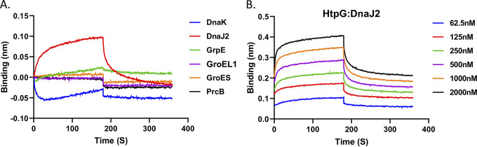
Protein folding is a crucial process in maintaining protein homeostasis, also known as proteostasis, in the cell. The requirement for the assistance of molecular chaperones in the appropriate folding of several proteins has already called into question the previously held view of spontaneous protein folding. These chaperones are highly ubiquitous cellular proteins, which not only help in mediating the proper folding of other nascent polypeptides but are also involved in refolding of the misfolded or the aggregated proteins. Hsp90 family proteins such as high-temperature protein G (HtpG) are abundant and ubiquitously expressed in both eukaryotic and prokaryotic cells. Although HtpG is known as an ATP-dependent chaperone protein in most organisms, function of this protein remains obscured in mycobacterial pathogens. Here, we aim to investigate significance of HtpG as a chaperone in the physiology of Mycobacterium tuberculosis. We report that M. tuberculosis HtpG (mHtpG) is a metal-dependent ATPase which exhibits chaperonin activity towards denatured proteins in coordination with the DnaK/DnaJ/GrpE chaperone system via direct association with DnaJ2. Increased expression of DnaJ1, DnaJ2, ClpX, and ClpC1 in a ΔhtpG mutant strain further suggests cooperativity of mHtpG with various chaperones and proteostasis machinery in M. tuberculosis. IMPORTANCE M. tuberculosis is exposed to variety of extracellular stressful conditions and has evolved mechanisms to endure and adapt to the adverse conditions for survival. mHtpG, despite being dispensable for M. tuberculosis growth under in vitro conditions, exhibits a strong and direct association with DnaJ2 cochaperone and assists the mycobacterial DnaK/DnaJ/GrpE (KJE) chaperone system. These findings suggest the potential role of mHtpG in stress management of the pathogen. Mycobacterial chaperones are responsible for folding of nascent protein as well as reactivation of protein aggregates. M. tuberculosis shows differential adaptive response subject to the availability of mHtpG. While its presence facilitates improved protein refolding via stimulation of the KJE chaperone activity, in the absence of mHtpG, M. tuberculosis enhances expression of DnaJ1/J2 cochaperones as well as Clp protease machinery for maintenance of proteostasis. Overall, this study provides a framework for future investigation to better decipher the mycobacterial proteostasis network in the light of stress adaptability and/or survival.

Keywords: Clp protease; DnaJ2; DnaK; HSP90; HtpG; Mycobacterium tuberculosis; chaperones; heat shock proteins; proteostasis.


Conflict of interest statement

The authors declare no conflict of interest.

Figures

FIG 1
FIG 1
Phylogenetic analysis of mHtpG. (A) Phylogenetic tree analysis of mHtpG. The phylogenetic tree was generated for mHtpG by using the phylomeDB database (http://phylomedb.org/). The interactive tree can be accessed by the following link: http://phylomedb.org/phylome_328?id=3&seqid=Phy001DWYA&phyid=328&method=LG&tree_features=motifs,lineage,support,best_name,spname&treeid=&sid=&seedid=001DWYA&snodes=&isPopUp=False&isExport=False&isPrincipal=True&snodes=#search_in_tree_box. The protein sequence of different bacterial species was retrieved from NCBI (https://www.ncbi.nlm.nih.gov/). (B) Evolutionary relationship of mHtpG with its counterpart from different mycobacterial species. Evolutionary analyses were conducted using MEGA X software (https://www.megasoftware.net/). The evolutionary relationship was inferred using the neighbor-joining method. The optimal tree with the sum of branch length 0.65537958 is shown. The evolutionary distances were computed using the Poisson correction method and are presented as the number of amino acid substitutions per site. This analysis involved 17 amino acid sequences. All ambiguous positions were removed for each sequence pair (pairwise deletion option). There were 674 positions in the final data set. Scales are shown in panels A and B for estimation of branch lengths between the nodes.
FIG 2
FIG 2
Analysis of ATPase activity of mHtpG. (A) Impact of different divalent cations on the ATPase activity. Shown is the effect of different metal ions such as Mg++, Ca++, Mn++, Zn++, Cd++, Co++, Ni++, Ba++, and Cu++ on the ATP hydrolysis activity of mHtpG. The assay was performed by using 1 μM mHtpG for 30 min, and the released Pi was estimated using a malachite green assay, as described in Materials and Methods. (B and C) Effect of temperature (B) and pH (C) on ATPase activity of mHtpG. Reactions were performed at various temperatures, such as 25°C, 37°C, 45°C, 50°C, 60°C, and 70°C, at pH 7.5 of the reaction buffer in panel B, and at different pHs of the reaction buffer, such as 4.5, 6.0, 7.0, 8.0, 9.0, and 10.0 at 50°C in panel C for 30 min, and the released Pi was estimated as described above. (D) Comparative analysis of the effect of Mg++ and Ca++ on kinetics of the ATPase activity of mHtpG. Reactions were performed with 1 μM mHtpG using different concentrations of ATP varying from 100 μM to 1,000 μM in the presence of Mg++ and Ca++ divalent cations. Kinetic parameters such as Km and Vmax were determined using GraphPad Prism 8.0 software (https://www.graphpad.com/scientific-software/prism/). All the reactions in panels A to D were performed in triplicate, and the mean ± standard deviation (SD) values are shown. Asterisks represent the level of significance, as determined by Student’s t test. ***, P ≤ 0.0005.
FIG 3
FIG 3
Analysis of refolding activity of mHtpG. (A) Dose-dependent effect of mHtpG on the refolding of denatured luciferase. The refolding activity of mHtpG was analyzed at different protein concentrations using 80 nM heat-denatured luciferase, as described in Materials and Methods. (B) Effect of mHtpG on luciferase refolding by the mycobacterial KJE chaperone. The refolding assay was performed with 2 μM mHtpG and DnaK/DnaJ2/GrpE (4 μM/400 nM/400 nM) proteins either alone or in combination to explore the synergistic effect of each of these components. (C) Dose-dependent effect of KJE on refolding in the absence and the presence of mHtpG. The refolding activity of the KJE chaperone system was examined using increasing concentrations of chaperones and constant mHtpG (2 μM). (D) Dose-dependent effect of mHtpG on refolding of denatured luciferase in the presence of the KJE chaperone system. The effect of the KJE chaperone system on the refolding activity of mHtpG was analyzed by using various concentrations of mHtpG and a fixed concentration (4 μM/400 nM/400 nM) of DnaK/DnaJ2/GrpE. All the reactions in (A to D) were performed in triplicate, and the mean ± SD values are shown. Asterisks represent the level of significance, as determined by Student’s t test. *, P < 0.05; **, P < 0.005; ***, P ≤ 0.0005.
FIG 4
FIG 4
ATPase activity is critical for the refolding activity of mHtpG. (A) Effect of single amino acid substitutions on ATPase activity of mHtpG. ATP hydrolyzing activity was investigated in wild-type and substitution mutants of mHtpG to understand the importance of specific amino acid residues, which reveals that all the N39D, G83A, and F130A residues are critical for its ATPase activity. (B) Analysis of the conformation of mutant mHtpG proteins. CD spectroscopy was performed to analyze the structural information of WT mHtpG and other substitution mutants, which reveals no effect on protein folding due to these substitutions. (C and D) Effect of mutant mHtpG on protein refolding, either alone (C) or with KJE (D). A luciferase refolding assay was performed using WT, N39D, G83A, and F130A mHtpG proteins (2 μM each) with 80 nM heat-denatured luciferase, which shows an ~50% reduction in the protein refolding due to any of these substitutions (C). Similarly, none of these mutant mHtpG variants are able to enhance the refolding activity of the KJE chaperone, indicating the importance of ATPase activity in mHtpG for protein refolding. The broken lines in panel D represent data presented in Fig. 3C that are used for reference purpose. All the reactions in panels (A, C, and D) were performed in triplicate, and the mean ± SD values are shown. Asterisks represent the level of significance, as determined by Student’s t test. **, P < 0.005; ***, P ≤ 0.0005.
FIG 5
FIG 5
Analysis of the interaction of mHtpG with other mycobacterial chaperones. (A) Interaction analysis between mHtpG and various chaperons. BLI-octet was used for analyzing the interaction between mHtpG and DnaK, DnaJ2, GrpE, GroEL1, GroES, and PrcB, as described in Materials and Methods. PrcB was used as a negative control. Based on the response, mHtpG appears to interact with only DnaJ2. (B) Analysis of binding kinetics of DnaJ2 with mHtpG. The interaction of mHtpG and DnaJ2 at different concentrations of DnaJ2 reveals strong binding with a dissociation constant (Kd) of ~200 nM. The Kd was estimated by using Octet software. Data are representative of two independent experiments in panels A and B.
FIG 6
FIG 6
Quantitative proteomic analysis of ΔhtpG. (A) Status of differentially regulated proteins in different biological replicates. The bar graph depicts downregulated (≤0.55-fold) (DN), unchanged (<1.83-fold to >0.55-fold) (no change), and upregulated (≥1.83-fold) (UP) proteins in the ΔhtpG strain of MtbH37Rv across four biological replicates (set 1 to set 4). (B) Analysis of differentially expressed proteins across replicate samples. The upregulated and downregulated proteins in four biological replicates are represented by the Venn diagrams to identify consistency in the expression pattern. The Venn diagrams were generated by using Venny 2.1 software. (C) Heat map analysis of differentially regulated proteins. Heat maps show both down- and upregulated proteins classified under different functional categories. The results reveal that loss of HtpG in M. tuberculosis leads to the downregulation of 164 proteins and upregulation of 221 proteins in at least 3 of the 4 biological replicates, attributed with different functional categories.
FIG 7
FIG 7
Graphical representation depicting maintenance of proteostasis in the wild-type and ΔhtpG strains of M. tuberculosis. In the wild-type cells, the newly synthesized protein (left) or the stress-induced protein aggregate (right) is recognized by the cochaperonin, DnaJ, which is transferred to the associated mHtpG or to some other chaperones. The mHtpG subsequently causes partial refolding of the substrate in an ATP-dependent manner, which is then passed on to DnaK in the presence of GrpE, resulting in proper folding. In the ΔhtpG strain, expression of DnaJ is enhanced to compensate for the loss of mHtpG, leading to direct transfer of the candidate protein to the DnaK chaperone, thus maintaining the homeostasis. Simultaneously, a fraction of protein, which remains in the aggregated form or which is unable to be folded properly, is taken care of by major protease machinery of the cell, such as ClpX or ClpC1-associated proteases, which are activated in the absence of mHtpG.

Similar articles

Cited by

References

    1. Lindquist S, Craig EA. 1988. The heat-shock proteins. Annu Rev Genet 22:631–677. doi:10.1146/annurev.ge.22.120188.003215. - DOI - PubMed
    1. Bukau B, Deuerling E, Pfund C, Craig EA. 2000. Getting newly synthesized proteins into shape. Cell 101:119–122. doi:10.1016/S0092-8674(00)80806-5. - DOI - PubMed
    1. Nover L, Hellmund D, Neumann D, Scharf K, Serfling E. 1984. The heat shock response of eukaryotic cells. Biologisches Zentralblatt 103:357–435.
    1. Kim YE, Hipp MS, Bracher A, Hayer-Hartl M, Hartl FU. 2013. Molecular chaperone functions in protein folding and proteostasis. Annu Rev Biochem 82:323–355. doi:10.1146/annurev-biochem-060208-092442. - DOI - PubMed
    1. Richter K, Haslbeck M, Buchner J. 2010. The heat shock response: life on the verge of death. Mol Cell 40:253–266. doi:10.1016/j.molcel.2010.10.006. - DOI - PubMed

Publication types

MeSH terms