. 2023 Apr 27;97(4):e0188922.
doi: 10.1128/jvi.01889-22. Epub 2023 Apr 6.

African Swine Fever Virus Envelope Glycoprotein CD2v Interacts with Host CSF2RA to Regulate the JAK2-STAT3 Pathway and Inhibit Apoptosis to Facilitate Virus Replication

Affiliations

African Swine Fever Virus Envelope Glycoprotein CD2v Interacts with Host CSF2RA to Regulate the JAK2-STAT3 Pathway and Inhibit Apoptosis to Facilitate Virus Replication

Qi Gao et al. J Virol. .

Abstract

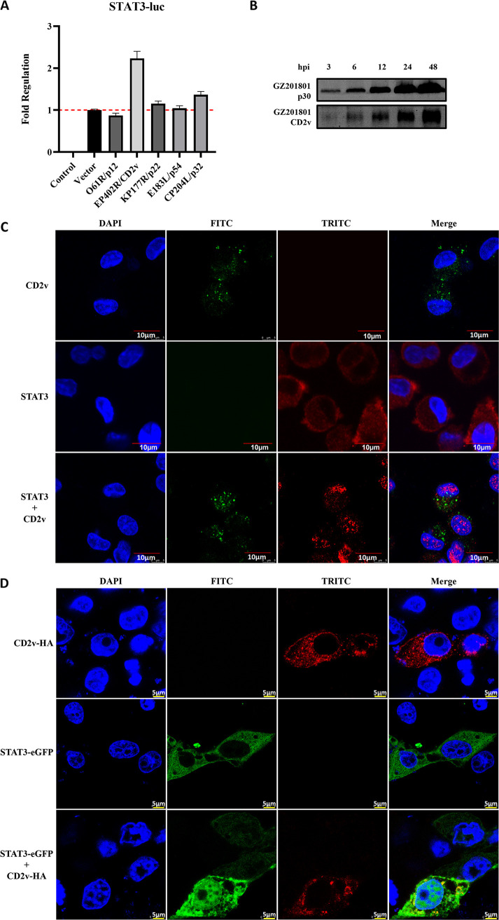
African swine fever (ASF) is a highly infectious disease caused by the African swine fever virus (ASFV) in swine. It is characterized by the death of cells in infected tissues. However, the molecular mechanism of ASFV-induced cell death in porcine alveolar macrophages (PAMs) remains largely unknown. In this study, transcriptome sequencing of ASFV-infected PAMs found that ASFV activated the JAK2-STAT3 pathway in the early stages and apoptosis in the late stages of infection. Meanwhile, the JAK2-STAT3 pathway was confirmed to be essential for ASFV replication. AG490 and andrographolide (AND) inhibited the JAK2-STAT3 pathway, promoted ASFV-induced apoptosis, and exerted antiviral effects. Additionally, CD2v promoted STAT3 transcription and phosphorylation as well as translocation into the nucleus. CD2v is the main envelope glycoprotein of the ASFV, and further investigations showed that CD2v deletion downregulates the JAK2-STAT3 pathway and promotes apoptosis to inhibit ASFV replication. Furthermore, we discovered that CD2v interacts with CSF2RA, which is a hematopoietic receptor superfamily member in myeloid cells and a key receptor protein that activates receptor-associated JAK and STAT proteins. In this study, CSF2RA small interfering RNA (siRNA) downregulated the JAK2-STAT3 pathway and promoted apoptosis to inhibit ASFV replication. Taken together, ASFV replication requires the JAK2-STAT3 pathway, while CD2v interacts with CSF2RA to regulate the JAK2-STAT3 pathway and inhibit apoptosis to facilitate virus replication. These results provide a theoretical basis for the escape mechanism and pathogenesis of ASFV. IMPORTANCE African swine fever is a hemorrhagic disease caused by the African swine fever virus (ASFV), which infects pigs of different breeds and ages, with a fatality rate of up to 100%. It is one of the key diseases affecting the global livestock industry. Currently, no commercial vaccines or antiviral drugs are available. Here, we show that ASFV replicates via the JAK2-STAT3 pathway. More specifically, ASFV CD2v interacts with CSF2RA to activate the JAK2-STAT3 pathway and inhibit apoptosis, thereby maintaining the survival of infected cells and promoting viral replication. This study revealed an important implication of the JAK2-STAT3 pathway in ASFV infection and identified a novel mechanism by which CD2v has evolved to interact with CSF2RA and maintain JAK2-STAT3 pathway activation to inhibit apoptosis, thus elucidating new information regarding the signal reprogramming of host cells by ASFV.

Keywords: ASFV; CD2v; CSF2RA; JAK2-STAT3 pathway; apoptosis.


Conflict of interest statement

The authors declare no conflict of interest.

Figures

FIG 1
FIG 1
Biological phenomena of ASFV-induced apoptosis. (A) Observation of CPE in PAMs infected with ASFV. Scale bar, 100 μm. (B) Projection electron microscopy images of the cell death of PAMs induced by ASFV infection. In the ASFV-infected PAMs, the white arrow represents viral factories, and the black arrows represent apoptotic bodies. (C to F) Cytotoxicity of z-VAD-FMK, Belnacasan, ferrostatin-1, and necrostatin-1 in PAMs. The red dotted line represents the 80% survival rate of cells treated with the drug. (G) The cell viability of ASFV-infected PAMs posttreatment with 2.5 μM z-VAD-FMK, 2.5 μM Belnacasan, 0.125 μM ferrostatin-1, and 1.25 μM necrostatin-1. Data are presented as mean ± SD. Asterisks indicate statistical difference (one-way ANOVA; n = 3; ***, P < 0.001; ****, P < 0.0001). (H) PAMs were stained with propidium iodide to observe the inhibitory effect of z-VAD-FMK on ASFV-induced cell death. ASFV, African swine fever virus (GZ201801 strain); CPEs, cytopathic effects; PAMs, porcine alveolar macrophages; PI, propidium iodide.
FIG 2
FIG 2
Characterization of ASFV infection-induced apoptosis of PAMs. (A) Western blotting was used to measure the expression of cleaved-caspase3, Bax, and Bcl-2 proteins in PAMs at 3, 6, 12, 24, 36, and 48 h after infection in the control and ASFV infection groups. β-Actin expression was used as a positive control. (B) Flow cytometry was used to detect apoptosis of PAMs induced by ASFV at 3, 6, 12, 24, and 48 h after infection. ASFV, African swine fever virus (GZ201801 strain); PAMs, porcine alveolar macrophages.
FIG 3
FIG 3
Transcriptomic analysis of PAMs infected with ASFV. (A, C, and E) Transcriptomic sequencing of PAMs infected with ASFV at MOI to 1. Volcanic map analysis of DEGs compared with that in the control group at 3, 12, and 48 h after infection. (B, D, and F) KEGG pathway enrichment analysis of DEGs compared with the control group at 3, 12, and 48 h after infection. (G) Analysis diagram of the JAK2-STAT3 signaling pathway. ASFV, African swine fever virus (GZ201801 strain); PAMs, porcine alveolar macrophages; MOI, multiplicity of infection; DEGs, differentially expressed genes.
FIG 4
FIG 4
ASFV infection activates the JAK2-STAT3 pathway. (A and B) Detection of the cytotoxicity of the JAK2-STAT3 pathway inhibitor AG490 (A) and AND (B) in PAMs. The red dotted line represents the 80% survival rate of cells treated with the drug. (C and D) IL-6 expression in PAMs infected with ASFV, with or without treatment with AG490 (C), and Western blotting of the expression of JAK2, P-JAK2, STAT3, and P-STAT3 proteins at 3, 6, 12, 24, 36, and 48 h after infection with ASFV with or without treatment with AG490 and AND (D). β-Actin expression was used as a positive control. ASFV, African swine fever virus (GZ201801 strain); PAMs, porcine alveolar macrophages; AND, andrographolide.
FIG 5
FIG 5
Effect of inhibition of the JAK2-STAT3 pathway on ASFV-induced apoptosis of PAMs. Western blotting of the expression of cleaved-caspase3, Bax, and Bcl-2 proteins at 3, 6, 12, 24, 36, and 48 h after infection with ASFV, with or without treatment with the JAK2-STAT3 pathway inhibitors AG490 and AND. β-Actin expression was used as a positive control. ASFV, African swine fever virus (GZ201801 strain); PAMs, porcine alveolar macrophages; AND, andrographolide; hpi, hours postinfection.
FIG 6
FIG 6
Antiviral activity of AG490 and AND against ASFV. (A, D, G) PAMs were infected with ASFV (MOI of 1) and then treated with 100 μM AG490 or 30 μM AND. The expression level of the ASFV B646L gene in PAMs was detected by real-time PCR analysis at 48 h. Each data point represents the results of three independent experiments (mean ± SD). (B, E, H) Western blotting of the expression of the ASFV p30 protein in PAMs at 3, 6, 12, 24, 36, and 48 h after infection with ASFV with or without treatment with AG490 and AND. β-Actin expression was used as a positive control. (C, F, I) Antiviral activity of AG490 and AND against ASFV in PAMs determined by IFA. Nuclei were counterstained with DAPI (blue). Each data point represents the results of three independent experiments (mean ± SD). The images represent three independent IFA trials with similar results. Asterisks indicate significant differences compared with the control group (***, P < 0.01). ASFV, African swine fever virus (GZ201801 strain); PAMs, porcine alveolar macrophages; AND, andrographolide; hpi, hours postinfection; IFA, immunofluorescence assay. The same image is used to represent the GZ201801 condition, to better depict and compare the various treatment conditions.
FIG 7
FIG 7
ASFV CD2v induces STAT3 phosphorylation and translocation into the nucleus. (A) Screening for capsular proteins that upregulate the STAT3 promoter through the dual luciferase reporting system. The red dotted line is the value of the Vector. (B) Western blot assay was used to detect changes in p30 and CD2v protein expression levels during ASFV infection. (C) Endogenous levels of the CD2v and STAT3 proteins detected in ASFV-infected PAMs, and the nuclear translocation of STAT3 observed through confocal microscopy. (D) The nuclear translocation of STAT3 protein observed by confocal microscopy in PK15 cells after transfection with the plasmid STAT3 alone or STAT3 plasmid + the common ASFV CD2v protein. Cell nuclei were stained with DAPI (blue). ASFV, African swine fever virus (GZ201801 strain); PAMs, porcine alveolar macrophages.
FIG 8
FIG 8
GZ201801-ΔCD2v downregulates the JAK2-STAT3 pathway to promote apoptosis. (A) Scheme representing the construction of GZ201801-ΔCD2v. (B) PCR validation of the CD2v deletion. (C and D) Validation of EGFP expression in PAMs infected with the GZ201801-ΔCD2v strain by fluorescence microscopy (C) and flow cytometry (D). The scale bar indicates 200 μm in C. (E) Growth curve replication of the GZ201801 and GZ201801-ΔCD2v strains in PAMs determined with the TCID50 assay (top, primary y axis). (F) Western blot assay used to detect the expression level of the p30 protein in the ASFV-GZ201801 wild strain and GZ201801-ΔCD2v deletion strain during the process of infection in PAMs. (G) Effect of GZ201801 and GZ201801-ΔCD2v infection on IL-6 expression in PAMs. (H) Western blotting of the expression of JAK2, P-JAK2, STAT3, and P-STAT3 proteins at 3, 6, 12, 24, and 48 hpi in GZ201801- and GZ201801-ΔCD2v-infected cells. β-Actin expression was used as a positive control. (I) Cell death induced by GZ201801-ΔCD2v infection observed by microscopy. (J) Number of dead cells post-GZ201801 and GZ201801-ΔCD2v infection detected by flow cytometry (24.67% and 29.03% of PAMs were killed by GZ201801 and GZ201801-ΔCD2v infection, respectively, after 48 h). (K) Western blotting of the expression of cleaved-caspase 3, Bax, and Bcl-2 proteins at 3, 6, 12, 24, 36, and 48 hpi with GZ201801 and GZ201801-ΔCD2v. The data are presented as the mean ± SD (n = 3; *, P < 0.05; **, P < 0.01; ***, P < 0.001; ****, P < 0.0001). GZ201801, wild-type strain of African swine fever virus; GZ201801-ΔCD2v; CD2v deletion strain of GZ201801; PAMs, porcine alveolar macrophages; hpi, hours postinfection. The results shown in H and K were obtained by probing the same membrane with the antibodies mentioned and the membrane was stripped and reprobed with additional antibodies in the figure. As a result, the blots labeled “β-actin” blots in H and K are identical.
FIG 9
FIG 9
ASFV CD2v interacts with the host CSF2RA protein to regulate the JAK2-STAT3 pathway. (A) PK-15 cells were cotransfected with HA-CD2v-expressing plasmids and EGFP-CSF2RA-expressing plasmids for 36 h, followed by subcellular localization analysis of ASFV CD2v and CSF2RA by immunofluorescence assay (IFA). Anti-CD2v (red) and anti-CSF2RA (green) antibodies and DAPI (blue) were used to stain the cells. (B) HEK-293T cells were cotransfected with HA-CD2v-expressing plasmids and EGFP-CSF2RA-expressing plasmids for 24 h. The cells were then lysed and subjected to immunoprecipitation (IP) using an anti-CD2v antibody. The IP complexes and WCLs were subjected to Western blotting using anti-CD2v or anti-EGFP antibodies. (C) PAMs were mock transfected or transfected with NC siRNA or CSF2RA siRNA for 24 h, followed by infection with ASFV at MOI of 1 for 24 h. The mRNA expression levels of CSF2RA were then measured by qPCR. The expression levels of CSF2RA and ASFV p30 proteins were detected by Western blotting. (D) Protein expression levels of P-JAK2, P-STAT3, cleaved-caspase3, Bcl-2, and ASFV p30 in PAMs infected with ASFV and treated with CSF2RA siRNA detected by Western blotting. Tubulin expression was used as a positive control. (E) Exploring the interaction between CSF2RA and KAP1 through confocal microscopy. (F) Exploring the interaction between CSF2RA and KAP1 by coimmunoprecipitation. The data are presented as the mean ± SD (n = 3; ***, P < 0.001; ****, P < 0.0001). ASFV, African swine fever virus (GZ201801 strain); PAMs, porcine alveolar macrophages; IP, immunoprecipitation; WCL, whole-cell lysate; NC, Negative Control.

Similar articles

Cited by

References

    1. Sánchez-Vizcaíno JM, Mur L, Martínez-López B. 2013. African swine fever (ASF): five years around Europe. Vet Microbiol 165:45–50. doi:10.1016/j.vetmic.2012.11.030. - DOI - PubMed
    1. Petrov A, Schotte U, Pietschmann J, Dräger C, Beer M, Anheyer-Behmenburg H, Goller KV, Blome S. 2014. Alternative sampling strategies for passive classical and African swine fever surveillance in wild boar. Vet Microbiol 173:360–365. doi:10.1016/j.vetmic.2014.07.030. - DOI - PubMed
    1. Gabriel C, Blome S, Malogolovkin A, Parilov S, Kolbasov D, Teifke JP, Beer M. 2011. Characterization of African swine fever virus Caucasus isolate in European wild boars. Emerg Infect Dis 17:2342–2345. doi:10.3201/eid1712.110430. - DOI - PMC - PubMed
    1. Zhou X, Li N, Luo Y, Liu Y, Miao F, Chen T, Zhang S, Cao P, Li X, Tian K, Qiu HJ, Hu R. 2018. Emergence of African swine fever in China, 2018. Transbound Emerg Dis 65:1482–1484. doi:10.1111/tbed.12989. - DOI - PubMed
    1. Rowlands RJ, Michaud V, Heath L, Hutchings G, Oura C, Vosloo W, Dwarka R, Onashvili T, Albina E, Dixon LK. 2008. African swine fever virus isolate, Georgia, 2007. Emerg Infect Dis 14:1870–1874. doi:10.3201/eid1412.080591. - DOI - PMC - PubMed

Publication types

MeSH terms

Substances