

Correlation between the IgG/IgA Antibody Response against PEDV Structural Protein and Virus Neutralization
- PMID: 37022185
- PMCID: PMC10269706
- DOI: 10.1128/spectrum.05233-22
Correlation between the IgG/IgA Antibody Response against PEDV Structural Protein and Virus Neutralization
Abstract
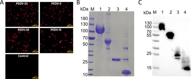
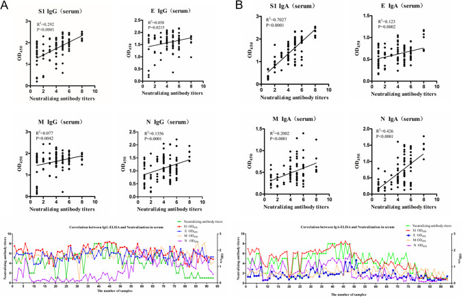
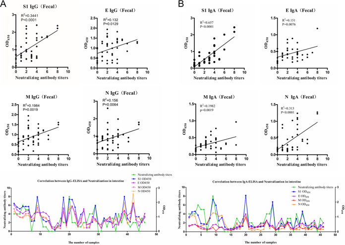
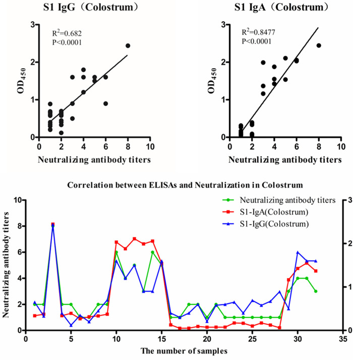
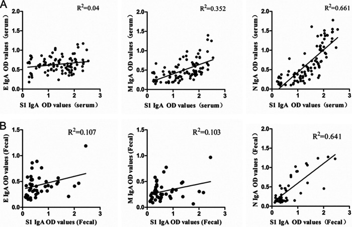
Porcine epidemic diarrhea (PED) is a highly contagious intestinal infectious disease caused by porcine epidemic diarrhea virus (PEDV). Large-scale outbreaks of PEDV have caused huge economic losses to the pig industry since 2010. Neutralizing antibodies play a pivotal role in protecting piglets from enteric infections. However, there has been no systematic report on the correlations between neutralizing antibody titers (NTs) and absorbance values of IgG or IgA to all PEDV individual structural proteins in clinical serum, fecal, and colostrum samples. In this study, the spike protein S1 domain (S1), membrane protein (M), envelope protein (E), and nucleocapsid protein (N) of the variant PEDV strain AH2012/12 were expressed and purified by using the human embryonic kidney (HEK) 293F expression system. A total of 92 clinical serum samples, 46 fecal samples, and 33 colostrum samples were collected, and the correlations between IgG or IgA absorbance values and NTs were analyzed. R2 values revealed that anti-S1 IgA absorbance values show the highest agreement with NTs in all serum, fecal, and colostrum samples, followed by the N protein. The correlations between anti-E or M IgA and NTs were very low. However, in the colostrum samples, both IgG and IgA to S1 showed high correlations with NTs. In addition, compared with E and M, the highest correlations of IgA absorbance values were with N and S1 in serum and fecal samples. Overall, this study revealed the highest correlation between NTs and IgA to PEDV S1 protein. Therefore, the diagnostic method with anti-S1 IgA can be used as a powerful tool for assessing the immune status of pigs. IMPORTANCE The humoral immune response plays an important role in virus neutralization. Against PEDV, both IgG and the mucosal immune component IgA play roles in virus neutralization. However, which plays a more prominent role and whether there are differences in different tissue samples are not clearly reported. Additionally, the relationship between IgG and IgA against individual structural proteins and viral neutralization remains unclear. In this study, we systematically determined the relationship between IgG and IgA against all PEDV structural proteins and viral neutralization in different clinical samples and found the highest correlation between neutralization activity and IgA to PEDV S1 protein. Our data have important guiding implications in the evaluation of immune protection.
Keywords: IgA; IgG; PEDV; S1; neutralizing antibody titers.
Conflict of interest statement
The authors declare no conflict of interest.
Figures
Similar articles
-
Humoral immune responses in piglets experimentally infected with a field strain of porcine epidemic diarrhea virus.Vet Microbiol. 2020 Jul;246:108742. doi: 10.1016/j.vetmic.2020.108742. Epub 2020 May 31. Vet Microbiol. 2020. PMID: 32605747
-
Characterization of anti-porcine epidemic diarrhea virus neutralizing activity in mammary secretions.Virus Res. 2016 Dec 2;226:85-92. doi: 10.1016/j.virusres.2016.06.002. Epub 2016 Jun 7. Virus Res. 2016. PMID: 27287711 Free PMC article.
-
S1 domain of the porcine epidemic diarrhea virus spike protein as a vaccine antigen.Virol J. 2016 Apr 1;13:57. doi: 10.1186/s12985-016-0512-8. Virol J. 2016. PMID: 27036203 Free PMC article.
-
Porcine epidemic diarrhea virus (PEDV): An update on etiology, transmission, pathogenesis, and prevention and control.Virus Res. 2020 Sep;286:198045. doi: 10.1016/j.virusres.2020.198045. Epub 2020 Jun 2. Virus Res. 2020. PMID: 32502552 Free PMC article. Review.
-
[Immunization against porcine epidemic diarrhea virus and vaccine development].Sheng Wu Gong Cheng Xue Bao. 2021 Aug 25;37(8):2603-2613. doi: 10.13345/j.cjb.200524. Sheng Wu Gong Cheng Xue Bao. 2021. PMID: 34472281 Review. Chinese.
Cited by
-
Feeding Sows with Multi-Species Probiotics During Late Pregnancy and the Lactating Period Influences IgA Concentration in Colostrum and Subsequently Increases the Survival Rate of Piglets in Porcine Epidemic Diarrhea Outbreak Herd.Animals (Basel). 2025 Jan 5;15(1):103. doi: 10.3390/ani15010103. Animals (Basel). 2025. PMID: 39795046 Free PMC article.
-
Effects of Low-Protein Amino Acid-Balanced Diets and Astragalus Polysaccharides on Production Performance, Antioxidants, Immunity, and Lipid Metabolism in Heat-Stressed Laying Hens.Animals (Basel). 2025 Aug 14;15(16):2385. doi: 10.3390/ani15162385. Animals (Basel). 2025. PMID: 40867713 Free PMC article.
-
The development of a lateral flow immunochromatographic test strip for measurement of specific IgA and IgG antibodies level against porcine epidemic diarrhea virus in pig milk.Vet Q. 2024 Dec;44(1):1-15. doi: 10.1080/01652176.2024.2429472. Epub 2024 Nov 21. Vet Q. 2024. PMID: 39568374 Free PMC article.
-
PEDV-spike-protein-expressing mRNA vaccine protects piglets against PEDV challenge.mBio. 2024 Feb 14;15(2):e0295823. doi: 10.1128/mbio.02958-23. Epub 2024 Jan 17. mBio. 2024. PMID: 38231557 Free PMC article.
-
Serum IgA antibody level against porcine epidemic diarrhea virus is a potential pre-evaluation indicator of immunization effects in sows during parturition under field conditions.Porcine Health Manag. 2024 Sep 3;10(1):32. doi: 10.1186/s40813-024-00382-w. Porcine Health Manag. 2024. PMID: 39228006 Free PMC article.
References
-
- Trujillo-Ortega ME, Beltrán-Figueroa R, García-Hernández ME, Juárez-Ramírez M, Sotomayor-González A, Hernández-Villegas EN, Becerra-Hernández JF, Sarmiento-Silva RE. 2016. Isolation and characterization of porcine epidemic diarrhea virus associated with the 2014 disease outbreak in Mexico: case report. BMC Vet Res 12:132. doi:10.1186/s12917-016-0763-z. - DOI - PMC - PubMed
-
- Gerber PF, Lelli D, Zhang J, Strandbygaard B, Moreno A, Lavazza A, Perulli S, Bøtner A, Comtet L, Roche M, Pourquier P, Wang C, Opriessnig T. 2016. Diagnostic evaluation of assays for detection of antibodies against porcine epidemic diarrhea virus (PEDV) in pigs exposed to different PEDV strains. Prev Vet Med 135:87–94. doi:10.1016/j.prevetmed.2016.11.005. - DOI - PMC - PubMed
-
- Jarvis MC, Lam HC, Zhang Y, Wang L, Hesse RA, Hause BM, Vlasova A, Wang Q, Zhang J, Nelson MI, Murtaugh MP, Marthaler D. 2016. Genomic and evolutionary inferences between American and global strains of porcine epidemic diarrhea virus. Prev Vet Med 123:175–184. doi:10.1016/j.prevetmed.2015.10.020. - DOI - PMC - PubMed
Publication types
MeSH terms
Substances
LinkOut - more resources
Full Text Sources
Miscellaneous
