. 2023 Jun;69(2-3):153-163.
doi: 10.1007/s00294-023-01266-2. Epub 2023 Apr 6.

Application of nanotags and nanobodies for live cell single-molecule imaging of the Z-ring in Escherichia coli

Affiliations

Application of nanotags and nanobodies for live cell single-molecule imaging of the Z-ring in Escherichia coli

Emma Westlund et al. Curr Genet. 2023 Jun.

Abstract

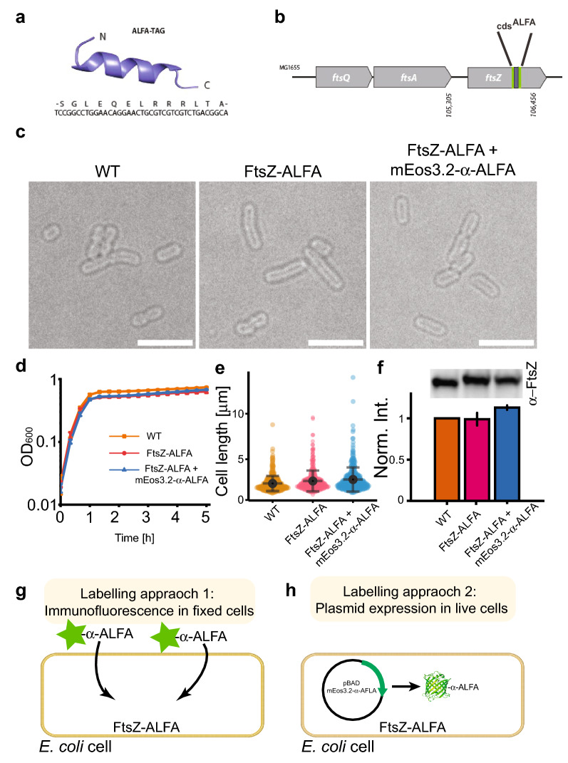
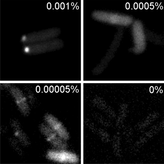
Understanding where proteins are localized in a bacterial cell is essential for understanding their function and regulation. This is particularly important for proteins that are involved in cell division, which localize at the division septum and assemble into highly regulated complexes. Current knowledge of these complexes has been greatly facilitated by super-resolution imaging using fluorescent protein fusions. Herein, we demonstrate with FtsZ that single-molecule PALM images can be obtained in-vivo using a genetically fused nanotag (ALFA), and a corresponding nanobody fused to mEos3.2. The methodology presented is applicable to other bacterial proteins.

Keywords: ALFA-tag; E. coli; FtsZ; Nanotags; PALM; STED.


Conflict of interest statement

The authors declare no competing financial interests.

Figures

Fig. 1
Fig. 1
Cell viability of the engineered FtsZ-ALFA strain and labelling approaches. a Structure and sequence of the ALFA-tag (Gotzke et al. 2019). b Locus on the chromosome in strain MG1655 where the ALFA-tag is incorporated. c Bright field images of strains used in this study. No morphological differences are noticeable. Scale bars = 5 m. d Growth curves of strains used (n = 3 for each strain). e Cell length measurements. WT = 2.34 ± 0.83 μm, FtsZ-ALFA = 2.61 ± 1.16 μm, FtsZ-ALFA + mEos3.2-α-ALFA = 2.78 ± 1.33 μm (n = 214 for each strain). Mean ± S.D. f Quantitative western blotting indicated that neither the ALFA-tag nor the additional expression of mEos3.2-α-ALFA altered the overall expression of FtsZ to any larger degree. WT = 1,FtsZ-ALFA = 0.99 ± 0.08,FtsZ-ALFA + mEos3.2-α-ALFA = 1.13 ± 0.03. Mean ± S.D. n = 3. Full length WB shown in Supplementary Fig. 3. g Approach 1: immunofluorescence labelling. Cells were fixed, membranes permeabilized followed by labelling with ATTO488 tagged α-ALFA nanobodies recognizing the ALFA-tag. h Approach 2: plasmid expression in live cells. Cells were transformed with a plasmid encoding for mEos3.2-α-ALFA nanobody. In both approaches, FtsZ-ALFA is labelled with fluorescently tagged nanobodies and imaged using super-resolution approaches (Figs. 2 and 4)
Fig. 2
Fig. 2
Super-resolution STED imaging of FtsZ-ALFA using an α-ALFA nanobody labelled with ATTO488. a Fixed cells immunolabelled and imaged using confocal and STED microscopy. Fluorescence signal overlayed with bright field images. Scale bar = 4 μm. b Longitudinal width (FWHM) of a representative FtsZ ring. Conf = Confocal. c Apparent widths (FWHM) were extracted from line scans as indicated by the white dotted line in the lower cells in the confocal image (A). The mean widths were 243 ± 26 nm (confocal) and 93 ± 12 nm (STED) (n = 54). Boxes indicate S.D., midline is the mean and whiskers incorporate 1-99% of the data range. d Schematic representation of cells lying on an agarose pad and trapped in a vertical position. e STED imaging of FtsZ-ALFA cells labelled with α-ALFA-ATTO488 trapped in a vertical position in micron holes. Representative FtsZ rings of various radii. r indicate radius. Scale bar = 500 nm
Fig. 3
Fig. 3
Efficient FtsZ-ALFA/mEos3.2-α-ALFA ring detection is concentration dependent. Epifluorescence imaging on FtsZ-ALFA tagged with mEos3.2-α-ALFA in live E. coli cells. mEos3.2 was excited and captured in the green state using a 488-laser line. Images of various conditions with different Arabinose concentrations, ranging from 0.001% to 0%. Too high induction (0.001% and above) produced large bright protein aggregates at the poles, 0.0005% produced too high background, while no induction resulted in essentially no fluorescence signal. As clearly can be seen, under our experimental conditions, 0.00005% Arabinose gave best results. Scale bar = 2 μm
Fig. 4
Fig. 4
Live cell single-molecule PALM imaging of FtsZ-ALFA using a co-expressed mEos3.2-α-ALFA nanobody. a A typical live cell imaged by bright field optics and using PALM. b Side-by-side comparison of FtsZ-ALFA rings using Epifluorescence and PALM. c Longitudinal width (FWHM) of a representative FtsZ-ALFA ring as determined by fitting a gaussian to fluorescence traces generated drawn over the FtsZ-ALFA rings (e.g., yellow line in (B)). EPI Epifluorescence. d Apparent widths (FWHM) from line scans as indicated by the yellow line in the lower cells in the epifluorescence image (B). The mean widths were 266 ± 22 nm (epifluorescence) and 110 ± 11 nm (PALM) (n = 109). Boxes indicate S.D., midline is the mean and whiskers incorporate 1–95% of the data range. e, Images from a typical time-lapse PALM sequence. Z-ring radius is clearly decreasing over time. Scale bars = 2 μm

References

    1. Akhuli D, Dhar A, Viji AS, Bhojappa B, Palani S. ALIBY: ALFA nanobody-based toolkit for imaging and biochemistry in yeast. mSphere. 2022;7:e0033322. doi: 10.1128/msphere.00333-22. - DOI - PMC - PubMed
    1. Attaibi M, den Blaauwen T. An updated model of the divisome: regulation of the septal peptidoglycan synthesis machinery by the divisome. Int J Mol Sci. 2022 doi: 10.3390/ijms23073537. - DOI - PMC - PubMed
    1. Betzig E, Patterson GH, Sougrat R, Lindwasser OW, Olenych S, Bonifacino JS, Davidson MW, Lippincott-Schwartz J, Hess HF. Imaging intracellular fluorescent proteins at nanometer resolution. Science. 2006;313:1642–1645. doi: 10.1126/science.1127344. - DOI - PubMed
    1. Bisson-Filho AW, Hsu YP, Squyres GR, Kuru E, Wu F, Jukes C, Sun Y, Dekker C, Holden S, VanNieuwenhze MS, Brun YV, Garner EC. Treadmilling by FtsZ filaments drives peptidoglycan synthesis and bacterial cell division. Science. 2017;355:739–743. doi: 10.1126/science.aak9973. - DOI - PMC - PubMed
    1. Bubeck P, Winkler M, Bautsch W. Rapid cloning by homologous recombination in vivo. Nucleic Acids Res. 1993;21:3601–3602. doi: 10.1093/nar/21.15.3601. - DOI - PMC - PubMed

MeSH terms

LinkOut - more resources