Review
. 2023 May;13(5):1520-1542.
doi: 10.1007/s13346-023-01328-5. Epub 2023 Apr 6.

Tumour-derived extracellular vesicle based vaccines for melanoma treatment

Affiliations
Review

Tumour-derived extracellular vesicle based vaccines for melanoma treatment

Lorena Gonzalez-Melero et al. Drug Deliv Transl Res. 2023 May.

Abstract

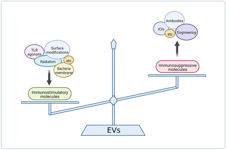
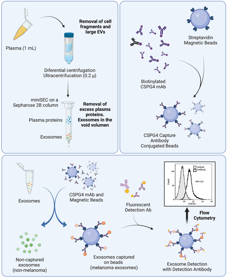
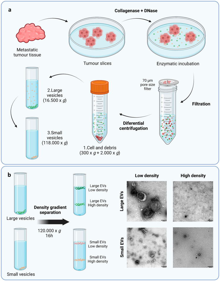
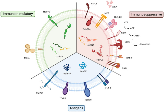
The interest of extracellular vesicles (EVs) in cancer immunotherapy is increasing every day. EVs are lipid bilayer vesicles released by most cells, which contain the molecular signature of their parent cell. Melanoma-derived EVs present antigens specific to this aggressive type of cancer, but they also exert immunomodulatory and pro-metastatic activity. Until now, most reviews focus on the immunoevasive characteristics of tumour-derived EVs, but do not help to overcome the issues related to them. In this review, we describe isolation methods of EVs from melanoma patients and most interesting markers to oversee their effect if they are used as antigen carriers. We also discuss the methods developed so far to overcome the lack of immunogenicity of melanoma-derived EVs, which includes EV modification or adjuvant co-administration. In summary, we conclude that EVs can be an interesting antigen source for immunotherapy development once EV obtaining is optimised and the understanding of the mechanisms behind their multiple effects is further understood.

Keywords: Exosomes; Extracellular vesicles; Immunostimulatory molecules; Immunotherapy; Melanoma; Tumour cells.


Conflict of interest statement

The authors declare no competing interests.

Figures

Fig. 1
Fig. 1
The duality in immune response of melanoma cell derived exosomes and the possible strategies to potentiate immunostimulatory abilities or reduce immunosuppressive ones. Among the strategies to potentiate immunostimulatory capacity, EV modification (such as surface modifications or cell radiation) or co-administration with adjuvants (like Toll-like receptor agonists (TLR) or bacteria membrane particles) are possible. On the other hand, immunosuppressive capacity can be obstructed blocking immunossupressive molecules with antibodies like immune checkpoint inhibitors (ICIs). Created with BioRender.com
Fig. 2
Fig. 2
Melanoma cell-derived exosome isolation process. a A schema for isolation of exosomes from plasma. The recovered exosomes are partly “purified” by removal of protein/high density lipoproteins (HDL) complexes on the miniSEC column. b Exosomes are captured on streptavidin-labelled magnetic beads using pre-titered biotin-labelled anti-CSPG4 mAb. c Exosomes are co-incubated with biotinylated, pre-tittered anti-CSPG4 Ab and are captured on streptavidin-coated magnetic beads, and the bead-bound melanoma exosome-Ab complexes are recovered using a magnet. The antigen (CSPG4) carried by the bead-bound exosomes is detected using a fluorochrome-labelled and pre-tittered detection anti-CSPG4 mAb. The flow cytometry-based detection provides the relative fluorescence intensity (RFI) value for exosomes carrying the antigen. RFI = MFI of detection Ab/MFI of isotype control Ab. Non-captured exosomes in the supernatant are recovered as well for subsequent profiling by flow cytometry. Adapted from [18] and created with BioRender.com
Fig. 3
Fig. 3
Schematic overview of the centrifugation-based protocol used to isolate vesicles from metastatic melanoma tissues, and posterior vesicle isolation through a density gradient separation. a The tumour tissues were dissected into smaller pieces that were incubated in cell culture media containing collagenase D and DNase I for 30 min allowing the tissue EVs to be released into the media. EVs were isolated from the media by differential centrifugation resulting in two subpopulations of EVs (large EVs (16,500 × gavg pellet) and small EVs (118,000 × gavg pellet)). b Isolated large and small EVs were bottom loaded on an iodixanol gradient (45–20%). From both populations of vesicles, two fractions were visible after the separation, with one fraction containing high density EVs (1.16–19 g/cm3) and one containing low density EVs (1.11–1.12 g/cm3). Ten micrograms of low and high density vesicles from both large and small EVs were loaded onto grids, negative stained and evaluated by transmission electron microscopy. Scale bars are 200 nm. Adapted from [19] and created with BioRender.com
Fig. 4
Fig. 4
Molecules expressed in melanoma-derived EVs. Molecules expressed in melanoma-derived EVs. In green, immunostimulatory molecules are represented; in red, immunosuppressive molecules; and in blue, antigens recognised by immune system. Abbreviations: microRNA, miRNA; major histocompatibility complex class I chain-related protein A, MICA; heat shock protein 70 KDa, HSP70; programmed cell death ligand 1, PD-L1; mesenchymal-epithelial transition factor, MET; hepatocyte growth factor, HGF; non-classical human leucocyte antigen G class I, HLA-G1; cluster of differentiation, CD; T cell immunoglobulin and mucin domain-containing-3, TIM-3; Fas ligand, FasL; heat shock protein 90 KDa, HSP90; very late antigen 4, VLA-4; glycoprotein 100, gp100; tyrosinase-related protein, TYRP; chondroitin sulphate proteoglycan 4, CSPG4; melanoma antigen gene family, MAGE; melanocyte antigen, Melan-A; adenosine tri-di-monophosphate, ATP, ADP, AMP. Created with BioRender.com
Fig. 5
Fig. 5
Immunisation with melanoma cell derived EVs (tEVs) combined with synthetic bacterial vesicles (SyBV) induces tumour-specific immunity. a, b Schematic diagram of SyBV preparation and tEV isolation from tumour tissue. a E. coli outer membranes were purified from culture. Peptidoglycan was removed and the resulting spheroplasts were pelleted and then sonicated. The unbroken cells were removed by centrifuging, and then whole membranes were pelleted from the supernatants. The membranes were incubated in 0.5% Sarkosyl, and the outer membranes were pelleted. Next, the pellets were incubated with high pH solution, then applied to a iodixanol density gradient in an ultracentrifuge tube (50%, 30% and 10% iodixanol). The layers formed between 10 and 30% iodixanol after ultracentrifugation were collected. Finally, the samples were sonicated for and considered as SyBV. b Small tumour pieces from humans or mice were incubated with collagenase D and DNase I to dissolve fibrotic structures. Cells and tissue debris were eliminated by centrifugation. Supernatants were centrifuged and ultracentrifuged to collect large and small vesicles, respectively. Only small vesicles were resuspended in PBS, and these were considered tEV. ce Total tumour-infiltrating lymphocytes were analysed by flow cytometry on day 17 following immunisation with tEV and/or SyBV (n = 4). c Average percentages of cells in tumour tissue. Mean percentages of d CD8 + T cells and e NK cells. Modified from [114] and created with BioRender.com
Fig. 6
Fig. 6
Melanoma cell-derived exosomes with immunostimulatory CpG DNA. a Schematic representation of the preparation of CpG DNA-modified exosomes. A plasmid DNA encoding a fusion protein of streptavidin (SAV), N-terminal secretion signal of lactadherin (LA) and C1C2 domain of LA (SAV-LA) was constructed. SAV-LA expressing exosomes (SAV-exo) were collected from the culture supernatants of B16BL6 cells transfected with the plasmid DNA. CpG DNA-modified exosomes (CpG SAV- exo) were prepared by mixing SAV-exo and biotinylated CpG DNA. be Two approaches were tested: immunitation pre- or post-tumour development. b, c Mice were intradermally immunised three times, at 3-day intervals, with the following formulations: PBS, CpG-SAV-exo, SAV-exo, Exo-CpG, or CpG-SAV-exo (1 pmol DNA and 1 µg exosomes in 50 µL PBS per mouse). At day 7 post-immunisation, mice were subcutaneously inoculated with B16BL6 cells (5 × 105 cells/mouse). d, e Mice were subcutaneously inoculated with B16BL6 cells to induce tumour development. When tumour volume exceeded 100 mm3, formulations were directly injected into tumours, at 3-day intervals. Graphics show daily measured values of b, d tumour volume and c, e mice survival. Results are expressed as the means ± standard deviations (n = 8). Data shown are representative of two independent experiments. fl The effect of the formulations in pulmonary metastasis was measured. B16BL6 cells were inoculated to mice, and then they were treated. At 1 day after the last immunisation, lungs were isolated and the number of pulmonary tumour nodules were counted. Photographs of lungs after the treatment with f phosphate-buffered saline (PBS), g CpG, h exosome (Exo), i SAV-LA-expressing exo (SAV-exo), j Exo- CpG, and k CpG DNA-modified exo (CpG-SAV-exo). l Number of pulmonary tumour nodules. Results are expressed as the means ± standard deviations (n = 4). *P < 0.05 compared with the Exo-CpG group. Adapted from [117] and created with BioRender.com
Fig. 7
Fig. 7
Schematic representation of PEG-NPs preparation and therapy. a B16F10 OVA cells are lysed via freeze–thaw cycling, sonicated to form nano-sized vesicles, collected after calcium-mediated aggregation and washed. PEGylation, removal of calcium with EDTA, and further wash steps are then performed, resulting in the formation of PEG-NPs. Finally, for mice immunisations, cholesterol-linked CpG is incorporated to nanoparticles by incubating both for 30 min at 37 °C. b Upon subcutaneous administration in tumour-bearing mice, PEG-NPs drain efficiently to lymph nodes where they are taken up by DCs for activation of antigen-specific cytotoxic CD8 + T lymphocytes (CTLs). After tumour-infiltration, CTLs recognise and kill cancer cells in synergy with anti-PD-1 immune checkpoint blockade, leading to tumour regression. Adapted from [124] and created with BioRender.com

Similar articles

Cited by

References

    1. Sung H, Ferlay J, Siegel RL, Laversanne M, Soerjomataram I, Jemal A, Bray F. Global cancer statistics 2020: GLOBOCAN estimates of incidence and mortality worldwide for 36 cancers in 185 countries. CA Cancer J Clin. 2021;71:209–249. doi: 10.3322/caac.21660. - DOI - PubMed
    1. Saginala K, Barsouk A, Aluru JS, Rawla P, Barsouk A. Epidemiology of melanoma. Med Sci (Basel) 2021;9:63. doi: 10.3390/medsci9040063. - DOI - PMC - PubMed
    1. Leiter U, Keim U, Garbe C. Epidemiology of skin cancer: update 2019. Adv Exp Med Biol. 2020;1268:123–139. doi: 10.1007/978-3-030-46227-7_6. - DOI - PubMed
    1. Cancer stat facts: melanoma of the skin. In: National Cancer Institute. https://seer.cancer.gov/statfacts/html/melan.html. Accessed 16 Jan 2023.
    1. Sood S, Jayachandiran R, Pandey S. Current Advancements and Novel Strategies in the Treatment of Metastatic Melanoma. Integr Cancer Ther. 2021;20. 10.1177/1534735421990078. - PMC - PubMed

Publication types

Substances