. 2023 May 22;8(10):e168793.
doi: 10.1172/jci.insight.168793.

Dissecting aortic aneurysm in Marfan syndrome is associated with losartan-sensitive transcriptomic modulation of aortic cells

Dissecting aortic aneurysm in Marfan syndrome is associated with losartan-sensitive transcriptomic modulation of aortic cells

Yifei Sun et al. JCI Insight. .

Abstract

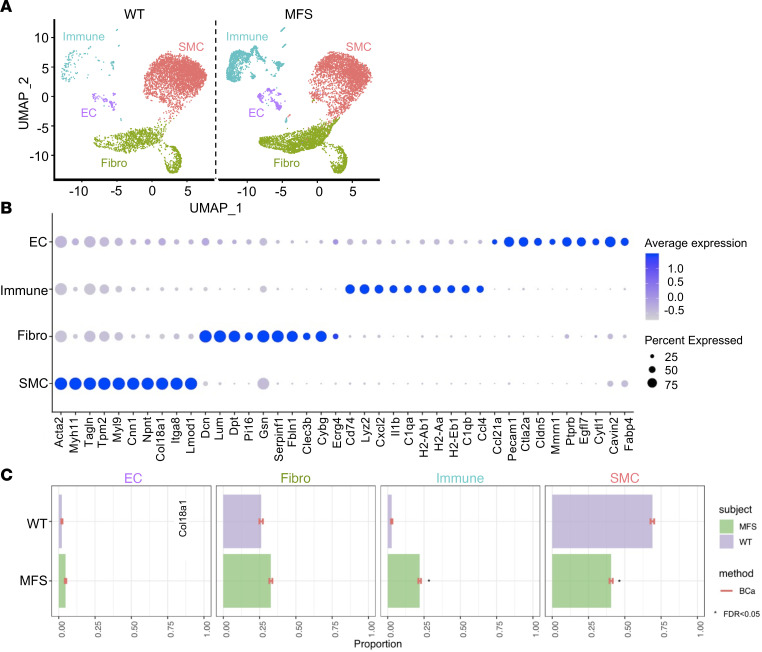
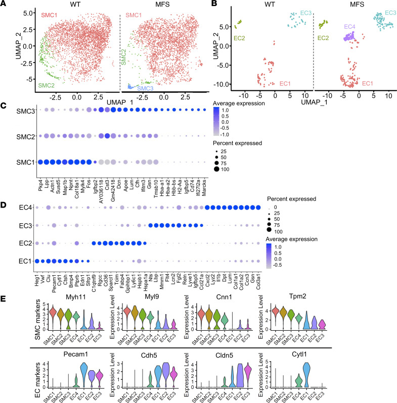
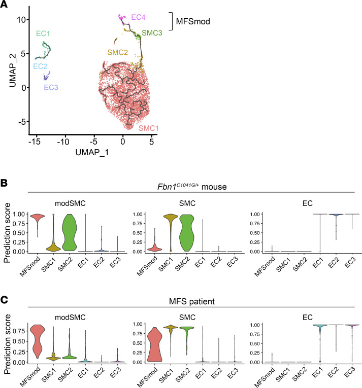
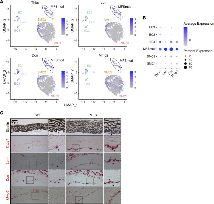
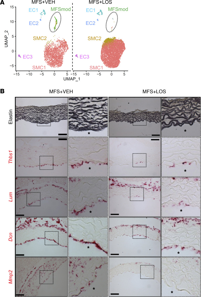
To improve our limited understanding of the pathogenesis of thoracic aortic aneurysm (TAA) that leads to acute aortic dissection, single-cell RNA sequencing (scRNA-seq) was employed to profile disease-relevant transcriptomic changes of aortic cell populations in a well-characterized mouse model of the most commonly diagnosed form of Marfan syndrome (MFS). As result, 2 discrete subpopulations of aortic cells (SMC3 and EC4) were identified only in the aorta of Fbn1mgR/mgR mice. SMC3 cells highly express genes related to extracellular matrix formation and nitric oxide signaling, whereas the EC4 transcriptional profile is enriched in smooth muscle cell (SMC), fibroblast, and immune cell-related genes. Trajectory analysis predicted close phenotypic modulation between SMC3 and EC4, which were therefore analyzed together as a discrete MFS-modulated (MFSmod) subpopulation. In situ hybridization of diagnostic transcripts located MFSmod cells at the intima of Fbn1mgR/mgR aortas. Reference-based data set integration revealed transcriptomic similarity between MFSmod- and SMC-derived cell clusters modulated in human TAA. Consistent with the angiotensin II type I receptor (At1r) contribution to TAA development, MFSmod cells were absent in the aorta of Fbn1mgR/mgR mice treated with the At1r antagonist losartan. Altogether, our findings indicate that a discrete dynamic alteration of aortic cell identity is associated with dissecting TAA in MFS mice and increased risk of aortic dissection in MFS patients.

Keywords: Cardiovascular disease; Expression profiling; Extracellular matrix; Genetics; Vascular Biology.


Figures

Figure 1
Figure 1. Transcriptomic atlas of ascending aorta cell types.
(A) Uniform manifold approximation and projection (UMAP) dimensional reduction of individual cells recovered from Fbn1mgR/mgR mice (n = 3) and WT littermates (n = 3) shows 4 identified major aortic cell types classified based on their enriched genes and annotation predicted by singleR package in R along with marker gene identification. (B) Dot plot shows top 10 genes highly enriched in major aortic cell populations. Dot size corresponds to the percentage of cells expressing each gene, and dot color corresponds to the level of expression. (C) Relative proportion of major cell clusters in MFS and WT aortas was evaluated with scDC (v0.1.0) using bias-corrected and accelerated (BCa) bootstrap confidence intervals, and FDR significance (<0.05) between groups was calculated after GLM fitting (22).
Figure 2
Figure 2. Disease-associated aortic cell types in MFS mice.
Uniform manifold approximation and projection (UMAP) dimensional reduction of (A) SMC and (B) EC clusters extracted from the major cell populations. Dot plots showing top 10 genes highly enriched in SMC (C) and EC subpopulations (D). Dot size corresponds to the percentage of cells expressing each gene, and dot color corresponds to the level of expression. (E) Violin plots showing expression of SMC marker genes (upper) and EC marker genes (lower) in each cell subpopulation of Fbn1mgR/mgR aortas.
Figure 3
Figure 3. Transcriptomic similarities between modulated aortic cells in MFS mice and patient.
(A) Trajectory analysis of EC and SMC subpopulations in both WT and Fbn1mgR/mgR samples indicating dynamic modulation between SMC3 and EC4, thus clustered together as MFSmod cells. (B and C) Violin plots showing the degree of transcriptomic homology of EC and SMC subpopulations in the ascending aorta of Fbn1mgR/mgR mice vs. either (B) the Fbn1C1041G/+ aortic root/ascending aorta or (C) the aortic root of an MFS patient (9).
Figure 4
Figure 4. RNAscope assays for MFSmod markers.
(A) Feature plots showing expression of genes that are highly enriched in MFSmod cells (highlighted by broken-line circle) and significantly less active in other EC and SMC subpopulations of Fbn1mgR/mgR aortas. Thbs1, thrombospondin-1; Lum, lumican; Dcn, decorin; Mmp2, matrix metalloproteinase 2. (B) Dot plots showing expression of the ECM-related genes in each cell subpopulation of Fbn1mgR/mgR aortas. Dot size corresponds to the percentage of cells expressing each gene, and dot color corresponds to the level of expression. (C) Representative images of Hart’s staining of elastin in WT and Fbn1mgR/mgR aortas (top) and in situ RNAscope hybridization (below) using probes specific for Thbs1, Lum, Dcn, and Mmp2 in both WT and MFS (n = 3 per genotype). Scale bar: 50 μm. Asterisks indicate the lumenal side.
Figure 5
Figure 5. MFSmod absence in losartan-treated mutant mice.
(A) Uniform manifold approximation and projection (UMAP) showing aortic EC and SMC subpopulations of Fbn1mgR/mgR mice treated with vehicle (VEH; n = 3) or losartan (LOS; n = 4). Broken-line circle indicates unique subpopulations of the mutant aorta. (B) Illustrative images of Hart’s staining of elastin (top) and in situ RNAscope hybridization (below) using probes specific for Thbs1, Lum, Dcn, and Mmp2 in the aortas of losartan-treated and untreated Fbn1mgR/mgR mice (n = 3 per group). Scale bars: 50 μm. Asterisks indicate the lumenal side.

References

    1. Milewicz DM, et al. Marfan syndrome. Nat Rev Dis Primers. 2021;7(1):64. doi: 10.1038/s41572-021-00298-7. - DOI - PMC - PubMed
    1. Wagenseil JE, Mecham RP. Vascular extracellular matrix and arterial mechanics. Amer Phys Soc. 2009;89:957–989. doi: 10.1152/physrev.00041.2008. - DOI - PMC - PubMed
    1. Asano K, et al. Pathophysiology and therapeutics of thoracic aortic aneurysm in Marfan syndrome. Biomolecules. 2022;12(1):128. doi: 10.3390/biom12010128. - DOI - PMC - PubMed
    1. Dobnikar L, et al. Disease-relevant transcriptional signatures identified in individual smooth muscle cells from healthy mouse vessels. Nat Commun. 2018;9:4567. doi: 10.1038/s41467-018-06891-x. - DOI - PMC - PubMed
    1. Kalluri AS, et al. Single-cell analysis of the normal mouse aorta reveals functionally distinct endothelial cell populations. Circulation. 2019;140(2):147–163. doi: 10.1161/CIRCULATIONAHA.118.038362. - DOI - PMC - PubMed

Publication types