Review
. 2023 Nov;39(13-15):957-979.
doi: 10.1089/ars.2022.0088. Epub 2023 Apr 5.

Glioblastomas: Hijacking Metabolism to Build a Flexible Shield for Therapy Resistance

Affiliations
Review

Glioblastomas: Hijacking Metabolism to Build a Flexible Shield for Therapy Resistance

Justine Bailleul et al. Antioxid Redox Signal. 2023 Nov.

Abstract

Significance: Glioblastomas (GBMs) are among the most lethal tumors despite the almost exclusive localization to the brain. This is largely due to therapeutic resistance. Radiation and chemotherapy significantly increase the survival for GBM patients, however, GBMs always recur, and the median overall survival is just over a year. Proposed reasons for such intractable resistance to therapy are numerous and include tumor metabolism, in particular, the ability of tumor cells to reconfigure metabolic fluxes on demand (metabolic plasticity). Understanding how the hard-wired, oncogene-driven metabolic tendencies of GBMs intersect with flexible, context-induced metabolic rewiring promises to reveal novel approaches for combating therapy resistance. Recent Advances: Personalized genome-scale metabolic flux models have recently provided evidence that metabolic flexibility promotes radiation resistance in cancer and identified tumor redox metabolism as a major predictor for resistance to radiation therapy (RT). It was demonstrated that radioresistant tumors, including GBM, reroute metabolic fluxes to boost the levels of reducing factors of the cell, thus enhancing clearance of reactive oxygen species that are generated during RT and promoting survival. Critical Issues: The current body of knowledge from published studies strongly supports the notion that robust metabolic plasticity can act as a (flexible) shield against the cytotoxic effects of standard GBM therapies, thus driving therapy resistance. The limited understanding of the critical drivers of such metabolic plasticity hampers the rational design of effective combination therapies. Future Directions: Identifying and targeting regulators of metabolic plasticity, rather than specific metabolic pathways, in combination with standard-of-care treatments have the potential to improve therapeutic outcomes in GBM. Antioxid. Redox Signal. 39, 957-979.

Keywords: glioblastoma; metabolism; plasticity; redox.


Conflict of interest statement

The authors have no conflict of interest.

Figures

FIG. 1.
FIG. 1.
Role of oncogenic mutations in promoting altered glucose metabolism. Glucose is transported into the cell via GLUTs. Once inside the cell, glucose is phosphorylated by HKs into G6P in the first, irreversible step of the glycolytic pathway, a modification that traps glucose inside the cell. G6P reversibly isomerizes into F6P, which is then converted into 1,6-FBP by PFK1. Alternatively, PFK2 can convert F6P to 2,6-FBP, which is a potent allosteric activator of PFK1. Opposing the PFKs, FBPs (FBP1 and FBP2) catalyze the dephosphorylation of 1,6-FBP/2,6-FBP back to F6P. These early glycolytic steps can be affected by mutations in the EGFR and TP53 pathways. There is evidence that mutant TP53 (TP53mut) can induce the expression of TIGAR, a TP53WT-regulated bisphosphatase. The first ATP-producing enzyme in glycolysis, PGK1, converts 1,3-PG to 3PG. 3PG is the initiating metabolite for the SBP. PTEN can dephosphorylate PGK1. Under a fine regulation by the EGFR/PTEN axis, PGK1 can translocate to the mitochondria where it regulates the TCA cycle via PDK1 activation and PDH inhibition. M2 tumor-associated macrophages can also phosphorylate and activate PGK1 via secretion of IL6. The PKM2, which catalyzes the last rate-limiting step in glycolysis converting PEP to PYR and making ATP in the process, is also regulated by phosphotyrosines on proteins phosphorylated via EGFR signaling, and by the TP53 pathway alterations, through HIF1α and mTORC1. PKM2 inhibition can lead to a bottleneck in glycolysis and rerouting of upstream metabolites into the oxidative and nonoxidative PPP. Enzymatically inactive PKM2 exhibits moonlighting functions and can regulate gene expression in the nucleus including genes responsible for DNA repair proteins. EGFR signaling represents the activation of EGFR and downstream signaling. Question marks refer to studies performed in tumor models other than GBM. Green arrows indicate activation, while red arrows indicate inhibition. 1,3-PG, 1,3-bisphosphoglycerate; 1,6-FBP, fructose-1,6-bisphosphate; 3PG, 3-phosphoglycerate; EGFR, epidermal growth factor receptor; F6P, fructose-6-phosphate; FBP, fructose bisphosphatase; G6P, glucose-6-phosphate; GBM, glioblastoma; GLUT, glucose transporter; HIF, hypoxia-inducible factor; HK, hexokinase; IL6, interleukin 6; mTORC1, mammalian target of rapamycin complex 1; PDH, pyruvate dehydrogenase complex; PDK1, pyruvate dehydrogenase kinase 1; PEP, phosphoenolpyruvate; PFK, phosphofructokinase; PGK1, phosphoglycerate kinase 1; PKM2, M2 isoform of pyruvate kinase; PPP, pentose phosphate pathway; PTEN, phosphatase and tensin homolog deleted on chromosome 10; PYR, pyruvate; SBP, serine biosynthesis pathway; TCA, tricarboxylic acid; TIGAR, TP53-induced glycolysis and apoptosis regulator; WT, wild-type.
FIG. 2.
FIG. 2.
Role of glycolysis and branching pathways in therapy resistance. As the cell oxidizes glucose via glycolysis, the resulting intermediates are often diverted away from glycolysis to fulfill other metabolic needs. For rapidly proliferating cancer cells, a priority is generating enough energy and building blocks to support cell growth and division while keeping in check the oxidative stress that accompanies enhanced metabolic activity. Oxidative stress is also one of the primary targets of RT. G6P feeds the oxidative PPP, which generates precursors (R5P) for nucleotide synthesis and DNA repair. PPP also generates NADPH, via G6PD and 6PGD. HK2, responsible for glucose phosphorylation, can also protect from oxidative stress through binding to the mitochondria membrane to prevent mitochondrial ROS production. 3PG feeds into the de novo SBP, which also generates building blocks for glutathione and nucleotide synthesis, and more NADPH. 3PG is converted into serine via three consecutive enzymatic reactions, catalyzed by PHGDH, PSAT1, and PSPH. In a reversible reaction, serine is converted to glycine via SHMT1/2. During the conversion of serine to glycine, SHMT concomitantly charges 1CM with one carbon units. 1CM couples the folate and methionine cycles, which generate amino acids, nucleotides, lipids, and reducing equivalents in the form of NADPH. Cancer cells can also take up serine and glycine from exogenous sources. PYR, produced by PK(M2), feeds into the TCA cycle or the FA synthesis pathway providing additional energy and building blocks for nucleic acids, proteins, and membranes. Glutamine, made by astrocytes, can be used by glioma cells and enters the cell via the amino acid antiporter ASCT2 and is converted to glutamate by GLS, which participates in GSH synthesis and antioxidant defenses. Glutamate can also be converted to α-KG by GDH1 thus replenishing the TCA cycle. Malate produced in the TCA cycle can be converted to PYR via the malic enzyme and NADPH is produced in the process. Glutamine can be converted back to glutamine via GS or exported outside the cell as an exchange for cystine (the oxidized dimer form of the amino acid cysteine) via the SLC7A11/xCT. 1CM, one-carbon metabolism; 6PGD, 6-phosphogluconate dehydrogenase; α-KG, α-ketoglutarate; ASCT2, Alanine, Serine, Cysteine Transporter 2; FA, fatty acid; G6PD, glucose-6-phosphate dehydrogenase; GDH1, glutamate dehydrogenase-1; GLS, glutaminase; GS, glutamine synthetase; GSH, reduced glutathione; NADPH, reduced nicotinamide adenine dinucleotide phosphate; PHGDH, phosphoglycerate dehydrogenase; PSAT1, phosphoserine aminotransferase 1; PSPH, phosphoserine phosphatase; R5P, ribose-5-phosphate; ROS, reactive oxygen species; RT, radiation therapy; SHMT, serine hydroxymethyl-transferase; SLC7A11/xCT, cystine/glutamate antiporter.
FIG. 3.
FIG. 3.
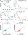
Gene expression levels of NADPH-producing enzymes in the PPP correlate with survival in GBM. TCGA data set analysis on GBMs reveals that gene expression levels of G6PD and 6PGD are highly correlated with the overall survival of LGG (A, B), whereas in GBM, the gene expression levels of only G6PD trend toward significance (p = 0.0762) for correlating with overall survival (C), but it significantly correlates with progression-free survival (D). Data sets were analyzed in GraphPad Prism software and p-values determined via log-rank (Mantel–Cox) test. LGG, low-grade gliomas; TCGA, The Cancer Genome Atlas.
FIG. 4.
FIG. 4.
Pathway alterations in the SBP in GBM. TCGA data set analysis on LGG and GBM reveals the presence of copy number gains and amplifications in all the SBP enzymes in both tumor types, but an increased frequency in GBM. 3PG, derived from glucose, is converted into serine via three consecutive enzymatic reactions, catalyzed by PHGDH, PSAT1, and PSPH. In a final reversible step, serine is converted to glycine via SHMTs, SHMT1 (cytoplasmic) or SHMT2 (mitochondrial). 3PHP, 3-phosphohydroxypyruvate, 3PS, 3-phosphoserine.
FIG. 5.
FIG. 5.
CNVs correlate with the overall survival in LGG and GBM. TCGA data set analysis of CNVs (include deletions) on LGG and GBM reveals that CNVs of PHGDH and PSPH correlate with overall survival, with PHGDH CNVs correlating with survival in only LGG and not GBM (A, B), whereas CNVs in the PSPH gene significantly correlate with overall survival in both LGG and GBM (C, D). CNVs are highly correlated with PSPH gene expression levels in both LGG and GBM (E, F). CNV, copy number variation.
FIG. 6.
FIG. 6.
Role of lipid metabolism in therapy resistance in GBM. Lipids exist in three main classes of esters—TAG (also referred to as triglycerides, three FAs individually esterified to each carbon of a glycerol molecule), PLs (two FAs joined to a phosphate group and an alcohol residue), and cholesteryl esters (an FA esterified to the hydroxyl group of cholesterol)—and can be obtained extracellularly from the diet or synthesized from glucose and glutamine carbons. Catabolism of glucose and glutamine via the TCA cycle provides citrate, which can be converted to Ac-CoA, a precursor for lipogenesis (synthesis of PL and TAG), as well as cholesterol synthesis via the mevalonate pathway, which can be activated by WTp53. Ac-CoA can also be generated from the breakdown of TAGs in the mitochondria, a process known as β-oxidation. This catabolic process generates NADH and FADH2, further utilized for ATP production. Within the cell, TAGs may also be stored inside lipid droplets, highly dynamic organelles that regulate the storage and hydrolysis of lipids. Lipid droplets are generally composed of a neutral lipid core encapsulated in a monolayer of PLs and can be broken down by lipophagy. They are ubiquitous in cells and often reflect their metabolic state. The mevalonate pathway is important for GSC self-renewal, in particular via HMGCR, the rate-limiting enzyme converting HMG-CoA to mevalonate. LXRs are nuclear receptors that regulate intracellular cholesterol levels by controlling the expression of genes related to cholesterol export. Upregulated EGFR signaling downregulates the expression of LXRα mRNA and leads to cholesterol accumulation. EGFR also induces the cleavage and nuclear translocation of SREBP, an important transcriptional regulator of FA and cholesterol synthesis. SREBP also increases cholesterol uptake by inducing the expression of LDLR. CYP46A1 converts cholesterol into 24(S)-hydroxycholesterol, thereby eliminating cholesterol. Its downregulation in GBM further increases intracellular levels of cholesterol, thus supporting growth. Upregulated EGFR signaling renders GBM cells dependent on plasma membrane remodeling, in an LPCAT1 and SMPD1-dependent manner. LPCAT1 controls membrane PL saturation, whereas SMPD1 converts sphingomyelin to ceramide and is crucial for lipid raft composition and clustering of membrane signaling molecules, such as EGFR. Ac-CoA, acetyl-CoA; CYP46A1, cholesterol 24-hydroxylase; FADH2, flavin adenine dinucleotide; GSC, GBM stem cell; HMGCR, 3-hydroxy-3-methyl-glutaryl-coenzyme A reductase; LDLR, low-density lipoprotein receptor; LPCAT1, lysophosphatidylcholine acyltransferase 1; LXR, liver X receptor; mRNA, messenger RNA; NADH, nicotinamide adenine dinucleotide; PL, phospholipid; SMPD1, sphingomyelin phosphodiesterase 1; SREBP, sterol regulatory element-binding protein; TAG, triacylglycerol.

Similar articles

References

    1. Altwairgi AK, Alghareeb WA, AlNajjar FH, et al. . Atorvastatin in combination with radiotherapy and temozolomide for glioblastoma: A prospective phase II study. Invest New Drugs 2021;39(1):226–231; doi: 10.1007/s10637-020-00992-5 - DOI - PubMed
    1. Anastasiou D, Poulogiannis G, Asara JM, et al. . Inhibition of pyruvate kinase M2 by reactive oxygen species contributes to cellular antioxidant responses. Science 2011;334(6060):1278–1283; doi: 10.1126/science.1211485 - DOI - PMC - PubMed
    1. Anastasiou D, Yu Y, Israelsen WJ, et al. . Pyruvate kinase M2 activators promote tetramer formation and suppress tumorigenesis. Nat Chem Biol 2012;8(10):839–847; doi: 10.1038/nchembio.1060 - DOI - PMC - PubMed
    1. Babic I, Anderson ES, Tanaka K, et al. . EGFR mutation-induced alternative splicing of Max contributes to growth of glycolytic tumors in brain cancer. Cell Metab 2013;17(6):1000–1008; doi: 10.1016/j.cmet.2013.04.013 - DOI - PMC - PubMed
    1. Bagga P, Behar KL, Mason GF, et al. . Characterization of cerebral glutamine uptake from blood in the mouse brain: Implications for metabolic modeling of 13C NMR data. J Cereb Blood Flow Metab 2014;34(10):1666–1672; doi: 10.1038/jcbfm.2014.129 - DOI - PMC - PubMed

Publication types

MeSH terms

Substances