. 2023 Apr 11;120(15):e2210808120.
doi: 10.1073/pnas.2210808120. Epub 2023 Apr 6.

African swine fever virus I73R is a critical virulence-related gene: A potential target for attenuation

Affiliations

African swine fever virus I73R is a critical virulence-related gene: A potential target for attenuation

Yingnan Liu et al. Proc Natl Acad Sci U S A. .

Abstract

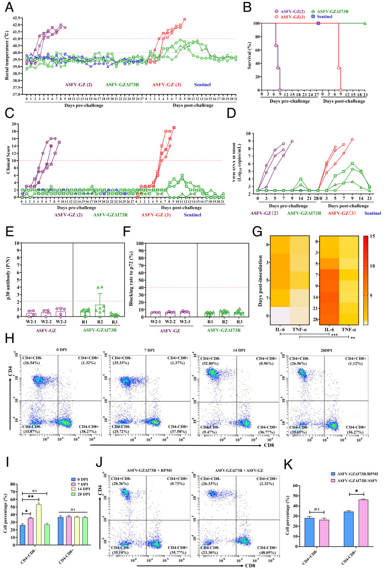
African swine fever virus (ASFV) is a large, double-stranded DNA virus that causes a fatal disease in pigs, posing a threat to the global pig industry. Whereas some ASFV proteins have been found to play important roles in ASFV-host interaction, the functional roles of many proteins are still largely unknown. In this study, we identified I73R, an early viral gene in the replication cycle of ASFV, as a key virulence factor. Our findings demonstrate that pI73R suppresses the host innate immune response by broadly inhibiting the synthesis of host proteins, including antiviral proteins. Crystallization and structural characterization results suggest that pI73R is a nucleic-acid-binding protein containing a Zα domain. It localizes in the nucleus and inhibits host protein synthesis by suppressing the nuclear export of cellular messenger RNA (mRNAs). While pI73R promotes viral replication, the deletion of the gene showed that it is a nonessential gene for virus replication. In vivo safety and immunogenicity evaluation results demonstrate that the deletion mutant ASFV-GZΔI73R is completely nonpathogenic and provides effective protection to pigs against wild-type ASFV. These results reveal I73R as a virulence-related gene critical for ASFV pathogenesis and suggest that it is a potential target for virus attenuation. Accordingly, the deletion mutant ASFV-GZΔI73R can be a potent live-attenuated vaccine candidate.

Keywords: ASFV; I73R; immune suppression; live-attenuated vaccine candidate; single-stranded RNA binding.


Conflict of interest statement

The authors declare no competing interest.

Figures

Fig. 1.
Fig. 1.
Characteristics of ASFV pI73R. I73R is a conserved gene and is transcribed and expressed in the early stage of the ASFV replication cycle. (A) Heatmap demonstrating the clustering of transcriptional patterns of the ASFV genome at various time points. The abundance of ASFV transcripts was expressed as fragments per kilobase million (FPKM) and indicated using a color key (blue and red correspond to decreased and increased transcriptional levels, respectively). Each column represents one sample, while each row represents the results of hierarchical clustering. (B) The transcript levels of the top 20 genes during ASFV replication. (C) The transcriptional levels of the I73R gene at various time points. The RNAs encoding the open reading frames of I73RB646L, and CP204L were isolated from BMDMs infected with ASFV-GZ at 3, 6, 9, 12, 15, and 18 h postinfection. (D) The protein expression levels at various time points. BMDMs were infected with ASFV-GZ at an m.o.i of 1 for 0, 3, 6, 9, 12, 15, and 18 h. The expression levels of pI73R, p72 and p30 were detected using anti-pI73R, anti-p72 and anti-p30 antibodies, respectively. β-actin was used as the loading control. (E) Subcellular localization of pI73R in BMDMs during ASFV infection. BMDMs were infected with ASFV-GZ (m.o.i = 1) for 6, 12, and 18 hpi and reacted with anti-pI73R monoclonal antibodies and Alexa 488-conjugated goat anti-mouse IgG secondary antibody (green). The nuclei were stained with DAPI (blue) (63×). Note: White arrows indicate the location of the cellular virus factories (VF) formed after ASFV infection. (F) Subcellular localization of pI73R in HeLa cells. HeLa cells were transfected with recombinant pI73R-Flag plasmid and probed with anti-Flag antibodies. The nucleus were stained with DAPI. Green fluorescent protein (GFP) (pseudocolored in green), DAPI (pseudocolored in blue) images were captured using a confocal microscope (63×).
Fig. 2.
Fig. 2.
Broad-spectrum inhibition of host protein synthesis by ASFV pI73R. (A) Luciferase (Rluc) assay was performed to detect the effects of pI73R on the Rluc reporter gene. At 24 h posttransfection, cell extracts were also subjected to IB (Bottom) and Rluc assay (Top). Error bars indicate SD of means from three independent experiments; (***P < 0.001 was considered extremely significant). (B) The effect of exogenous pI73R on Rluc mRNA production was determined using qRT-PCR at 12 and 24 h when the cells were pretreated with or without CHX. Error bars indicate SD of means from three independent experiments. Means between different groups were compared using two-tailed Student’s t tests; **P < 0.01; ***P < 0.001. (C) HEK-293T (Left), PAM (Middle), and BMDM (Right) cells were transfected with the pCAGGS or pI73R-Flag plasmid. The cells were pulsed with 3 μM puromycin for 1 h at 24 hpt, and whole-cell lysates were obtained and subsequently subjected to IB with anti-puromycin and anti-Flag antibodies. (D) BMDMs were transfected with pI73R mRNA for 24 h, followed by infected with the ASFV-GZ strain for 6, 12, and 18 h and subsequently subjected to IB with anti-p30 and anti-HA antibodies. (E) BMDMs were transfected with pI73R mRNA for 24 h, followed by ASFV-GZ infection for 6, 12, 24, 36, or 48 h. ASFV titers were determined using the median tissue culture infectious dose (TCID50) method. Data represent mean ± SDs. The sensitivity limit of virus detection was 102.45 TCID50/mL. (F) Schematic representation of the I73R gene deletion/insertion region in ASFV-GZ. All transfer vectors were designed to have 1.2-kb homology arms on both sides of the deletion/insertion cassette (represented as dashed lines), which induced deletion/insertion in the indicated viruses in the area between the dashed lines. (G) In vitro growth characteristics of recombinant mutants and parental ASFV-GZ virus. PAMs or BMDMs were infected with the viruses at an m.o.i of 0.1. The virus yields were titrated at the indicated time points postinfection. Data represent means and SDs. The sensitivity limit of virus detection was 102.45 TCID50/mL. (H) Cellular protein synthesis during ASFV and ASFV-GZΔI73R infection. PAM (Left) or BMDM (Right) cells were infected with ASFV-GZ (m.o.i = 1) or ASFV-GZΔI73R (m.o.i = 10). After 18 hpi, the cells were labeled with 3 mM puromycin for an additional hour. The whole-cell lysates were subjected to IB analysis.
Fig. 3.
Fig. 3.
ASFV pI73R is a high-affinity nucleic acid-binding protein. (A) Crystal structure of pI73R. The monomer structure of pI73R is shown as a colored cartoon (Left). The alpha-helices and beta-sheets are colored green and purple, respectively. The topology diagram of pI73R is also shown (Right). (B and C) Structural alignment of pI73R (gray) with human ADAR1 (cyan, PDB ID 2GXB), mouse ZBP1 (green, PDB ID 1J75), poxvirus E3L (yellow, PDB ID 1SFU), and herpesvirus ORF112 (brown, PDB ID 4HOB). (rmsd values = 1.40, 1.45, 1.93, and 2.56 Å, respectively). (D) EMSA was performed for analysing the binding between ASFV pI73R and nucleic acids. pI73R was assayed at various concentrations (0, 20, 40, 80, and 160 μM) with 1 μM 5′-Cy5-labeled ssRNA fragments. Arrow indicates the mobility shift of Cy5-labeled oligos. (E) BLI was performed for analyzing the binding kinetics between pI73R and nucleic acids. The pI73R protein was diluted to a concentration of 10 µg/mL in kinetics buffer, and loaded onto HIS1K biosensors, and incubated with twofold serially diluted nucleic acids (ssRNA, dsRNA, ssDNA, or dsDNA).
Fig. 4.
Fig. 4.
ASFV pI73R binds specifically to host mRNAs, especially to ssRNA with a high GC content. (A) Flowchart of the RNA immunoprecipitation (RIP) assay. (B) RIP assay was performed with lysates from BMDMs infected with the ASFV-GZ-mI73RHA strain. Whole-cell extracts were subjected to immunoprecipitation with the endogenous target anti-HA mAb, with a matched IgG mAb served as the control. RNA yields were determined using a NanoDrop spectrophotometer (Thermo Fisher Scientific). Data are presented as mean ± SD. Means were compared using unpaired t tests; ***P < 0.001, n = 3. (C) Venn diagram demonstrating the specific binding of cellular mRNA to ASFV pI73R. (D) Venn diagram demonstrating the specific binding of viral mRNA to ASFV pI73R. (E) Finding of predicted pI73R-binding motifs from the RIP-Seq data. (F) EMSA was performed with purified pI73R and 5′-Cy5-labeled with high-GC and GC-free ssRNA. Arrow indicates mobility shift of Cy5-labeled oligos. (G) RIP and RT-PCR for detecting the interaction between pI73R and TNF-α mRNA (Upper: qRT-PCR for TNF-α mRNA, lower panels: immunoblotting for pI73R). (H) MS2/MCP reporter system for detecting the migration of TNF-α mRNA (Upper: Schematic describing the constructs used in this approach. The system comprises two components, a reporter mRNA and a MS2-GFP fusion protein. The reporter mRNA containing 12x MS2-binding sites was introduced after the stop codon of TNF-α mRNA sequences. Lower panel: Re Representative images of cells expressing the pMS2-GFP and the TNF-α-12x MS2 reporter mRNA.) (I) ISH images revealed that in contrast to the empty vector control, pI73R retained the TNF-α mRNA in the nucleus. (J) Representative images of cells transfected with plasmids of pI73R fused with various nuclear export signals (pI73R/Wild, pI73R/SMAD4NES, pI73R/MEK1NES, pI73R/CPEB4NES). The nuclei were stained with DAPI. Red fluorescence protein (RFP) (pseudocolored in red) and DAPI (blue) images were captured using a confocal microscope (63×). (K) HEK-293T cells were transfected with the pCAGGS or pI73R/Wild, pI73R/SMAD4NES, pI73R/MEK1NES, and pI73R/CPEB4NES for 24 h. Next, the cells were pulsed with 3 μM puromycin for 1 h, and whole-cell lysates were prepared and subjected to immunoblotting with anti-puromycin and anti-Flag antibodies.
Fig. 5.
Fig. 5.
Evaluation of the ASFV-GZΔI73R deletion mutant in vivo. (A) The rectal temperature of pigs inoculated with 103.0 or 105.0 TCID50 of ASFV-GZΔI73R (green and blue, respectively) or the parental ASFV-GZ strain (dark red). (B) The clinical score was calculated after pigs were intramuscularly (i.m.) inoculated with 103.0 or 105.0 TCID50 of ASFV-GZΔI73R or the parental ASFV-GZ strain. (C) Survival outcomes of the pigs after inoculation with ASFV-GZΔI73R during the 28-d observation period. (D) Viremia (shown as viral DNA copies) in all groups of pigs after inoculation. (E and F) Viral shedding in oral and anal swabs were detected via qPCR against the p72 gene after inoculation. (G) Pathogenic scores were calculated in all groups of pigs and compared with those of the placebo control group. (H) Viral DNA copies in the tonsil, liver, spleen, lung, inguinal lymph node (LN), kidney, mesenteric LN, and thymus in the ASFV-GZΔI73R group were compared with those in the parental ASFV-GZ group. The sensitivity of viral DNA detection was ≥log102.45 copies/mL.
Fig. 6.
Fig. 6.
Protective efficacy of ASFV-GZΔI73R against challenge with parental ASFV-GZ. (A) The rectal temperature of pigs inoculated with 103.0 TCID50 of ASFV-GZΔI73R (green), sentinel (blue), or 104.0 TCID50 of ASFV-GZ (purple, ASFV-GZ 2) before and after challenge with 104.0 TCID50 of ASFV-GZ virus (red, ASFV-GZ 3). (B) Survival outcomes of pigs after intramuscular inoculation and challenge. (C) The clinical score of pigs inoculated with 103.0 TCID50 of ASFV-GZΔI73R (green), Sentinel (blue), or 104.0 TCID50 of ASFV-GZ (purple, ASFV-GZ 2) before and after challenge with 104.0 TCID50 of ASFV-GZ virus (red, ASFV-GZ 3). (D) Viremia (shown as viral DNA copies) in all groups of pigs after inoculation and challenge. (E and F) The anti-p30 and anti-p72 antibody response of pigs inoculated with ASFV-GZΔI73R or ASFV-GZ. (G) Proinflammatory cytokines associated with TNF signaling pathway (IL-6 and TNF-α) in pigs inoculated with 103.0 TCID50 of ASFV-GZΔI73R or 104.0 TCID50 of ASFV-GZ, respectively. The heatmap was drawn using the median of each group. Each small square represents cytokine expression, and the depth of the color represents the level of expression. Means between the two groups were compared using two-tailed Student’s t tests; **P < 0.01; ***P < 0.001. (H) Representative data from pigs on different days after immunized with ASFV-GZΔI73R. (I) Mean percentages of CD3+CD4+CD8 T cells and CD3+CD4CD8+ T cells in PBMCs of pigs immunized with ASFV-GZΔI73R. (J) Representative data from pigs on D 28 postinoculation followed by stimulation with ASFV-GZ for 60 h in vitro. (K) Mean proliferation percentages of PBMCs after stimulation with RPMI-1640 medium (control group) or ASFV-GZ.

References

    1. Gaudreault N. N., Madden D. W., Wilson W. C., Trujillo J. D., Richt J. A., African Swine fever virus: An emerging DNA arbovirus. Front. Vet. Sci. 7, 1–17 (2020). - PMC - PubMed
    1. Galindo I., Alonso C., African swine fever virus: A review. Viruses 9, 1255 (2017). - PMC - PubMed
    1. Alonso C., et al. , ICTV virus taxonomy profile: Asfarviridae. J. Gen. Virol. 99, 613–614 (2018). - PubMed
    1. Zhou X., et al. , Emergence of African Swine fever in China, 2018. Transbound. Emerg. Dis. 65, 1482–1484 (2018). - PubMed
    1. Tao D., et al. , One year of African swine fever outbreak in China. Acta Trop. 211, 105602 (2020). - PubMed

Publication types

Substances

LinkOut - more resources