. 2023 Apr 19;145(15):8583-8592.
doi: 10.1021/jacs.3c01003. Epub 2023 Apr 6.

Environmentally Ultrasensitive Fluorine Probe to Resolve Protein Conformational Ensembles by 19F NMR and Cryo-EM

Affiliations

Environmentally Ultrasensitive Fluorine Probe to Resolve Protein Conformational Ensembles by 19F NMR and Cryo-EM

Yun Huang et al. J Am Chem Soc. .

Abstract

Limited chemical shift dispersion represents a significant barrier to studying multistate equilibria of large membrane proteins by 19F NMR. We describe a novel monofluoroethyl 19F probe that dramatically increases the chemical shift dispersion. The improved conformational sensitivity and line shape enable the detection of previously unresolved states in one-dimensional (1D) 19F NMR spectra of a 134 kDa membrane transporter. Changes in the populations of these states in response to ligand binding, mutations, and temperature correlate with population changes of distinct conformations in structural ensembles determined by single-particle cryo-electron microscopy (cryo-EM). Thus, 19F NMR can guide sample preparation to discover and visualize novel conformational states and facilitate image analysis and three-dimensional (3D) classification.


Conflict of interest statement

The authors declare no competing financial interest.

Figures

Figure 1
Figure 1
Synthesis of the mFE probes and site-specific protein labeling. Synthesis of deuterated and protonated mFE probes (a) TsSCD2CD2F and (b) TsSCH2CH2F. (c) Cysteine-specific labeling. (d) Structure of the GltPh trimer with scaffold domains colored tan and the transport domains colored blue. Colored spheres show the sites of single-cysteine mutations A380C (cyan), A381C (magenta), and M385C (yellow). Dashed line indicates the distance between the 19F labels. (e) Elevator transition of the GltPh transport domain from the OFS (left) to IFS (right). Single protomers are shown in the membrane plane, represented by the dotted lines. The bound substrate aspartate is shown as lime spheres. ACN: acetonitrile; DAST: diethylaminosulfur trifluoride; DCM: dichloromethane; X: hydrogen or deuterium; and Ts: toluenesulfonyl.
Figure 2
Figure 2
mFED-labeled GltPh variants show wide chemical shift dispersion. 19F NMR spectra of (a, b) GltPh-M385C and (c, d) GltPh-A380C labeled with (a, c) tFE and (b, d) mFED. From top to bottom, (a, b) WT and RSMR mutants in 100 mM Na+ and 2 mM aspartate (Asp) and the RSMR mutant in 100 mM Na+ and 0.6 mM TFB-TBOA, and (c, d) WT in 400 mM Na+ and 100 mM Na+ and 2 mM aspartate. All spectra were recorded at 25 °C. Raw data are black, fitted spectra are pink, and deconvoluted Lorentzian peaks are blue. The asterisk denotes a signal from the CF3 group of TFB-TBOA. S1–S5 denote resolved resonances of GltPh-M385C-mFED. Δδ is the largest chemical shift difference observed for the labeling site. All 19F NMR spectra here and elsewhere were recorded at least twice on independently prepared protein samples, producing similar results.
Figure 3
Figure 3
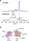
Identification and structural elucidation of new transporter conformations. (a) 19F NMR spectra of WT-M385C-mFED (upper panel) and RSMR-M385C-mFED (bottom panel) in the presence of 100 mM NaCl and 2 mM TBOA. The resonances occurring with similar chemical shifts to those in Figure 2 are labeled S2, S3, and S4. (b) Cryo-EM density maps of TBOA-bound RSMR GltPh mutant protomers in the IFS (left) and OFS (right) structural classes. The corresponding populations are below the maps.
Figure 4
Figure 4
Assignment of IFS and OFS conformations based on solvent PRE effects. 19F NMR spectra (left) were recorded, and T1 relaxation times (right) were measured in 300 mM NaCl and 2 mM Asp at 15 °C. Relaxation data were fitted to monoexponential functions (solid lines). The fitted T1 values are (a) 1.36 ± 0.20, 1.41 ± 0.02, 1.26 ± 0.33, 1.56 ± 0.16, and 1.26 ± 0.09 s for S2, S3, S4, S5, and S6, respectively, in the absence of Gd-DPTA–BMA and (b) 1.15 ± 0.20, 1.04 ± 0.08, 0.39 ± 0.08, 0.42 ± 0.06, and 1.01 ± 0.06 s in the presence of 20 mM Gd-DPTA–BMA. The error bars, estimated as described in the Materials and Methods Section, are too small to see.
Figure 5
Figure 5
State populations observed in 3D classifications of protomers in cryo-EM parallel NMR measurements. (a) NMR spectra of RSMR-M385C-mFED were recorded in the presence of 500 mM NaCl and 2 mM Asp at 4 (top), 15 (middle), and 30 °C (bottom). (b) Representative maps of the four structural classes identified during cryo-EM particle 3D classifications of RSMR-M385C-mFED prepared at 4 °C. (c) Populations of protomers’ 3D classes in samples preincubated at 4, 15, and 30 °C (left). Open circles are results obtained using different tau values and numbers of classes during 3D classifications in RELION (see the Materials and Methods Section for details). The state populations measured in 19F NMR experiments at the same temperatures are shown in the right panel. Errors are from multiple-peak deconvolutions of spectra in OriginPro 2019.

References

    1. Lee Y.; Lazim R.; Macalino S. J. Y.; Choi S. Importance of protein dynamics in the structure-based drug discovery of class A G protein-coupled receptors (GPCRs). Curr. Opin. Struct. Biol. 2019, 55, 147–153. 10.1016/j.sbi.2019.03.015. - DOI - PubMed
    1. Peng J. W. Communication Breakdown: Protein Dynamics and Drug Design. Structure 2009, 17, 319–320. 10.1016/j.str.2009.02.004. - DOI - PubMed
    1. Sekhar A.; Kay L. E. An NMR View of Protein Dynamics in Health and Disease. Annu. Rev. Biophys. 2019, 48, 297–319. 10.1146/annurev-biophys-052118-115647. - DOI - PubMed
    1. Palmer A. G. NMR Characterization of the Dynamics of Biomacromolecules. Chem. Rev. 2004, 104, 3623–3640. 10.1021/cr030413t. - DOI - PubMed
    1. Lerner E.; Cordes T.; Ingargiola A.; Alhadid Y.; Chung S.; Michalet X.; Weiss S. Toward dynamic structural biology: Two decades of single-molecule Förster resonance energy transfer. Science 2018, 359, 28810.1126/science.aan1133. - DOI - PMC - PubMed

Publication types