. 2023 May:91:104546.
doi: 10.1016/j.ebiom.2023.104546. Epub 2023 Apr 4.

B4GALNT3 regulates glycosylation of sclerostin and bone mass

Affiliations

B4GALNT3 regulates glycosylation of sclerostin and bone mass

Sofia Movérare-Skrtic et al. EBioMedicine. 2023 May.

Abstract

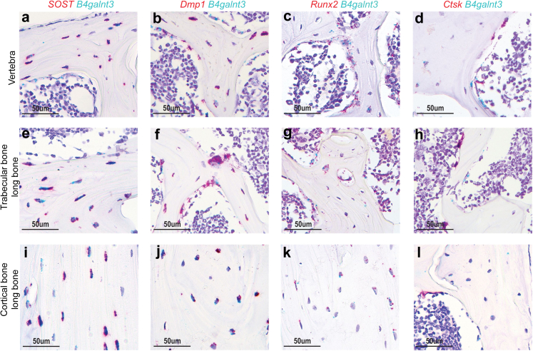
Background: Global sclerostin inhibition reduces fracture risk efficiently but has been associated with cardiovascular side effects. The strongest genetic signal for circulating sclerostin is in the B4GALNT3 gene region, but the causal gene is unknown. B4GALNT3 expresses the enzyme beta-1,4-N-acetylgalactosaminyltransferase 3 that transfers N-acetylgalactosamine onto N-acetylglucosaminebeta-benzyl on protein epitopes (LDN-glycosylation).

Methods: To determine if B4GALNT3 is the causal gene, B4galnt3-/- mice were developed and serum levels of total sclerostin and LDN-glycosylated sclerostin were analysed and mechanistic studies were performed in osteoblast-like cells. Mendelian randomization was used to determine causal associations.

Findings: B4galnt3-/- mice had higher circulating sclerostin levels, establishing B4GALNT3 as a causal gene for circulating sclerostin levels, and lower bone mass. However, serum levels of LDN-glycosylated sclerostin were lower in B4galnt3-/- mice. B4galnt3 and Sost were co-expressed in osteoblast-lineage cells. Overexpression of B4GALNT3 increased while silencing of B4GALNT3 decreased the levels of LDN-glycosylated sclerostin in osteoblast-like cells. Mendelian randomization demonstrated that higher circulating sclerostin levels, genetically predicted by variants in the B4GALNT3 gene, were causally associated with lower BMD and higher risk of fractures but not with higher risk of myocardial infarction or stroke. Glucocorticoid treatment reduced B4galnt3 expression in bone and increased circulating sclerostin levels and this may contribute to the observed glucocorticoid-induced bone loss.

Interpretation: B4GALNT3 is a key factor for bone physiology via regulation of LDN-glycosylation of sclerostin. We propose that B4GALNT3-mediated LDN-glycosylation of sclerostin may be a bone-specific osteoporosis target, separating the anti-fracture effect of global sclerostin inhibition, from indicated cardiovascular side effects.

Funding: Found in acknowledgements.

Keywords: Fracture risk; Mendelian randomization; Osteoblasts; Osteoporosis.


Conflict of interest statement

Declaration of interests CO has two patents/patent applications in the field of probiotics and bone health. All other authors declare that they have no conflict of interest.

Figures

Fig. 1
Fig. 1
B4galnt3−/−mice have increased levels of total serum sclerostin but decreased levels of LDN-glycosylated sclerostin in serum. a) Schematic illustration of the B4galnt3 inactivated mouse model. The B4galnt3 gene has 20 exons and the two LoxP sites are located in the introns upstream of exons 8 and 10. b) mRNA expression analyses of B4galnt3 in the cortical bone of tibia and aorta in 13-wk-old female B4galnt3+/+ mice (n = 11) and B4galnt3−/− homozygote (n = 12) mice. Individual values are presented with the mean as horizontal lines and ±SEM as vertical lines. Statistical analyses were performed using two-sided Student's t test. ND = not detectable. c–e) Total serum sclerostin levels in female + male (c), female (d), and male (e) B4galnt3+/+ and B4galnt3−/− mice (female B4galnt3+/+ n = 11; female B4galnt3−/− n = 11; male B4galnt3+/+ n = 11; male B4galnt3−/− n = 11) as measured by ELISA. Values are given as medians and interquartile range. Statistical analyses were performed using two-sided Student's t test. f–h) Normalized LDN-glycosylated sclerostin protein levels from serum of female + male (f), female (g), and male (h) B4galnt3+/+ and B4galnt3−/− mice (female B4galnt3+/+ n = 11; female B4galnt3−/− n = 11; male B4galnt3+/+ n = 11; male B4galnt3−/− n = 11) after precipitation with Wisteria floribunda agglutinin (WFA) agarose beads and analysed by Western blot. On each Western blot (see example in Supplemental Figure S2a and b), serum from one random B4galnt3+/+ and one random B4galnt3−/− mice with and without WFA in the precipitation were included and the levels in B4galnt3−/− mouse were normalized to the levels in the B4galnt3+/+ mouse set to 1. Values are given as medians and interquartile range. Statistical analyses were performed on normalized data using Mann–Whitney U-test. i–k) Ratio of LDN-glycosylated/total sclerostin levels in female + male (i), female (j), and male (k) B4galnt3+/+ and B4galnt3−/− mice (female B4galnt3+/+ n = 11; female B4galnt3−/− n = 11; male B4galnt3+/+ n = 11; male B4galnt3−/− n = 11). The ratio in a B4galnt3−/− mouse was normalized to the ratio in the B4galnt3+/+ mouse analysed on the same Western blot. Values are given as medians and interquartile range. Statistical analyses were performed on normalized data using Mann–Whitney U-test. l) Quantification of the intensity of sclerostin immunoreactivity per osteocyte in tibia. Results are expressed as mean intensity per osteocyte (male B4galnt3+/+ mice n = 6; B4galnt3−/− mice n = 6). Individual values are presented with the mean as horizontal lines and ±SEM as vertical lines. Statistical analyses were performed using two-sided Student's t test. The results refer to 13-week-old B4galnt3+/+ and B4galnt3−/− mice. LDN = LacdiNAc.
Fig. 2
Fig. 2
B4galnt3 is expressed in osteoblasts and osteocytes but not in osteoclasts. a–l) Representative in situ hybridization images in mice. Transverse sections across lumbar vertebra 5 (a–d) and longitudinal sections of femur or tibia (e–l) in mice demonstrate the mRNA expression of B4galnt3 (blue; a–l) and Sost (red; a, e, i), Dmp1 (red; b, f, j), Runx2 (red; c, g, k) or Ctsk (red; d, h, l). B4galnt3 mRNA could be observed in Sost-, Dmp1-, and Runx2-expressing osteocytes and osteoblast- lineage cells on the bone surface. In contrast, B4galnt3 was not detectable in Ctsk-expressing osteoclasts. Scale bar 50 μm. Images are representative of at least three independent experiments.
Fig. 3
Fig. 3
B4GALNT3 contributes to post-translational modification by regulating LDN-glycosylation of sclerostin in human osteoblast-like cells. a and b) B4GALNT3 (a) and SOST (b) mRNA expression in Saos-2 cells following transfection with negative control siRNA (Neg.si) or B4GALNT3 siRNA (B4GN3si) (n = 6 per group). Values are given as means ± SEM. Statistical analyses were performed using two-sided Student's t test. c) Representative Western blot showing LDN-glycosylated sclerostin protein levels after precipitation with WFA agarose beads or negative control (Neg. CTR) agarose beads from Saos-2 cells following transfection with negative control siRNA (Neg.si) or B4GALNT3 siRNA (B4GN3si). d) Normalized quantification of LDN-glycosylated sclerostin protein levels after precipitation with WFA agarose beads from Saos-2 cells following transfection with negative control siRNA (Neg.si) or B4GALNT3 siRNA (B4GN3si) and detected by Western blot analysis (n = 5 per group). Values are given as means ± SEM. Statistical analyses were performed on normalized data using two-sided Student's t test. e and f) B4GALNT3 (e) and SOST (f) mRNA expression in Saos-2 cells following transfection with empty vector (V) or a construct encoding human B4GALNT3 (n = 6 per group). Values are given as means ± SEM. Statistical analyses were performed using Mann–Whitney U-test for (e) and two-sided Student's t test for (f). g) Representative Western blot showing LDN-glycosylated sclerostin protein levels after precipitation with WFA agarose beads or negative control (Neg. CTR) agarose beads from Saos-2 cells following transfection with empty vector (V) or a construct encoding human B4GALNT3. h) Normalized quantification of LDN-glycosylated sclerostin protein levels after precipitation with WFA agarose beads from Saos-2 cells following transfection with empty vector (V) or a construct encoding human B4GALNT3 and detected by Western blot analysis (n = 8 per group). Values are given as means ± SEM. Statistical analyses were performed on normalized data using two-sided Student's t test. LDN = LacdiNAc, PD = precipitation, WFA = Wisteria floribunda agglutinin, a.u. = arbitrary units.
Fig. 4
Fig. 4
Hormonal regulation of B4galnt3 expression in bone and reduced cortical bone mass and strength in B4galnt3−/−mice. a–d) Cortical area of femur (a), B4galnt3 mRNA expression in cortical bone (b), total serum sclerostin levels (c), and Sost mRNA expression in cortical bone (d), in 16-wk-old wildtype female mice treated with glucocorticoids (GC; n = 9, open circle) or vehicle (Veh; n = 9, filled circle) for four weeks. e) B4galnt3 mRNA expression in primary calvarial osteoblastic cells cultured in osteogenic media for 48 h, with and without 100 nM dexamethasone (DEX). f) Proposed mechanism of hormonal regulation of B4GALNT3 and effects on LDN-glycosylation of sclerostin (LDN-Sclerostin). g–i) Areal bone mineral density (BMD) of total body (g), lumbar spine (L2–L5) (h), and whole femur (i), of 5-wk-old female B4galnt3−/− (n = 11) and B4galnt3+/+ (n = 10) mice. j–n) Cortical content (j), cortical area (k), cortical thickness (l), periosteal circumference (m), and endosteal circumference (n), of tibia in 13-wk-old female B4galnt3−/− (n = 12) and B4galnt3+/+ (n = 11) mice. o and p) Trabecular bone volume over total volume (BV/TV) of vertebra (o) and tibia (p) in 13-wk-old female B4galnt3−/− (n = 12) and B4galnt3+/+ (n = 11) mice. q) Maximal load at failure (N) of humerus as measured by three-point-bending in 13-wk-old female B4galnt3−/− (n = 11) and B4galnt3+/+ (n = 9) mice. Statistical analyses were performed using two-sided Student's t test. Values are given as means ± SEM.

References

    1. Hernlund E., Svedbom A., Ivergard M., et al. Osteoporosis in the European Union: medical management, epidemiology and economic burden. A report prepared in collaboration with the International Osteoporosis Foundation (IOF) and the European Federation of Pharmaceutical Industry Associations (EFPIA) Arch Osteoporos. 2013;8(1):136. - PMC - PubMed
    1. Johnell O., Kanis J.A. An estimate of the worldwide prevalence and disability associated with osteoporotic fractures. Osteoporos Int. 2006;17(12):1726–1733. - PubMed
    1. Bovijn J., Krebs K., Chen C.Y., et al. Evaluating the cardiovascular safety of sclerostin inhibition using evidence from meta-analysis of clinical trials and human genetics. Sci Transl Med. 2020;12(549) - PMC - PubMed
    1. Cosman F., Crittenden D.B., Adachi J.D., et al. Romosozumab treatment in postmenopausal women with osteoporosis. N Engl J Med. 2016;375(16):1532–1543. - PubMed
    1. McClung M.R., Grauer A., Boonen S., et al. Romosozumab in postmenopausal women with low bone mineral density. N Engl J Med. 2014;370(5):412–420. - PubMed

Substances