. 2023 May 25;64(3):520-529.
doi: 10.1093/jrr/rrad016.

DAP3-mediated cell cycle regulation and its association with radioresistance in human lung adenocarcinoma cell lines

Affiliations

DAP3-mediated cell cycle regulation and its association with radioresistance in human lung adenocarcinoma cell lines

Yoshiaki Sato et al. J Radiat Res. .

Abstract

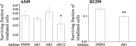
Mitochondria play important roles in the cellular response to various types of stress, including that triggered by ionizing radiation. We have previously reported that the mitochondrial ribosomal protein death-associated protein 3 (DAP3) regulates the radioresistance of human lung adenocarcinoma (LUAD) cell lines A549 and H1299. However, the underlying mechanism of this regulation remains to be elucidated. To this end, we have herein investigated the role of DAP3 in the cell cycle regulation after irradiation. Notably, the DAP3 knockdown attenuated the radiation-induced increase of the G2/M cell population. Furthermore, western blotting analysis has revealed that the DAP3 knockdown decreased the expression of proteins related to the G2/M arrest, such as those of the phosphorylated cdc2 (Tyr15) and the phosphorylated checkpoint kinase 1 (Ser296), in irradiated A549 cells and H1299 cells. Moreover, by using a chk1 inhibitor, we were able to demonstrate that chk1 is involved in the radiation-induced G2/M arrest in both A549 and H1299 cells. Notably, the chk1 inhibitor was able to enhance the radiosensitivity of H1299 cells, while both chk1 inhibitor-abolished G2 arrest and inhibition of chk2-mediated events such as downregulation of radiation-induced p21 expression were required for enhancing radiosensitivity of A549 cells. Collectively, our findings reveal a novel role of DAP3 to regulate G2/M arrest through pchk1 in irradiated LUAD cells and suggest that chk1-mediated G2/M arrest regulates the radioresistance of H1299 cells, whereas both the chk1-mediated G2/M arrest and the chk2-mediated events contribute to the radioresistance of A549 cells.

Keywords: in vitro; G2 arrest; checkpoint kinase 1; death-associated protein 3; lung adenocarcinoma; radioresistance.


Conflict of interest statement

There are no conflicts of interest to declare.

Figures

Fig. 1
Fig. 1
Involvement of DAP3 in the radiation-induced G2/M arrest of human LUAD cell lines. (A) Human LUAD cell lines (A549 and H1299) transfected with control or DAP3 siRNA were harvested, and the DAP3 protein expression was analyzed through western blotting. A representative image of an immunoblot is shown. GAPDH was used as the loading control. (B, C) Human LUAD cell lines transfected with control or DAP3 siRNA were treated with radiation. After 8 or 24 h of culturing, the cells were harvested for cell cycle analysis. (Left) Representative histograms and the obtained data are presented. Double-headed arrows indicate the G2/M population, whereas the inset number in the figure indicates the G2/M proportion in the total cells. (Right) The net increase in G2/M populations as a result of the undertaken irradiation is shown. Data are presented as the mean ± SD of three independent experiments. Symbols used: *P < 0.05 and **P < 0.01; both versus control siRNA.
Fig. 2
Fig. 2
Effect of DAP3 knockdown on radiation-induced mitotic catastrophe in human LUAD cell lines. (A, B) Human LUAD cell lines transfected with control or DAP3 siRNA were treated with radiation. After 72 h of culturing, the cells were harvested for the analysis of mitotic catastrophe. (Top) Representative pictures of mitotic catastrophe are shown. Micronuclei, multilobed nuclei and fragmented nuclei were counted as indicative of the occurring mitotic catastrophe. Arrows indicate the cells undergoing mitotic catastrophe. (B) The percentages of the cells affected by mitotic catastrophe are shown. Data are presented as the mean ± SD of three independent experiments. Symbols used: *P < 0.05 and **P < 0.01; both versus control siRNA.
Fig. 3
Fig. 3
Effects of DAP3 knockdown on the expression of G2 arrest regulators in irradiated human LUAD cell lines. (A, B) A549 (A) and H1299 (B) cells transfected with control or DAP3 siRNA were treated with 4 Gy irradiation and were cultured for 8 or 24 h. The cells were harvested for the undertaking of western blot analysis. A representative image of an immunoblot is shown. Actin was used as the loading control. The relative values of the pcdc2/actin and the cyclin B1/actin ratios are presented, where pcdc2 indicates the phosphorylated-cdc2. Data are presented as the mean ± SD of three independent experiments. Symbols used: *P < 0.05 and **P < 0.01; both versus control siRNA.
Fig. 4
Fig. 4
Effects of DAP3 knockdown on the expression of checkpoint kinases in irradiated human LUAD cell lines. (A, B) A549 (A) and H1299 (B) cells transfected with control or DAP3 siRNA were treated with 4 Gy irradiation and were cultured for 0.5 or 3 h. The cells were harvested for the undertaking of western blot analysis. A representative image of an immunoblot is shown. Actin was used as the loading control. The relative values of the pchk1/actin and the pchk2/actin ratios are presented, where pchk1 and pchk2 indicate the phosphorylated-chk1 and the phosphorylated-chk2, respectively. Data are presented as the mean ± SD of three independent experiments. Symbols used: *P < 0.05 and *P < 0.01; both versus control siRNA.
Fig. 5
Fig. 5
Involvement of chk1 in radiation-induced G2/M arrest in human LUAD cell lines. (A) Human LUAD cell lines were incubated with DMSO or with a chk1 inhibitor. After incubation for 1 h, the cells were irradiated (4 Gy). After 8 h of culturing, the cells were harvested for the undertaking of cell cycle analysis. (Left) Representative histograms and the obtained data are presented. Double-headed arrows indicate the G2/M population, whereas the inset number in the figure indicates the G2/M proportion in the total cells. (Right) The net increase in G2/M populations as a result of the undertaken irradiation is shown. Data are presented as the mean ± SD of three independent experiments. Symbols used: *P < 0.05 and **P < 0.01; both versus DMSO. (B) A549 cells treated with DMSO or with a chk2 inhibitor were cultured for 8 h. The cells were harvested for the undertaking of cell cycle analysis. Representative histograms and the obtained data are presented. Double-headed arrows indicate the G2/M population, whereas the inset number in the figure indicates the G2/M proportion in the total cells.
Fig. 6
Fig. 6
Involvement of checkpoint kinases in the radioresistance of human LUAD cell lines. (Left) A549 cells were incubated with DMSO or with a chk1 and/or a chk2 inhibitor and (Right) H1299 cells were incubated with DMSO or with a chk1 inhibitor. After an incubation for 1 h, the cells were irradiated (6 Gy). After 24 h of culturing, the cells were harvested and seeded for the undertaking of colony formation assays. The results are shown as the surviving fraction of human LUAD cell lines. Data are presented as the mean ± SD of three independent experiments. Symbols used: *P < 0.05 and **P < 0.01; both versus DMSO.
Fig. 7
Fig. 7
Effects of checkpoint kinase inhibitors on the radiation-induced p21 expression in A549 cells. A549 cells were incubated with DMSO or with a chk1 and/or a chk2 inhibitor. After an incubation for 1 h, the cells were irradiated (8 Gy). The cells were harvested for the undertaking of western blot analysis. A representative image of an immunoblot is shown. Actin was used as the loading control. The relative value of the p21/actin ratio is presented. Data are presented as the mean ± SD of three independent experiments. Symbols used: *P < 0.05 and **P < 0.01; both versus DMSO.

References

    1. Siegel RL, Miller KD, Fuchs HE, Jemal A. Cancer statistics, 2021. CA Cancer J Clin 2021;71:7–33. - PubMed
    1. Butnor KJ. Controversies and challenges in the histologic subtyping of lung adenocarcinoma. Transl Lung Cancer Res 2020;9:839–46. - PMC - PubMed
    1. Luo Y-H, Luo L, Wampfler JA et al. 5-year overall survival in patients with lung cancer eligible or ineligible for screening according to US preventive services task force criteria: a prospective, observational cohort study. Lancet Oncol 2019;20:1098–108. - PMC - PubMed
    1. Huang G, Li H, Zhang H. Abnormal expression of mitochondrial ribosomal proteins and their encoding genes with cell apoptosis and diseases. Int J Mol Sci 2020;21:8879. - PMC - PubMed
    1. Malladi S, Parsa KVL, Bhupathi D et al. Deletion mutational analysis of BMRP, a pro-apoptotic protein that binds to Bcl-2. Mol Cell Biochem 2011;351:217–32. - PubMed