Review
. 2023 May;44(5):602-610.
doi: 10.3174/ajnr.A7837. Epub 2023 Apr 6.

MELAS: Phenotype Classification into Classic-versus-Atypical Presentations

Affiliations
Review

MELAS: Phenotype Classification into Classic-versus-Atypical Presentations

C A P F Alves et al. AJNR Am J Neuroradiol. 2023 May.

Abstract

Background and purpose: An increased number of pathogenic variants have been described in mitochondrial encephalomyopathy lactic acidosis and strokelike episodes (MELAS). Different imaging presentations have emerged in parallel with a growing recognition of clinical and outcome variability, which pose a diagnostic challenge to neurologists and radiologists and may impact an individual patient's response to therapeutic interventions. By evaluating clinical, neuroimaging, laboratory, and genetic findings, we sought to improve our understanding of the sources of potential phenotype variability in patients with MELAS.

Materials and methods: This retrospective single-center study included individuals who had confirmed mitochondrial DNA pathogenic variants and a diagnosis of MELAS and whose data were reviewed from January 2000 through November 2021. The approach included a review of clinical, neuroimaging, laboratory, and genetic data, followed by an unsupervised hierarchical cluster analysis looking for sources of phenotype variability in MELAS. Subsequently, experts identified "victory-variables" that best differentiated MELAS cohort clusters.

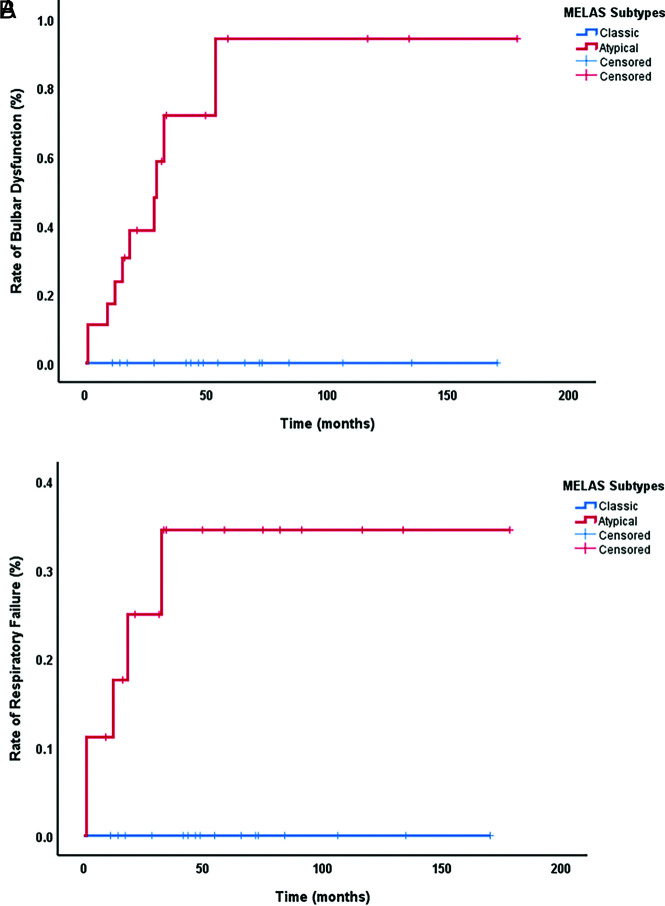
Results: Thirty-five patients with a diagnosis of mitochondrial DNA-based MELAS (median age, 12 years; interquartile range, 7-24 years; 24 female) were eligible for this study. Fifty-three discrete variables were evaluated by an unsupervised cluster analysis, which revealed that two distinct phenotypes exist among patients with MELAS. After experts reviewed the variables, they selected 8 victory-variables with the greatest impact in determining the MELAS subgroups: developmental delay, sensorineural hearing loss, vision loss in the first strokelike episode, Leigh syndrome overlap, age at the first strokelike episode, cortical lesion size, regional brain distribution of lesions, and genetic groups. Ultimately, 2-step differentiating criteria were defined to classify atypical MELAS.

Conclusions: We identified 2 distinct patterns of MELAS: classic MELAS and atypical MELAS. Recognizing different patterns in MELAS presentations will enable clinical and research care teams to better understand the natural history and prognosis of MELAS and identify the best candidates for specific therapeutic interventions.


Figures

FIG 1.
FIG 1.
A, Biopsy of the cerebellum showing a subacute infarct involving the folia (H&E stain, original magnification ×40). B, Higher magnification of the subacute infarct. There is rarefaction, marked vascular proliferation, and macrophage collections within the molecular layer as well as loss of Purkinje neurons (H&E stain, original magnification ×100). C, CD68 immunostain demonstrates numerous macrophages within the molecular layer (CD68 immunostain, original magnification ×40). D, Glial fibrillary acidic protein (GFAP) highlights reactive gliosis (GFAP immunostain, original magnification ×40). E, Postmortem tissue from a second, classic MELAS patient demonstrates the cortex with an infarct involving the gyral crest (H&E stain, original magnification ×40). F, Higher magnification image of the infarct shows neuronal loss, tissue rarefaction, and gliosis (H&E stain, original magnification ×200).
FIG 2.
FIG 2.
Work flow diagram. yo indicates years of age.
FIG 3.
FIG 3.
Unsupervised cluster analysis of the binary data based on the modified variable list (y-axis) using the Ward hierarchic clustering method. Each column represents 1 patient (x-axis). Red and blue squares show the presence and absence of the findings in each patient, respectively. At the top of the heatmap, the height of the dendrogram for main clusters demonstrates the high distance between clusters, which confirms the 2 distinct patterns of disease.
FIG 4.
FIG 4.
Classic MELAS. A large unique cortical lesion involves the right occipital and temporal lobes (posterolateral region of the brain) with FLAIR and T2 hyperintensity (A and B) and components of restricted diffusion in the medial occipital lobe confirmed in the ADC MAP (C and D). Atypical MELAS. Scattered, multiple, small cortical lesions involving both frontal and parietal lobes with FLAIR hyperintensity (E–H). Atypical MELAS with LS overlap. Multiple scattered cortical lesions with subcortical extension in association with basal ganglia involvement, configuring LS overlap, with FLAIR hyperintensity (F) and some areas of restricted diffusion (I) confirmed in the ADC MAP, not shown. Atypical MELAS with predominant cerebellar involvement. Cerebellar FLAIR hyperintensity (G) and components of restricted diffusion (J) were confirmed in the ADC MAP, not shown.
FIG 5.
FIG 5.
A, Clinical outcome (rates of bulbar dysfunction between MELAS subgroups). B, Clinical outcome (rates of respiratory failure between MELAS subgroups).

References

    1. Finsterer J. Mitochondrial metabolic stroke: phenotype and genetics of stroke-like episodes. J Neurol Sci 2019;400:135–41 10.1016/j.jns.2019.03.021 - DOI - PubMed
    1. Lu JQ, Tarnopolsky MA. Mitochondrial neuropathy and neurogenic features in mitochondrial myopathy. Mitochondrion 2021;56:52–61 10.1016/j.mito.2020.11.005 - DOI - PubMed
    1. Chakrabarty S, Govindaraj P, Sankaran BP, et al. . Contribution of nuclear and mitochondrial gene mutations in mitochondrial encephalopathy, lactic acidosis, and stroke-like episodes (MELAS) syndrome. J Neurol 2021;268:2192–207 10.1007/s00415-020-10390-9 - DOI - PMC - PubMed
    1. Wei X, Du M, Li D, et al. . Mutations in FASTKD2 are associated with mitochondrial disease with multi-OXPHOS deficiency. Hum Mutat 2020;41:961–72 10.1002/humu.23985 - DOI - PubMed
    1. Yoo DH, Choi YC, Nam DE, et al. . Identification of FASTKD2 compound heterozygous mutations as the underlying cause of autosomal recessive MELAS-like syndrome. Mitochondrion 2017;35:54–58 10.1016/j.mito.2017.05.005 - DOI - PubMed

Substances