. 2023 Apr 6;14(1):1936.
doi: 10.1038/s41467-023-37336-9.

Factor Xa cleaves SARS-CoV-2 spike protein to block viral entry and infection

Affiliations

Factor Xa cleaves SARS-CoV-2 spike protein to block viral entry and infection

Wenjuan Dong et al. Nat Commun. .

Abstract

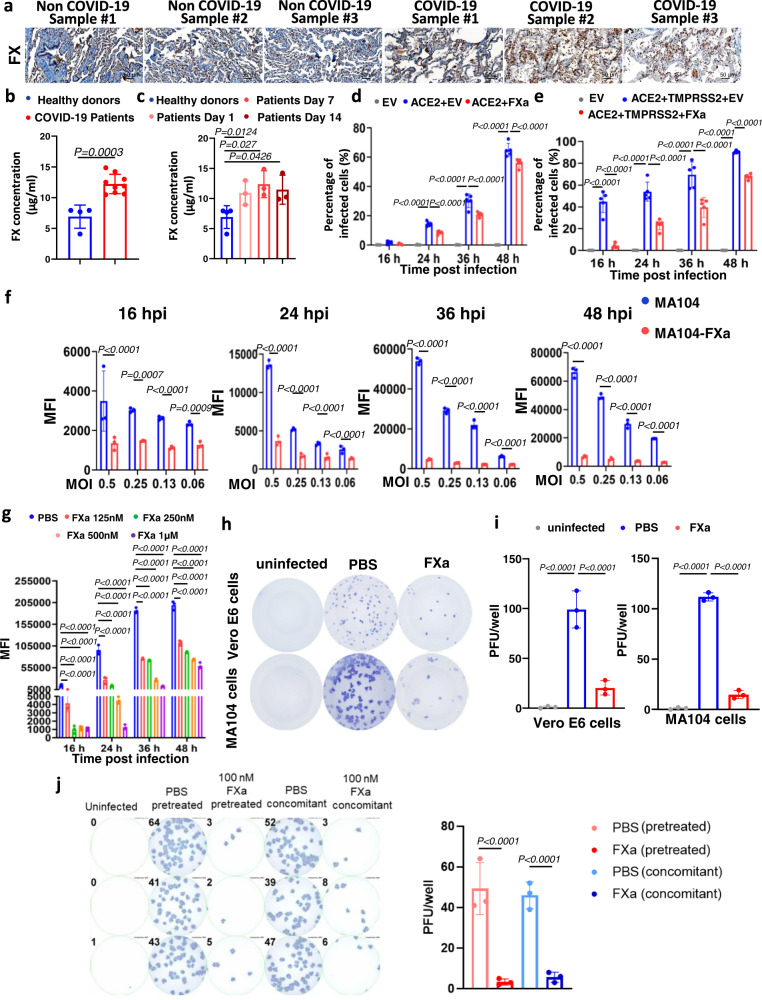
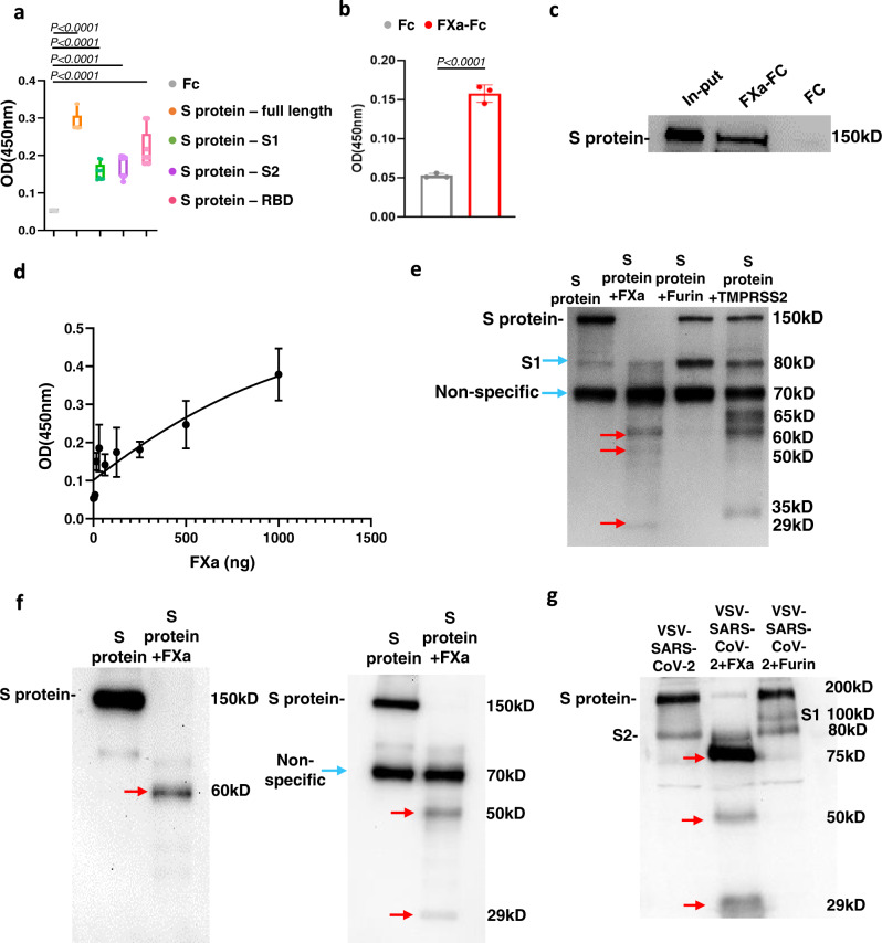
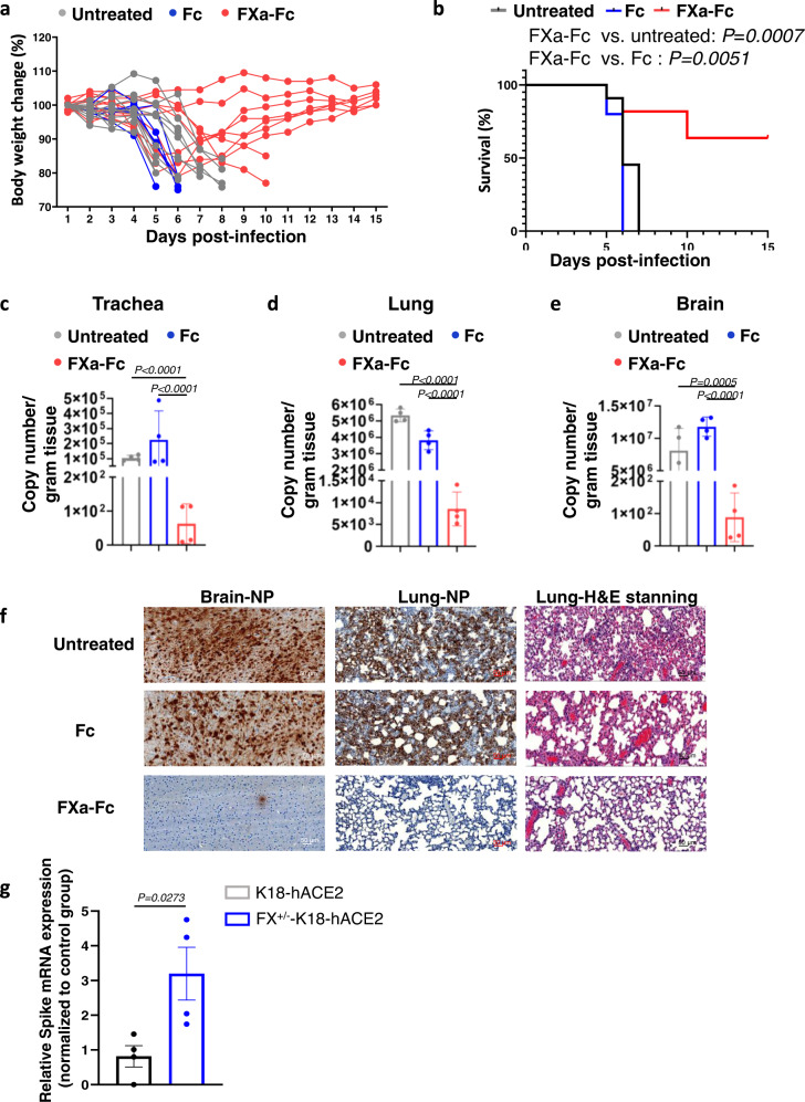
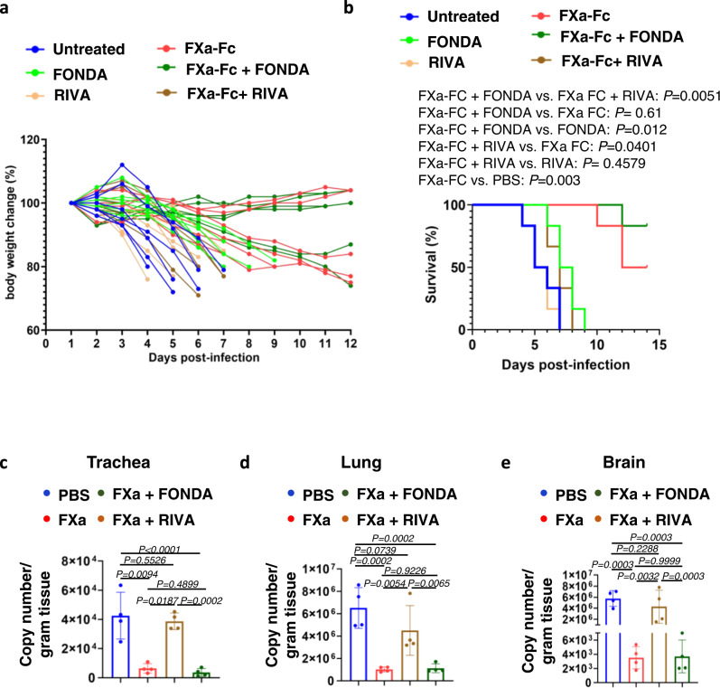
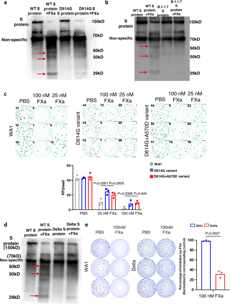
Serine proteases (SP), including furin, trypsin, and TMPRSS2 cleave the SARS-CoV-2 spike (S) protein, enabling the virus to enter cells. Here, we show that factor (F) Xa, an SP involved in blood coagulation, is upregulated in COVID-19 patients. In contrast to other SPs, FXa exerts antiviral activity. Mechanistically, FXa cleaves S protein, preventing its binding to ACE2, and thus blocking viral entry and infection. However, FXa is less effective against variants carrying the D614G mutation common in all pandemic variants. The anticoagulant rivaroxaban, a direct FXa inhibitor, inhibits FXa-mediated S protein cleavage and facilitates viral entry, whereas the indirect FXa inhibitor fondaparinux does not. In the lethal SARS-CoV-2 K18-hACE2 model, FXa prolongs survival yet its combination with rivaroxaban but not fondaparinux abrogates that protection. These results identify both a previously unknown function for FXa and an associated antiviral host defense mechanism against SARS-CoV-2 and suggest caution in considering direct FXa inhibitors for preventing or treating thrombotic complications in COVID-19 patients.


Conflict of interest statement

The authors declare no competing interests.

Figures

Fig. 1
Fig. 1. FXa inhibits wild-type SARS-CoV-2 infection by targeting viral particles.
a, b FX protein levels in lungs (a) or plasma (b) of COVID-19 patients vs. non-COVID-19 donors, using IHC (a) and ELISA (b), respectively. The staining results shown in a are representative of at least two independent experiments with similar results. Scale bar, 50 μm. b n = 9 for COVID-19 patients and n = 4 for non-COVID-19 donors. c Post-diagnosis concentrations of FX in plasma of COVID-19 patients (n = 3) compared to non-COVID-19 donors (n = 3), as measured by ELISA. d, e HEK293T cells co-transfected with a plasmid encoding ACE2 and the other plasmid encoding FXa or an empty vector (EV) in the absence (d) or presence (e) of another plasmid encoding TMPRSS2 were infected by VSV-SARS-CoV-2. Infectivity of the cells was quantified by flow cytometry at 16, 24, 36, and 48 hpi (n = 4 or 5 biologically independent samples). f MA104 cells transduced with the plasmid encoding FXa (MA104-FXa) or an empty vector (MA104-EV) were infected with VSV-SARS-CoV-2. Infectivity of the cells was quantified by flow cytometry at 16, 24, 36, and 48 hpi (n = 3 biologically independent samples). g VSV-SARS-CoV-2 was pre-incubated with FXa at the indicated concentrations 1 hour before infection (n = 3 biologically independent samples). h MA104 and Vero E6 cells were infected with live wild-type SARS-CoV-2. At 24 hpi, infectivity was measured with an immuno-plaque assay. i Summary of data from h (n = 3 independent experiments for each cell line). j A549-ACE2 cells were infected with either authentic WT SARS-CoV-2 preincubated with 100 nM FXa for 1 hour, or the cells were treated with FXa at the time of viral infection. Infectivity was measured by immuno-plaque assays 24 hours post-infection. Representative infection and the summary data are presented at the left and right, respectively (n = 3 biologically independent samples). Data in bg and i, j are presented as mean values ± standard deviation (SD) and statistical analyses were performed by two-sided Student’s t tests (b), one-way ANOVA models (c, i) and two-way ANOVA models (dg, j). MFI data were log2 transformed before running the statistical models. Source data are provided as a Source Data file.
Fig. 2
Fig. 2. FXa suppresses viral entry by binding to and cleaving the SARS-CoV-2 S protein.
a The binding affinity of FXa with full-length wild-type S protein, subunit S1, subunit S2, or RBD was quantified by ELISA (n = 9 biologically independent samples). Bounds of box is from 25% percentile to 75% percentile, horizontal bar indicates median, and whiskers indicate data ranges. b The binding affinity of FXa to VSV-SARS-CoV-2 viral particles was quantified by ELISA (n = 3 biologically independent samples). c The interaction between FXa protein and full-length S protein was examined with a pull-down assay. d The binding affinity at indicated concentrations of FXa was measured by ELISA (n = 3 biologically independent samples). e The cleavage of S protein by furin, TMPRSS2, and FXa after 3-hour incubation was analyzed by immunoblotting using an anti-S protein antibody (40591-T62, Sino Biological). f S protein was cleaved by FXa, followed by immunoblotting with an anti-RBD antibody (MAB10540-100, R&D) (left) and an anti-S2 antibody (MA5-35946, Invitrogen) (right). g The cleavage of VSV-SARS-CoV-2 by FXa or furin was analyzed by immunoblotting using an anti-S protein antibody (40591-T62, Sino Biological). The larger size of S protein from VSV-SARS-CoV-2 (~200 kD) compared to recombinant S protein (~150 kD) may be due to the glycosylation of the former. All data are representative of at least three independent experiments. a, b, d, data are presented as mean values ± SD. Statistical analyses were performed by two-sided Student’s t test (b) or one-way ANOVA models (a). Source data are provided as a Source Data file.
Fig. 3
Fig. 3. FXa cleavage reduces the binding between S protein and ACE2.
a The binding between ACE2 with S protein or FXa-pre-treated S protein was measured by ELISA (n = 9 biologically independent samples). b The binding between S protein or FXa-pre-treated S protein and ACE2 expressed on A549 human lung cancer cells was measured by flow cytometry (left, representative flow cytometry histogram; right, summary data) (n = 4 independent experiments). c The binding of FXa with S protein, S protein–ACE2 complex, or PBS control was measured by ELISA (n = 9 biologically independent samples). d The binding of membrane-bound (mb) FXa with S protein or the binding of mb FXa with S protein–ACE2 complex on 293 T cells was measured by flow cytometry (left, representative flow cytometry histogram; right, summary data) (n = 3 independent experiments). PBS served as control for S protein and the S protein–ACE2 complex. e The effect of RIVA or FONDA on the binding of FXa with S protein was measured by ELISA (n = 3 independent experiments). f, g Infectivity of VSV-SARS-CoV-2 in MA104 cells that were pre-treated or not treated with FXa in the presence or absence of RIVA or FONDA was examined with fluorescent microscopy (f) and flow cytometry (g, n = 3 biologically independent samples). h S protein cleavage by FXa was examined by immunoblotting 3 hours after their incubation in the presence or absence of RIVA or FONDA, using an anti-S protein antibody (40591-T62, Sino Biological). i, j FXa pre-treated or not treated with RIVA or FONDA was incubated with S protein; then the binding capability of those S proteins with ACE2 coated on a plate (i, n = 9 biologically independent samples) or ACE2 expressed on 293 T cells (j) was assessed. k Infectivity of live SARS-CoV-2 in A549-ACE2 cells pre-treated or not treated with FXa in the presence or absence of RIVA or FONDA was examined using an immuno-plaque assay (n = 3 independent experiments). a, c, i, the top and the bottom bound of box are 75th percentile and 25th percentile, respectively; horizontal bar indicates median; and whiskers indicate data ranges. Experiments in h and j are representative of three independent experiments with similar data. For all panels, data are presented as mean values ± SD, and statistical analyses were performed by one-way ANOVA models. MFI data were log2 transformed before running the statistical models. Source data are provided as a Source Data file.
Fig. 4
Fig. 4. The in vivo effect of FXa protein and heterozygous knockout on WT SARS-CoV-2 infection in a K18-hACE2 mouse model of COVID-19.
a, b Body weight (a) and survival (b) of mice infected with 5 × 103 PFU SARS-CoV-2 WT strain and treated with or without 200 µg FXa-Fc fusion protein. Fc-protein alone was used as control. N = 11 mice for PBS and FXa-Fc group. N = 5 mice for Fc group. ce Relative viral copy numbers in the tracheas (c), lungs (d), and brains (e) of mice treated with or without FXa-Fc fusion protein were assessed by qPCR. Fc protein alone served as control. N = 4 biologically independent mice per group. f SARS-CoV-2 was detected in the brain and lung of mice treated with FXa-Fc or Fc-protein by using IHC staining with an antibody against SARS-CoV-2 nucleocapsid protein (NP). Pathological analysis by H&E staining of lungs of untreated mice and mice treated with FXa-Fc or Fc-protein alone. The experiment was repeated with four mice per group with similar results. Scale bar, 50 μm. The untreated and Fc-protein-treated mice were sacrificed on day 5 post infection given their declining health; as the FXa-Fc group survived for more than 2 weeks, their tissues were collected on day 15 post infection (e, f). g S protein mRNA levels in lungs of FXa+/−-K18-hACE2 mice and WT K18-hACE2 mice (littermate control) were assessed by qPCR. N = 4 biologically independent mice per group. Data are presented as mean values ± SD and statistical analyses were performed by one-way ANOVA models (ce), two-sided Student’s t test (g), or log-rank test (b). Source data are provided as a Source Data file.
Fig. 5
Fig. 5. Effect of the direct FXa inhibitor RIVA and the indirect inhibitor FONDA on FXa-mediated protection of K18-hACE2 mice from WT SARS-CoV-2 infection.
a, b Body weight (a) and survival (b) of mice infected with 5 × 103 PFU of SARS-CoV-2 (WA1) and treated with or without FXa-Fc in the presence or absence of RIVA or FONDA. N = 6 mice for each group. ce Relative viral copy numbers in the trachea (c), lung (d), and brain (e) of mice treated with or without FXa-Fc in the presence or absence of RIVA or FONDA were assessed by qPCR. All the mice were sacrificed on day 5 post infection. N = 4 mice for each group. ce Data are presented as mean values ±SD. Statistical analyses were performed by one-way ANOVA models (ce) or log-rank test (b). Copy number (ce) was log2 transformed before running statistical models. Source data are provided as a Source Data file.
Fig. 6
Fig. 6. FXa is less effective in blocking infection of the SARS-CoV-2 B.1.1.7 variant in vitro and in vivo.
a A549-ACE2 cells were preincubated or not preincubated with 100 nM FXa for 1 h, and then infected with either live SARS-CoV-2 WA1 or the SARS-CoV-2 B.1.1.7 variant. Infectivity was measured with an immuno-plaque assay 24 hours post infection and the infection inhibition ratio induced by FXa was summarized (right panel). N = 3 biologically independent samples. b Vero E6 cells were pre-treated with FXa and then infected with live SARS-CoV-2 WA1 or the SARS-CoV-2 B.1.1.7 variant at various MOIs. At 24 hpi, infectivity was measured with an immuno-plaque assay (left panel), and the infection inhibition ratio induced by FXa at different MOIs was summarized (right panel). N = 3 biologically independent samples. c, d Body weight (c) and survival (d) of mice infected with 5 × 103 PFU wild-type SARS-CoV-2 or B.1.1.7 variant and treated with or without FXa-Fc fusion protein. e, f Viral load in the lung (e) and brain (f) of mice treated with or without FXa-Fc fusion protein, was assessed by qPCR. All the mice were sacrificed at day 5 post infection. N = 5 biologically independent mice (cf). Data in a, b, e, f are presented as mean values ± SD. Statistical analyses were performed by two-way ANOVA models (a, b), one-way ANOVA models (e, f), or log-rank test (d). Copy number (e, f) was log2 transformed before running statistical models. Source data are provided as a Source Data file.
Fig. 7
Fig. 7. FXa is less effective in blocking infection of the SARS-CoV-2 variants with the D614G mutation.
a Cleavage of WT WA1 S protein or D614G S protein by FXa after 1-hour incubation was analyzed by immunoblotting using an anti-S protein antibody (40591-T62, Sino Biological). b Cleavage of WT WA1 S protein or B.1.1.7 S protein by FXa after 1-hour incubation was analyzed by immunoblotting using the same antibody in a. c Live SARS-CoV-2 WT WA1, the D614G variant, and the D614G variant engineered with an A570D mutation (D614G + A570D) were treated with 100 nM or 25 nM FXa in A549-ACE2 cells. Infectivity was measured with an immuno-plaque assay 24 hours post infection. d Cleavage of WT WA1 S protein or Delta S protein by FXa after 1-hour incubation was analyzed by immunoblotting using the same antibody in a. e Live SARS-CoV-2 WT WA1 and the Delta variant were treated with 100 nM FXa in A549-ACE2 cells. Infectivity was measured with an immuno-plaque assay 24 hours post infection) (left) and then summarized in graphical form (right). N = 3 biologically independent samples c, e. Immunoblotting data are representative of three independent experiments (a, b, d). Data in c and e are presented as mean values ± SD and statistical analyses were performed by two-way ANOVA models (c) or two-sided Student’s t test (e). Source data are provided as a Source Data file.

References

    1. Hu, B., Guo, H., Zhou, P. & Shi, Z. L. Characteristics of SARS-CoV-2 and COVID-19. Nat. Rev. Microbiol. 10.1038/s41579-020-00459-7 (2020). - PMC - PubMed
    1. Worldometer. Worldometer’s COVID-19 data. <https://www.worldometers.info/coronavirus/country/us/> (2020).
    1. Parums DV. Editorial: current status of oral antiviral drug treatments for SARS-CoV-2 infection in non-hospitalized patients. Med. Sci. Monit. 2022;28:e935952. doi: 10.12659/MSM.935952. - DOI - PMC - PubMed
    1. Tregoning JS, Flight KE, Higham SL, Wang Z, Pierce BF. Progress of the COVID-19 vaccine effort: viruses, vaccines and variants versus efficacy, effectiveness and escape. Nat. Rev. Immunol. 2021;21:626–636. doi: 10.1038/s41577-021-00592-1. - DOI - PMC - PubMed
    1. Pia, L. & Rowland-Jones, S. Omicron entry route. Nat. Rev. Immunol. 10.1038/s41577-022-00681-9 (2022). - PMC - PubMed

Publication types