. 2023 Apr 6;13(1):5632.
doi: 10.1038/s41598-023-28996-0.

Prognostic factors and Doxorubicin involved in malignant progression of meningioma

Affiliations

Prognostic factors and Doxorubicin involved in malignant progression of meningioma

Xulei Huo et al. Sci Rep. .

Abstract

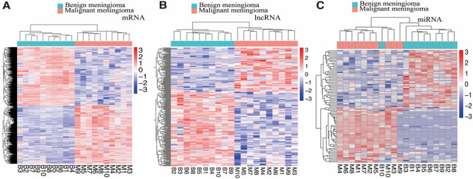
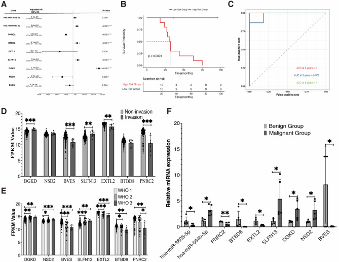
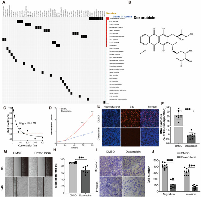
Meningioma was the most primary intracranial tumor, but the molecular characteristics and the treatment of malignant meningioma were still unclear. Nine malignant progression-related genes based prognostic signatures were identified by transcriptome analysis between benign meningioma and malignant meningioma. The external dataset GEO136661 and quantitative Real-time Polymerase Chain Reaction were used to verify the prognostic factors. has-miR-3605-5p, hsa-miR-664b-5p, PNRC2, BTBD8, EXTL2, SLFN13, DGKD, NSD2, and BVES were closed with malignant progression. Moreover, Doxorubicin was identified by Connectivity Map website with the differential malignant progression-related genes. CCK-8 assay, Edu assay, wound healing assay, and trans-well experiment were used to reveal that Doxorubicin could inhibit proliferation, migration and invasion of IOMM-Lee Cells.


Conflict of interest statement

The authors declare no competing interests.

Figures

Figure 1
Figure 1
MRI of meningioma patients with benign progression and with malignant progression. (AB, C) Same meningioma patient with WHO 1 pathology; (DE, F) Same meningioma patient with WHO 3 pathology and the tumor penetrate the dura mater and invade into brain. Kaplan–Meier survival analysis of meningioma patients. (G) Meningioma patients with malignant progression have shorter overall survival time than patients with benign progression (P < 0.0001); (H) Meningioma patients with malignant progression have shorter progression-free survival time than patients with benign progression (P < 0.0001). Log-rank test was used as the method.
Figure 2
Figure 2
LncRNA, mRNA and miRNA expression profile changes between malignant group and benign group in meningioma. (A, B, C) Hierarchical clustering of all samples revealed the nonrandom partitioning of samples into two major group: one group containing ten malignant progression penetration samples and another group containing ten benign progression samples. Each column represents one sample and each row represents one lncRNA, mRNA and miRNA set. “ComplexHeatmap” R package (version 3.15) was used to graph the result.
Figure 3
Figure 3
Functional pathway enrichment of malignant progression-related mRNAs in meningioma (A, B, C) Biological process, cellular component, and molecular function pathway enrichment (GO); (D) Kyoto Encyclopedia of Genes and Genomes pathway enrichment; (E, F) GSEA pathway enrichment of cancer markers and immunologic markers.
Figure 4
Figure 4
Identification and validation of nine MPRGs-based prognostic factors. (A) Forest plot of the impact of MPRGs-based prognostic factors on overall survival (OS); (B) Kaplan–Meier OS curve of high- and low-risk groups based on the risk score in the group; (C) Time-dependent ROC curves in our cohort; (D, E) The expression profile of seven MPRGs-based mRNAs between invasion group samples and no invasion group or among WHO grades samples in 157 meningioma tumors from the GEO136661 dataset. (F) Expression level of the nine MPRGs-based prognostic factors between malignant group samples and benign group samples were analyzed using qRT-PCR, U6 and GAPDH was set as internal control. Error bars represent the standard errors of independent samples. *P < 0.05, **P < 0.01, ***P < 0.001.
Figure 5
Figure 5
Doxorubicin inhibits proliferation, migration and invasion of meningioma. (A) Correlation of compound with differently expressed mRNA: Heatmap showing compounds (top) from the CMap shares mechanisms of action and the mechanisms of action was sorted by descending number of compounds (right); “ComplexHeatmap” R package (version 3.15) was used to graph the result. (B) The molecular structure of Doxorubicin; (C) The dose-curve of Doxorubicin in IOMM-Lee cell line; (D) CCK-8 assay of the cell proliferation ability in IOMM-Lee cells upon Doxorubicin or DMSO treatment; (E) EdU assay of the cell proliferation ability in IOMM-Lee cells upon Doxorubicin or DMSO treatment. Scale bar: 125 μm; (F) Statistical analysis of the EdU-positive cell ratio; (G) Wound healing assay showed delayed closure of the wound gap upon Doxorubicin treatment. (H) Statistical analysis of the cell migration in the scratch wound healing assays. (I) Transwell invasion and migration assay of IOMM-Lee cells upon Doxorubicin or DMSO treatment. (J) Statistical analysis of the cell numbers passing through the transwell chamber. All data were the means ± SD. Results triplicate and repeated at least three times. Results are expressed as mean ± SEM. *P < 0.05, **P < 0.01, ***P < 0.001.

Similar articles

References

    1. Ostrom QT, et al. CBTRUS statistical report: Primary brain and other central nervous system tumors diagnosed in the United States in 2010–2014. Neuro Oncol. 2017;19(suppl_5):v1–v88. doi: 10.1093/neuonc/nox158. - DOI - PMC - PubMed
    1. Achey RL, et al. Nonmalignant and malignant meningioma incidence and survival in the elderly, 2005–2015, using the Central Brain Tumor Registry of the United States. Neuro Oncol. 2019;21(3):380–391. doi: 10.1093/neuonc/noy162. - DOI - PMC - PubMed
    1. Perry A, et al. "Malignancy" in meningiomas: A clinicopathologic study of 116 patients, with grading implications. Cancer. 1999;85(9):2046–2056. - PubMed
    1. Sun SQ, et al. Management of atypical cranial meningiomas, part 2: Predictors of progression and the role of adjuvant radiation after subtotal resection. Neurosurgery. 2014;75(4):356–363. doi: 10.1227/NEU.0000000000000462. - DOI - PubMed
    1. Brokinkel B, Hess K, Mawrin C. Brain invasion in meningiomas-clinical considerations and impact of neuropathological evaluation: a systematic review. Neuro Oncol. 2017;19(10):1298–1307. doi: 10.1093/neuonc/nox071. - DOI - PMC - PubMed

Publication types