. 2023 May;26(5):820-829.
doi: 10.1038/s41593-023-01291-x. Epub 2023 Apr 6.

Prefrontal engrams of long-term fear memory perpetuate pain perception

Affiliations

Prefrontal engrams of long-term fear memory perpetuate pain perception

Alina Stegemann et al. Nat Neurosci. 2023 May.

Abstract

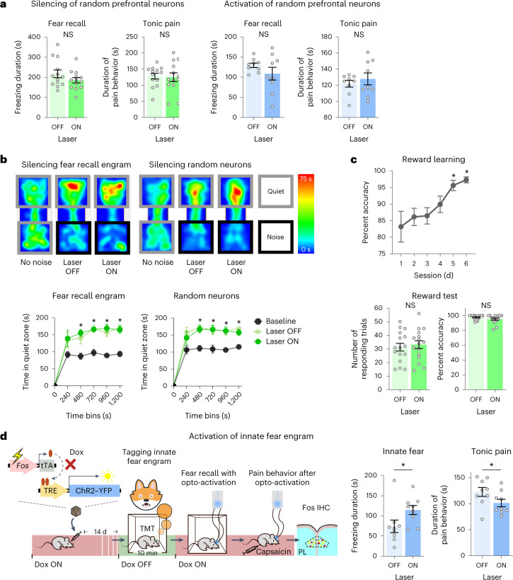
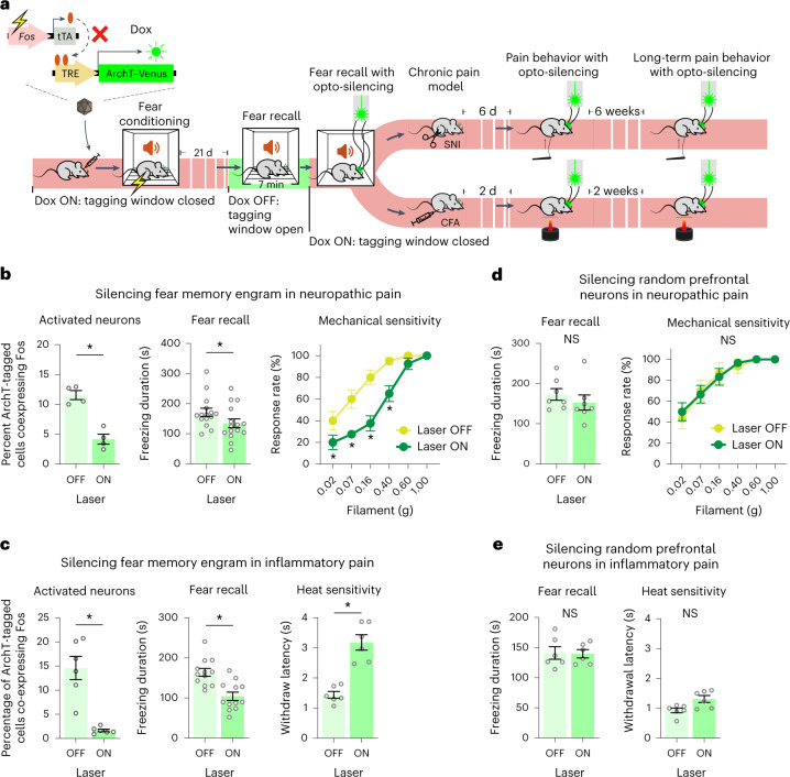
A painful episode can lead to a life-long increase in an individual's experience of pain. Fearful anticipation of imminent pain could play a role in this phenomenon, but the neurobiological underpinnings are unclear because fear can both suppress and enhance pain. Here, we show in mice that long-term associative fear memory stored in neuronal engrams in the prefrontal cortex determines whether a painful episode shapes pain experience later in life. Furthermore, under conditions of inflammatory and neuropathic pain, prefrontal fear engrams expand to encompass neurons representing nociception and tactile sensation, leading to pronounced changes in prefrontal connectivity to fear-relevant brain areas. Conversely, silencing prefrontal fear engrams reverses chronically established hyperalgesia and allodynia. These results reveal that a discrete subset of prefrontal cortex neurons can account for the debilitating comorbidity of fear and chronic pain and show that attenuating the fear memory of pain can alleviate chronic pain itself.


Conflict of interest statement

The authors declare no competing interests.

Figures

Fig. 1
Fig. 1. Anatomical prefrontal substrates and functional interrogation of the interplay between long-term memory and tonic pain.
a, Viral-mediated, Dox-controlled expression of protein tags under the Fos promoter, leading to activity-dependent tagging of prelimbic neurons with mCherry following painful foot shock or capsaicin or during recall of fear memory 28 d after cued fear conditioning. b, Experimental scheme (top left), typical examples (bottom left) showing successive labeling of neuronal populations over discrete behavioral states with mCherry and Fos immunohistochemistry (arrowheads: double-labeled neurons; scale bar, 50 µm) and quantitative summary (top right) of overlapping activated neuronal populations; total numbers of positive/tagged cells are shown underneath. Data were analyzed by one-way ANOVA with Tukey’s multiple comparisons test; *P < 0.05; foot shock followed by foot shock and fear recall followed by fear recall, n = 4 mice per group; fear recall followed by capsaicin, n = 5 mice per group; capsaicin followed by fear recall, n = 6 mice per group; IHC, immunohistochemistry. c, Characterization of labeled prefrontal neurons in fear recall engrams or by tonic pain using markers of excitatory and GABAergic neurons. Typical examples (left) and a quantitative summary (right) are shown; scale bar, 50 µm. Data were analyzed by two-way ANOVA with a Šidák correction for multiple comparisons test; *P < 0.05; n = 3 mice per group; PV, parvalbumin. df, Experimental scheme (d) for activity-dependent tagging of long-term fear engrams with optogenetic actuators ArchT or ChR2 and testing effects of light-induced silencing (e) or activation (f) of fear engrams on prelimbic Fos immunohistochemistry, fear recall behavior and capsaicin-evoked tonic pain-related behavior are shown. In e and f, an average of 4,280 neurons per mm3 were labeled with ArchT and 3,894 neurons per mm3 were labeled with ChR2, respectively; light-induced silencing and activation: unpaired t-test, n = 7 mice per group (e) and laser off n = 7 mice per group and laser on n = 8 mice per group (f); fear recall behavior: paired t-test, n = 17 mice (e) and n = 17 mice (f); tonic pain-related behavior: paired t-test, n = 18 mice (e) and n = 15 mice (f). All P and F values are shown in Supplementary Table 1. Source data
Fig. 2
Fig. 2. In vivo tetrode recordings to study encoding of fear recall and tonic pain in prefrontal excitatory neurons and inhibitory neurons.
a, Schematic representation of experiments on electrophysiological recordings using tetrodes implanted in the prelimbic cortex (PL) during fear recall, neutral context or capsaicin-induced tonic pain. b, Summary of prelimbic units (n = 261 units from seven animals) demonstrating significant rate changes during fear recall or tonic pain over the neutral context. c, Delineation of global rate-coding units for fear recall and tonic pain into putative principal (excitatory) neurons (PN; left; n = 231 units) and interneurons (IN; right; n = 30 units). Data were analyzed by two-way ANOVA with a Šidák correction for multiple comparisons test; *P < 0.05; n = 7 mice per group. Data are shown as mean ± s.e.m. d, Example heat map illustrations (left) and quantitative summary (right) of data from in vivo prelimbic tetrode recordings showing rate-coding units with temporal specificity for episodes (superimposed on units) of fear-related freezing behavior or pain-related nocifensive behaviors, activity common to both or no association with either behavior. The proportion of behavior-specific firing-increased neurons, detected with a permutation test for mean difference between binned firing rates (P < 0.05, effect size > 0.5; n = 146 units over seven mice), was similar in fear recall and tonic pain states (P > 0.5, unpaired t-test). Shaded horizontal lines represent state-specific behavioral episodes. All data are shown as mean ± s.e.m. All P and F values are shown in Supplementary Table 1. Source data
Fig. 3
Fig. 3. Analysis of specificity of the interplay between long-term fear memory and pain.
a, Impact of optogenetic silencing or activation of a randomly targeted prelimbic neuronal population on long-term fear recall and capsaicin-induced nocifensive behaviors; an average of 6,919 neurons per mm3 were labeled with ArchT and 3,689 neurons per mm3 were labeled with ChR2. Data were analyzed by paired t-test; n = 13 mice for the silencing group and 9 mice for the activation group. b, Impact of optogenetic silencing of the fear engram or random prefrontal neurons on avoidance of non-painful, aversive white noise, shown in the form of example body heat maps (above) and quantitative overview (below). Data were analyzed by two-way ANOVA with a Šidák correction for multiple comparisons; *P < 0.05 compared to baseline; silencing fear recall neurons: n = 10 mice per group; silencing random neurons: n = 11 mice per group. c, Impact of optogenetic silencing of prefrontal fear engram neurons on appetitive reward learning behavior. For reward learning, a one-way ANOVA with a Dunnett correction for multiple comparisons was performed. For the number of responding trials, a paired t-test was performed and for percent accuracy a Wilcoxon test was performed; *P < 0.05; n = 16 mice per group. d, Experimental scheme for labeling prefrontal neurons activated during recall of innate fear and impact of their optogenetic activation on fear recall and capsaicin-induced tonic pain behavior. Data were analyzed by paired t-test; *P < 0.05; n = 9 mice per group; NS, not significant. All data are shown as mean ± s.e.m. All P and F values are shown in Supplementary Table 1. Source data
Fig. 4
Fig. 4. Plasticity of prefrontal representation of fear memory, nociception and touch and their connectivity in inflammatory and neuropathic pain.
a, Experimental scheme (top) and impact of fear conditioning on withdrawal responses of the contralateral paw to heat or to graded intensities of mechanical stimulation in naive mice (baseline) and mice with unilateral paw inflammation (CFA; n = 6 mice per group) or nerve injury (SNI; n = 8 mice per group). Data were analyzed by two-way ANOVA with a Tukey’s multiple comparisons test; *P < 0.05; FC, fear conditioning. b, Quantitative summary of overlapping prefrontal neurons commonly activated by a heat ramp (Fos) and long-term fear memory (ArchT–Venus) under baseline conditions (average Fos+ cells = 5,935 cells per mm3; average Venus+ cells = 3,028 cells per mm3) and after paw inflammation (average Fos+ cells = 7,306 cells per mm3; average Venus+ cells = 2,612 cells per mm3). Data were analyzed by unpaired t-test; *P < 0.05; n = 5 mice per group. c, Quantitative summary of overlapping prefrontal neuronal populations commonly activated by tactile stimulation (Fos) and long-term fear memory (ArchT–Venus) under sham (average Fos+ cells = 5,400 cells per mm3; average Venus+ cells = 4,656 cells per mm3) and neuropathic conditions (average Fos+ cells = 5,011 cells per mm3; average Venus+ cells = 5,319 per mm3). Data were analyzed by unpaired t-test; *P < 0.05; n = 4 mice per group. df, Scheme for labeling projections (d) and analysis of projection intensity (e) from prefrontal neurons tagged during tactile stimulation under baseline conditions (sham) and following SNI in mice subjected to fear conditioning or not. Projection density is shown in the form of heat maps of tactile-responsive prefrontal neurons (groups 2–5) that were either not exposed to fear conditioning (groups 2 and 4) or following fear conditioning (groups 3 and 5) under sham conditions (groups 2 and 3) and after SNI (groups 4 and 5); SNI FR, projection maps from prefrontal fear engram neurons labeled during long-term fear recall (group 1); n = 3 mice per group. PAG, periaqueductal gray. Data in f show average values of projection intensity in the rostral anterior cingulate cortex and mediodorsal thalamic nucleus. Data were analyzed by two-way ANOVA with a Šidák test for multiple comparisons; *P < 0.05. All data are shown as mean ± s.e.m. All P and F values are shown in Supplementary Table 1. Source data
Fig. 5
Fig. 5. Disrupting long-term fear memory reduces established nociceptive hypersensitivity in inflammatory and neuropathic pain.
a, Scheme of tagging prefrontal long-term fear memory engram neurons with ArchT and impact of optogenetic silencing of the fear memory engram on CFA-induced inflammatory pain and SNI-induced neuropathic pain. b,c, Activity-dependent expression of ArchT in prefrontal long-term fear engram neurons for light-induced silencing (left) of the specific neuronal population is shown via Fos immunohistochemistry; impact on fear recall (middle) and neuropathic mechanical hypersensitivity (at day 6 after SNI; b) or thermal hypersensitivity after paw inflammation (at day 2 after CFA; c) is shown. In b and c, an average of 4,844 neurons per mm3 and 4,084 neurons per mm3 were labeled with ArchT, respectively. d,e, Impact of optogenetically silencing a randomly targeted prelimbic neuronal population on fear recall and neuropathic mechanical hypersensitivity (d) or inflammatory thermal hypersensitivity (e). In d and e, an average of 3,147 neurons per mm3 and 2,575 neurons per mm3 were labeled with ArchT, respectively; *P < 0.05. Data for von Frey behavior were analyzed by two-way ANOVA with a Tukey’s multiple comparisons test. A paired t-test was used for freezing behavior and heat sensitivity, and an unpaired t-test was used for comparing overlapping populations. All data are shown as mean ± s.e.m. All P and F values are shown in Supplementary Table 1. Source data
Extended Data Fig. 1
Extended Data Fig. 1. Activity labeling system for labelling prefrontal engrams for long term fear recall and neurons responsive to foot shock stimulation and tonic pain.
(a) Doxycycline-controlled, activity-evoked expression of protein tags under c-fos promoter following viral injection of tagging constructs in the murine prelimbic cortex. Shown is an example of neurons tagged with mCherry expression in an activity-dependent manner. (b) Control of tagging by presence of Doxycyline (Dox ON, 1 mg/mL Dox for 16 days) in comparison to Doxycline-free time window (Dox OFF) is demonstrated in baseline home-cage conditions. Example images show mCherry reporter expression (red) on the background of Hoechst-stained cell nuclei (blue). Magnified images are shown in lower panels. Quantitative summary is shown on the right; unpaired t-test; * p < 0.05; Dox OFF: n = 2 mice/group, Dox ON: n = 4 mice/group. (c, d) Examples (panel c) and quantitative summary (panel d) of prelimbic neuronal labeling upon foot shock, fear memory recall at 28 days post-conditioning or paw injection of capsaicin. In panel d, one-way ANOVA with Dunnet’s multiple comparisons; * p < 0.05; Homecage: n = 7 mice/group, Fear Recall: n = 9 mice/group, Capsaicin: n = 6 mice/group, Footshock: n = 4 mice/group. (e) Typical examples showing dual labeling of neuronal activity over two discrete events of the same behavioral state or two distinct behavioral states via mCherry and Fos immunohistochemistry. Arrow-heads: double-labelled neurons. Scale bars represent 0.5 mm in panel a, 250 µm in upper images in panel b and 50 µm lower images in panel b, 100 µm in panel c and 50 µm in panel e. (f) Distribution of labelled neurons across layers of the prelimbic cortex. All data are shown as mean + /− S.E.M. All p and F values are shown in Supplementary Table 1. Source data
Extended Data Fig. 2
Extended Data Fig. 2. Control experiments for optogenetic activation or silencing of prefrontal fear memory engram.
(a) Optimization of the time course of Doxycycline (Dox) withdrawal to uncover conditions yielding a significant increase (* p < 0.05) in the number of cells labelled with optogenetic actuators, when compared to homecage controls (two-way ANOVA with Sidak’s test for multiple comparisons); Capsaicin: n = 4 injections/group, 72 h Homecage: n = 6 injections/group, 24 h Capsaicin/72 h Capsaicin: n = 8 injections/group. (b) Typical examples of labeling prefrontal remote fear memory engram neurons with Channelrhodopsin (ChR2-YFP) or Archeorhodopsin (ArchT-Venus) and validation of optogenetic activation or silencing, respectively, via increase or decrease in immunohistochemical detection of expression of activity marker Fos respectively, upon laser light stimulation in vivo; arrowheads indicate double-labelled neurons and scale bars represent 50 µm. (c, d) Behavior associated with locomotion in open field (in the same cohort of mice with (Laser ON) or without (Laser OFF)) upon optogenetic silencing (c) or activation (d) of the prefrontal fear memory engram; paired t-test; In panel c, distance travelled: n = 18 mice/group, speed: n = 15 mice/group; in panel d, n = 10 mice/group; n.s.: non-significant. All data are shown as mean + /− S.E.M. All p and F values are shown in Supplementary Table 1. Source data
Extended Data Fig. 3
Extended Data Fig. 3. In vivo tetrode recordings to study encoding of fear recall and tonic pain in prefrontal excitatory neurons and inhibitory neurons.
(a) Example plots showing state-specific changes in firing rates of units. Insets show spike waveforms (mean ± SD, 1.2 ms) for each unit. (b) Example heat map illustration of binned firing rates normalized to activity in neutral context for units showing increased activity during fear recall or tonic pain. Source data
Extended Data Fig. 4
Extended Data Fig. 4. Experiments for testing specificity of interaction between fear memory and pain.
(a) Experimental scheme for labelling of a random population of prelimbic neurons with optogenetic actuators ArchT and ChR2 using the murine synapsin promoter to drive expression, and scheme for testing effect of their activation or silencing on remote fear recall and capsaicin-induced pain behaviors. (b) Examples of reporter expression driven by random labelling of neurons using the synapsin promoter or expression driven by labelling of the fear recall engram in the prelimbic cortex; scale bars represent 200 µm. The injection was repeated and verified in n = 6 mice in the Synapsin promotor-driven expression group and n = 8 in the fear recall-driven expression group. (c) Experimental scheme for testing impact of optogenetic silencing of the prefrontal fear memory engram on pain-unrelated aversion to white noise. (d) Typical examples of labeling prefrontal neurons responding to innate fear upon exposure to compound TMT from fox urine with Channelrhodopsin (ChR2-YFP) and validation of optogenetic activation via increase in immunohistochemical detection of Fos upon laser light stimulation in vivo; arrowheads indicate double-labelled neurons and scale bars represent 50 µm. Source data
Extended Data Fig. 5
Extended Data Fig. 5. Impact of cued fear conditioning on nociceptive and tactile sensitivity and prefrontal basis of interplay between fear and pain.
(a) Impact of fear conditioning on paw withdrawal responses to graded mechanical von Frey force across noxious and non-noxious intensities in naïve mice (baseline); n = 16 mice/group. (b) Impact of fear conditioning on withdrawal responses to von Frey hairs after nerve injury; shown are data from the ipsilateral paw; n = 8 mice/group. (c) Impact of fear conditioning on withdrawal responses to thermal nociceptive stimuli (Hargreaves plantar test) in baseline condition and longitudinally after induction of unilateral paw inflammation (CFA); shown are data from the ipsilateral paw; n = 6 mice/group. In all panels, two-way ANOVA with Tukey’s multiple comparisons test was performed. (d) Experimental scheme for labelling prefrontal long-term fear memory engram and testing its overlap with prefrontal neurons responsive to a heat ramp or tactile von Frey stimulation in baseline conditions and changes thereof after induction of paw inflammation (CFA) or nerve injury (SNI). (e) Typical examples fear engram-labelled neurons (Venus) and overlap (arrows) with neurons activated by light touch or heat (Fos+Venus); the expression was verified and quantified in n = 4 mice in the Sham group and n = 4 mice in the SNI group; scale bars represent 50 µm; n.s.: non-significant. Data are shown as mean + /− S.E.M. All p and F values are shown in Supplementary Table 1. Source data
Extended Data Fig. 6
Extended Data Fig. 6. Analysis of neural circuit mechanisms of interactions between long-term fear memory and chronic pain.
(a) Analysis of neurochemical identity of prelimbic neurons that are activated commonly between long-term fear recall engram (mCherry-expressing) and mechanical pain (Fos-expressing) in mice 4 days after nerve injury (SNI) or sham surgery (sham). Shown is their classification based upon immunohistochemical detection of marker proteins; n = 3 mice/group; two-way ANOVA with Tukey’s multiple comparisons test was performed. Data are shown as mean + /− S.E.M. (b) More detailed representation of projection density shown in main Fig. 3e; here, data are shown for individual mice in form of heat maps for tactile-responsive prefrontal neurons (groups 2–5) that were either not exposed to fear conditioning (groups 2 and 4) or following fear conditioning (groups 3 and 5) in physiological conditions (sham, groups 2 and 3) and after nerve injury (SNI, groups 4 and 5); projection heat maps from prefrontal fear engram neurons labelled in mice with long-term fear recall (group 1) are included here comparison; n = 3 mice/group. (c) Average values of projection intensity in the indicated areas; this figure extends the data shown in main Fig. 3f. n = 3 mice/group. Two-way ANOVA with Sidak’s test for multiple comparisons, * p < 0.05; n.s.: non-significant. All data are shown as mean + /− S.E.M. All p and F values are shown in Supplementary Table 1. Source data
Extended Data Fig. 7
Extended Data Fig. 7. Experimental scheme and analysis strategy for connectivity analysis of specific functional groups of prefrontal neurons.
(a) Details of viral constructs employed for activity-dependent labeling of prefrontal neurons and their projections. (b) Steps undertaken for measuring intensity of projections of labelled neurons to specific brain areas and conversion to heat maps for representation. Source data
Extended Data Fig. 8
Extended Data Fig. 8. Comparison of spatial placement of prelimbic neurons projecting to diverse brain areas via retrograde labelling from projection areas.
In panels a-e, the left panels show schematics of injections of YFP- or mCherry-expressing retro-AAV virions either in the indicated projection targets of prelimbic cortex (PL). Middle panels show the injection areas and the two panels on the right show example images from confocal microscopy revealing the juxtaposition or intermingling of PL neurons that project to these discrete and distant areas. The viral expression was bilaterally verified in n = 2 mice per group. Abbreviations: MD: mediodorsal thalamus; PAG: periaqueductal gray; AI: anterior insula; BLA: basolateral amygdala; ACC: rostral anterior cingulate cortex; PI: posterior insula. Scare bars represent 200 µm in the middle panels and 50 µm in panels on the right. Source data
Extended Data Fig. 9
Extended Data Fig. 9. Controls for optogenetic manipulations and testing impact on pain-unrelated aversion in pathological pain.
(a, b) Typical examples of Fos immunohistochemistry showing validation of optogenetic silencing of prefrontal fear memory engram in mice with nerve injury (a) or inflammatory pain (b, left panel) and silencing of random neurons by synapsin promoter-mediated expression of ArchT in a random population (b, right panel); the expression was verified and quantified in n = 4 mice/group; arrowheads indicate double-labelled neurons and scale bars represent 50 µm. (c, d) Impact of optogenetic silencing of prefrontal long-term fear memory engram on chronic stages of neuropathic mechanical hypersensitivity (6 weeks post-SNI, panel c) and inflammatory heat hyperalgesia (2 weeks post-CFA, panel d); two-way ANOVA with Tukey’s multiple comparisons test for von Frey behavior and paired t-test for heat sensitivity; * p < 0.05; n = 6 mice/group for CFA experiments. All data are shown as mean + /− S.E.M. All p and F values are shown in Supplementary Table 1. Source data
Extended Data Fig. 10
Extended Data Fig. 10. Prefrontal mechanistic basis of chronic pain-fear interaction.
Model based on results of this study to explain the cellular basis of how long-term fear memory induced by a painful episode can alter the perception of tonic pain as well as nociceptive hypersensitivity in chronic neuropathic or inflammatory pain conditions at a remote point in life. Source data

Comment in

  • Fear memory engram is the mind-killer.
    Pouget C, Vetere G. Pouget C, et al. Nat Neurosci. 2023 May;26(5):729-731. doi: 10.1038/s41593-023-01292-w. Nat Neurosci. 2023. PMID: 37024574 No abstract available.

References

    1. Baliki MN, Apkarian AV. Nociception, pain, negative moods, and behavior selection. Neuron. 2015;87:474–491. doi: 10.1016/j.neuron.2015.06.005. - DOI - PMC - PubMed
    1. Vlaeyen JW, Linton SJ. Fear-avoidance and its consequences in chronic musculoskeletal pain: a state of the art. Pain. 2000;85:317–332. doi: 10.1016/S0304-3959(99)00242-0. - DOI - PubMed
    1. Olango WM, Finn DP. Neurobiology of stress-induced hyperalgesia. Curr. Top. Behav. Neurosci. 2014;20:251–280. doi: 10.1007/7854_2014_302. - DOI - PubMed
    1. Grotle M, Vollestad NK, Veierod MB, Brox JI. Fear-avoidance beliefs and distress in relation to disability in acute and chronic low back pain. Pain. 2004;112:343–352. doi: 10.1016/j.pain.2004.09.020. - DOI - PubMed
    1. Koenen LR, et al. Greater fear of visceral pain contributes to differences between visceral and somatic pain in healthy women. Pain. 2017;158:1599–1608. doi: 10.1097/j.pain.0000000000000924. - DOI - PubMed

Publication types