. 2023 Apr 6;21(1):124.
doi: 10.1186/s12957-023-03009-6.

Prognostic values of tissue-resident CD8+T cells in human hepatocellular carcinoma and intrahepatic cholangiocarcinoma

Affiliations

Prognostic values of tissue-resident CD8+T cells in human hepatocellular carcinoma and intrahepatic cholangiocarcinoma

Lujun Chen et al. World J Surg Oncol. .

Abstract

Background: Tissue-resident CD8+T cells (CD103+CD8+T cells) are the essential effector cell population of anti-tumor immune response in tissue regional immunity. And we have reported that IL-33 can promote the proliferation and effector function of tissue-resident CD103+CD8+T cells. As of now, the immunolocalization and the prognostic values of tissue-resident CD8+T cells in human hepatocellular carcinoma (HCC) and intrahepatic cholangiocarcinoma (ICC) still remain to be illustrated.

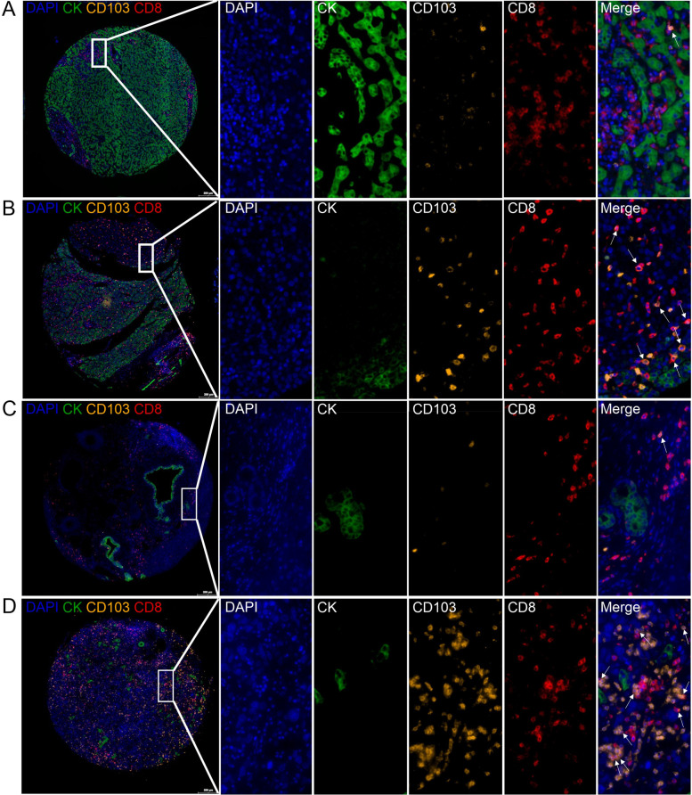
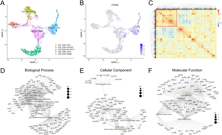
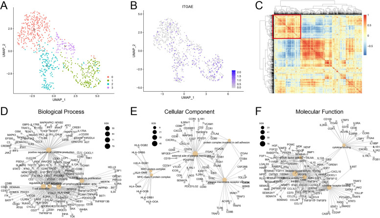
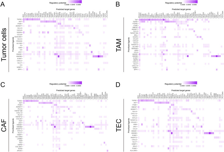
Methods: In our present study, we used the tissue microarrays of HCC and ICC, the multicolor immunohistochemistry (mIHC), and imaging analysis to characterize the tissue-resident CD8+T cells in HCC and ICC tissues. The prognostic values and clinical associations were also analyzed. We also studied the biological functions and the cell-cell communication between tumor-infiltrating CD103+CD8+T cells and other cell types in HCC and ICC based on the published single-cell RNA sequencing (scRNA-seq) data.

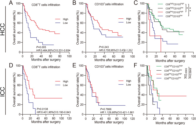
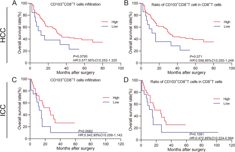
Results: Our work unveiled the expressions of CD8 and CD103 and immunolocalization of tissue-resident CD8+T cells in human HCC and ICC. Elevated CD8+T cells indicated a better overall survival (OS) rate, implying that tumor-infiltrating CD8+T cells in HCC and ICC could serve as an independent prognostic factor. Moreover, the number of CD103+CD8+T cells was increased in HCC and ICC tissues compared with adjacent normal tissues. HCC patients defined as CD8highCD103high had a better OS, and the CD8lowCD103low group tended to have a poorer prognosis in ICC. Evaluation of the CD103+CD8+T-cell ratio in CD8+T cells could also be a prognostic predictor for HCC and ICC patients. A higher ratio of CD103+CD8+T cells over total CD8+T cells in HCC tissues was negatively and significantly associated with the advanced pathological stage. The percentage of higher numbers of CD103+CD8+T cells in ICC tissues was negatively and significantly associated with the advanced pathological stage. In contrast, the higher ratio of CD103+CD8+T cells over total CD8+T cells in ICC tissues was negatively and significantly associated with the advanced pathological stage. In addition, single-cell transcriptomics revealed that CD103+CD8+T cells were enriched in genes associated with T-cell activation, proliferation, cytokine function, and T-cell exhaustion.

Conclusion: The CD103+ tumor-specific T cells signified an important prognostic marker with improved OS, and the evaluation of the tissue-resident CD103+CD8+T cells might be helpful in assessing the on-treatment response of liver cancer.

Keywords: Hepatocellular carcinoma; Intrahepatic Cholangiocarcinoma; Multicolor immunohistochemistry; Prognosis; Tissue-resident CD103+CD8+T cells.


Conflict of interest statement

The authors declare no competing interests.

Figures

Fig. 1
Fig. 1
Characterization of the expressions and localizations of CD8 and CD103 in human HCC and ICC tissues by mIHC. An enlarged subsection of the core was highlighted, showing each of the individual markers in the composite image after spectral unmixing, together with the DAPI nuclear marker (pseudo-colored blue) and the autofluorescence signal (pseudo-colored black). A The expressions of CD8 (membrane, pseudo-colored red) and CD103 (membrane, pseudo-colored brown) in adjacent normal tissues of HCC identified by CK (membrane, pseudo-colored green), together with the DAPI nuclear marker (pseudo-colored blue). B Expressions of CD8 and CD103 in tumor tissues of HCC. C Expressions of CD8 and CD103 in adjacent normal tissues of ICC. D Expressions of CD8 and CD103 in tumor tissues of ICC. Scale bar for the first image of A, B, C, or D, 200 μm
Fig. 2
Fig. 2
Prognostic values of CD8 and CD103 expressions in tumor tissues for the patients with HCC or ICC. Kaplan–Meier survival analysis was performed to predict the prognostic values of tumor-infiltrating CD8+T cells and CD103+ immune cells in human HCC or ICC tissues. A, B, D, and E Patients were stratified according to the infiltration intensities of tumor-infiltrating CD8+T cells or CD103+ immune cells (high infiltration in red and low infiltration in blue). C and F Patients were categorized into four subpopulations as CD8high CD103high (green), CD8high CD103low (black), CD8low CD103high (red), and CD8low CD103low (blue). Cutoffs for low and high expressions were defined with the Cutoff Finder method. *P < 0.05, **P < 0.01
Fig. 3
Fig. 3
Prognostic values of tissue-resident CD103+CD8+T cells in tumor tissues for the patients with HCC or ICC. Kaplan–Meier survival analysis was performed to predict the prognostic values of tumor-infiltrating tissue-resident CD103+CD8+T cells in human HCC or ICC tissues. A and C Patients with high intensity of tissue-resident CD103+CD8+T cells (red) showed a better OS rate than those with low intensity of tissue-resident CD103+CD8+T cells (blue), P = 0.0795 and P = 0.0582 in HCC and ICC, respectively. B and D Patients with a high ratio of CD103+CD8+T cells over CD8+T cells (red) showed a better OS rate than those with a low ratio of CD103+CD8+T cells in CD8+T cells (blue), P = 0.071 and P = 0.1091 in HCC and ICC, respectively
Fig. 4
Fig. 4
Identification and characterization of CD103+CD8+T cells in HCC. A UMAP plot of sub-clustered CD103+CD8+T cells labeled with cell annotations provided by the article [14]. B UMAP showed ITGAE expression in CD8+TILs. C Heatmap showed correlations between ITGAE and other immune-related genes in CD103+CD8+TILs. D, E, and F GO enrichment analysis of ITGAE-related immune genes (clustered with ITGAE in C), and three network plots showed biological process, cellular component, and molecular function
Fig. 5
Fig. 5
Identification and characterization of CD103+CD8+T cells in ICC. A UMAP plot of scRNA-seq data colored by Seurat cluster. B UMAP showed ITGAE expression in CD8+TILs. C Heatmap showed correlations between ITGAE and other immune-related genes in CD103+CD8+TILs. D, E, and F GO enrichment analysis of CD103-related immune genes (clustered with CD103 in C) and three network plots showed biological process, cellular component, and molecular function
Fig. 6
Fig. 6
Cell–cell communication between CD8+TILs and other cell types in HCC and ICC. Heatmaps showed the regulatory potential of the prioritized ligands in tumor cells, TAMs, CAFs, and TECs that interact with signature genes of CD8+CD103+T cells

References

    1. Xue R, Chen L, Zhang C, Fujita M, Li R, Yan SM, Ong CK, Liao X, Gao Q, Sasagawa S, et al. Genomic and transcriptomic profiling of combined hepatocellular and intrahepatic cholangiocarcinoma reveals distinct molecular subtypes. Cancer Cell. 2019;35(6):932–947.e938. doi: 10.1016/j.ccell.2019.04.007. - DOI - PMC - PubMed
    1. Tang M, Zhao Y, Zhao J, Wei S, Liu M, Zheng N, Geng D, Han S, Zhang Y, Zhong G, et al. Liver cancer heterogeneity modeled by in situ genome editing of hepatocytes. Science Adv. 2022;8(25):eabn5683. doi: 10.1126/sciadv.abn5683. - DOI - PMC - PubMed
    1. Llovet JM, Ricci S, Mazzaferro V, Hilgard P, Gane E, Blanc JF, de Oliveira AC, Santoro A, Raoul JL, Forner A, et al. Sorafenib in advanced hepatocellular carcinoma. N Engl J Med. 2008;359(4):378–390. doi: 10.1056/NEJMoa0708857. - DOI - PubMed
    1. Sangro B, Sarobe P, Hervás-Stubbs S, Melero I. Advances in immunotherapy for hepatocellular carcinoma. Nat Rev Gastroenterol Hepatol. 2021;18(8):525–543. doi: 10.1038/s41575-021-00438-0. - DOI - PMC - PubMed
    1. Ostroumov D, Duong S, Wingerath J, Woller N, Manns MP, Timrott K, Kleine M, Ramackers W, Roessler S, Nahnsen S, et al. Transcriptome profiling identifies TIGIT as a marker of T-cell exhaustion in liver cancer. Hepatology (Baltimore, MD) 2021;73(4):1399–1418. doi: 10.1002/hep.31466. - DOI - PubMed

MeSH terms