. 2023 Apr 6;23(1):142.
doi: 10.1186/s12886-023-02892-7.

Bacillary layer detachment with malignant choroidal tumors: a case series

Affiliations

Bacillary layer detachment with malignant choroidal tumors: a case series

Yousef Ahmed Fouad et al. BMC Ophthalmol. .

Abstract

Purpose: To study the incidence and characteristics of bacillary layer detachment (BALAD) occurring with the two most common choroidal malignancies, choroidal metastasis and choroidal melanoma.

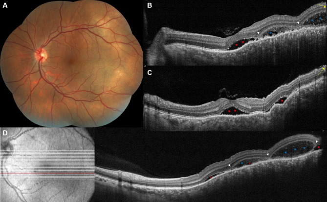
Methods: A retrospective multicentric record analysis. Eyes with a diagnosis of choroidal melanoma or choroidal metastasis that had good-quality fundus photography and spectral domain optical coherence tomography (OCT) scans of the macular and tumor regions allowing for delineation of the retinal layers were included for analysis. Qualitative image evaluation was done by two independent graders for the presence, location, and OCT features of BALAD, as well as any associated intraretinal or subretinal fluid. Demographic and clinical data were also retrieved.

Results: Of the 11 eyes with choroidal metastasis and 7 eyes with choroidal melanoma that were included in the final analysis, 6 (54.5%) and 1 (14.3%) had BALAD, respectively. The BALAD co-localized with the subretinal fluid in all cases and with the intraretinal fluid in 1/3 cases (33.3%), was foveal in location in 3 eyes (42.9%), was overlying the tumor in 6 eyes (85.7%), and varied in number and size. Reflectivity within the BALAD was consistently higher than the vitreous and adjacent subretinal fluid, and discernable suspended hyperreflective particles were noted in 5 eyes (71.4%).

Conclusion: BALAD is relatively common with choroidal metastasis. The OCT features described supplement our recognition of this new entity.

Keywords: BALAD; Bacillary Layer detachment; Choroidal Melanoma; Choroidal Metastasis; Choroidal tumors.


Conflict of interest statement

The authors declare no competing interests.

Figures

Fig. 1
Fig. 1
Choroidal melanoma with BALAD. Montaged fundus photography of a left eye in panel (A) depicts a lower temporal macular amelanotic mass with overlying pigment deposition and cystic changes. On B-scan ultrasonography the tumor is dome shaped with low to moderate internal reflectivity (B). Optical coherence tomography line scan passing through the fovea and the nasal edge of the tumor (C) shows localized SRF (red asterisk), diffuse cystoid edema over sloping edge of the tumor (green asterisks), and BALAD (blue asterisks). The BALAD is noted in the foveal region, adjacent to the tumor margin, and co-localizing with other fluid compartments. Hyperreflective material can be seen at the floor of the BALAD obscuring part of the EZ and RPE underneath it
Fig. 2
Fig. 2
A case of choroidal metastasis with BALAD in a 40-year-old female patient with breast cancer. Fundus examination revealed multiple temporal choroidal masses with overlying SRF involving the foveal region (A). Radial scans centered over the fovea (B, C) and a raster scan over the lower macula (D) show SRF accumulation (red asterisks) involving the fovea and temporal macula, and BALAD (blue asterisks) localized to the lower parafoveal and lower temporal macular areas. White arrowheads point towards the site of splitting between the EZ and the ELM. Compared to SRF which appear as clear hyporeflective spaces, BALAD consistently demonstrates moderately hyperreflective material (turbidity) within the lumen. Yellow arrows point to the direction of the radial OCT B scan
Fig. 3
Fig. 3
Representative cases of OCT appearance of BALAD in eyes with choroidal metastasis. A 67-year-old female patient with multiple choroidal metastases in both eyes from breast cancer. The right eye scans (A,B) show SRF beneath the fovea and nasally (red asterisks), parafoveal IRF (green asterisk), and a single small BALAD in the nasal macula within the peripapillary region (blue asterisk). The left eye scans (C,D) show nasal SRF (red asterisk) and a single large BALAD in the upper peripapillary region (blue asterisk). A 51-year-old with choroidal metastasis in the left eye from breast cancer, with OCT scans (E,F) showing extensive SRF accumulation (red asterisk) and a single large BALAD in the upper perifoveal region (blue asterisk). Multiple vertical hyperreflective lines are seen traversing the thickness of the BALAD. Yellow arrows point to the direction of the OCT B scan

References

    1. Ramtohul P, Engelbert M, Malclès A, Gigon E, Miserocchi E, Modorati G, et al. BACILLARY LAYER DETACHMENT: MULTIMODAL IMAGING AND HISTOLOGIC EVIDENCE OF a NOVEL OPTICAL COHERENCE TOMOGRAPHY TERMINOLOGY: literature review and proposed theory. Retina. 2021;41:2193–207. doi: 10.1097/IAE.0000000000003217. - DOI - PubMed
    1. Yordi S, Sarici K, Cetin H, Lunasco LM, Le TK, Sevgi DD et al. Bacillary Detachment in Neovascular Age-Related Macular Degeneration: Incidence, Clinical Features, and Response to Anti-VEGF Therapy. Ophthalmol Retin. 2022. - PMC - PubMed
    1. Jung JJ, Soh YQ, Yu DJG, Rofagha S, Lee SS, Freund KB et al. Bacillary Layer Detachment Because of Macular Neovascularization. Retina. 2021;41:2106–14. - PMC - PubMed
    1. Ramtohul P, Malclès A, Gigon E, Freund KB, Introini U, Bandello F, et al. Long-term outcomes of Bacillary Layer detachment in Neovascular Age-Related Macular Degeneration. Ophthalmol Retin. 2022;6:185–95. doi: 10.1016/j.oret.2021.09.010. - DOI - PubMed
    1. Kim JH, Kim JW, Kim CG, BACILLARY LAYER DETACHMENT IN A KOREAN COHORT WITH NEOVASCULAR AGE-RELATED MACULAR DEGENERATION. Retina. 2022;42:1028–37. doi: 10.1097/IAE.0000000000003437. - DOI - PubMed