. 2023 Apr 7;21(1):73.
doi: 10.1186/s12915-023-01576-4.

The UAS thioredoxin-like domain of UBXN7 regulates E3 ubiquitin ligase activity of RNF111/Arkadia

Affiliations

The UAS thioredoxin-like domain of UBXN7 regulates E3 ubiquitin ligase activity of RNF111/Arkadia

Sadek Amhaz et al. BMC Biol. .

Abstract

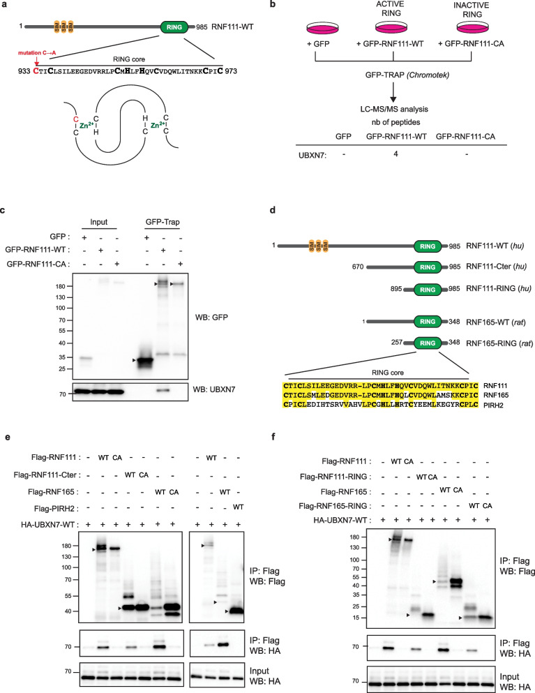
Background: E3 ubiquitin ligases play critical roles in regulating cellular signaling pathways by inducing ubiquitylation of key components. RNF111/Arkadia is a RING E3 ubiquitin ligase that activates TGF-β signaling by inducing ubiquitylation and proteasomal degradation of the transcriptional repressor SKIL/SnoN. In this study, we have sought to identify novel regulators of the E3 ubiquitin ligase activity of RNF111 by searching for proteins that specifically interacts with its RING domain.

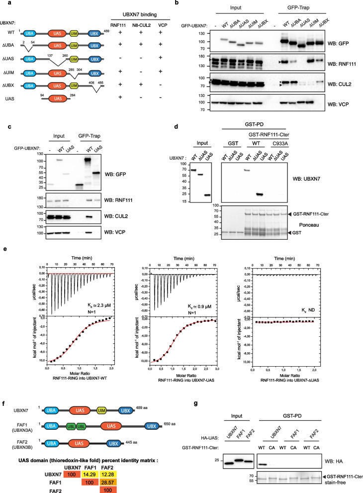
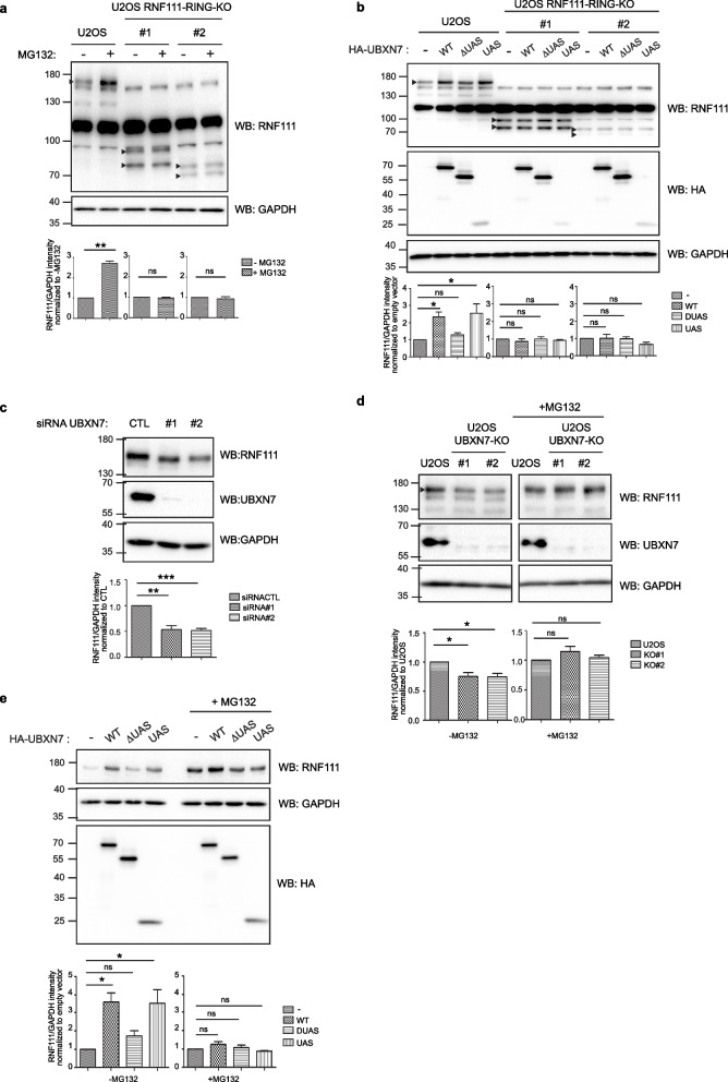
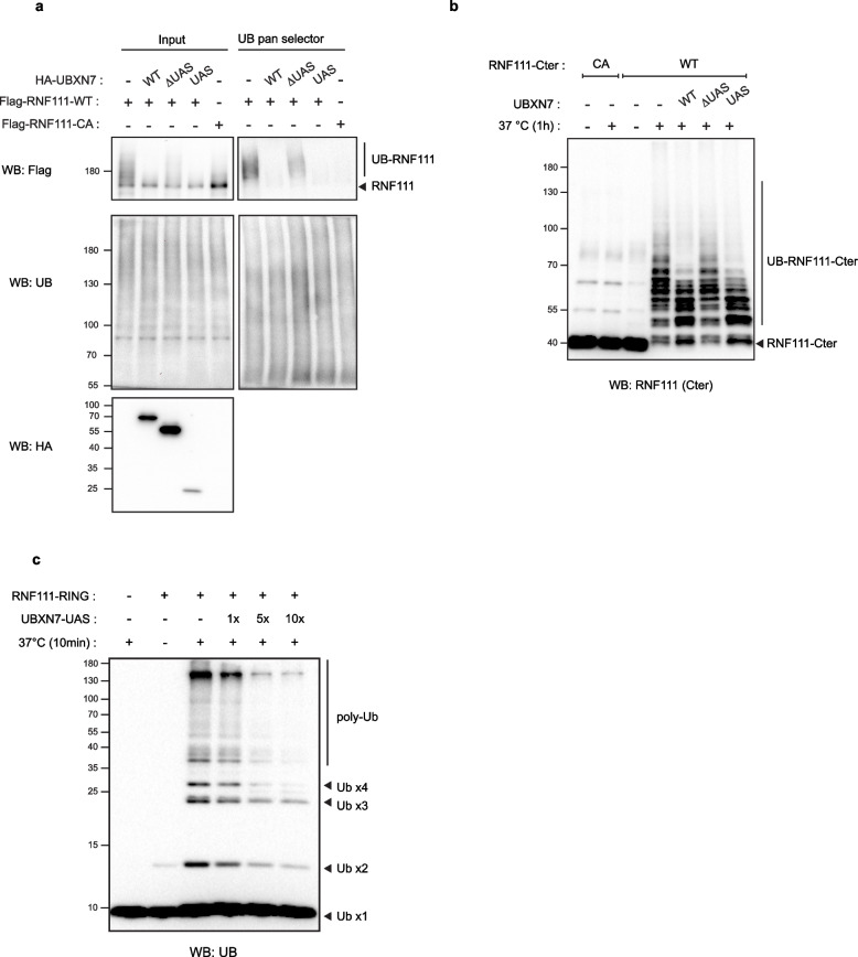
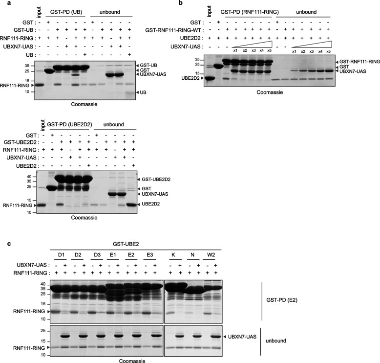
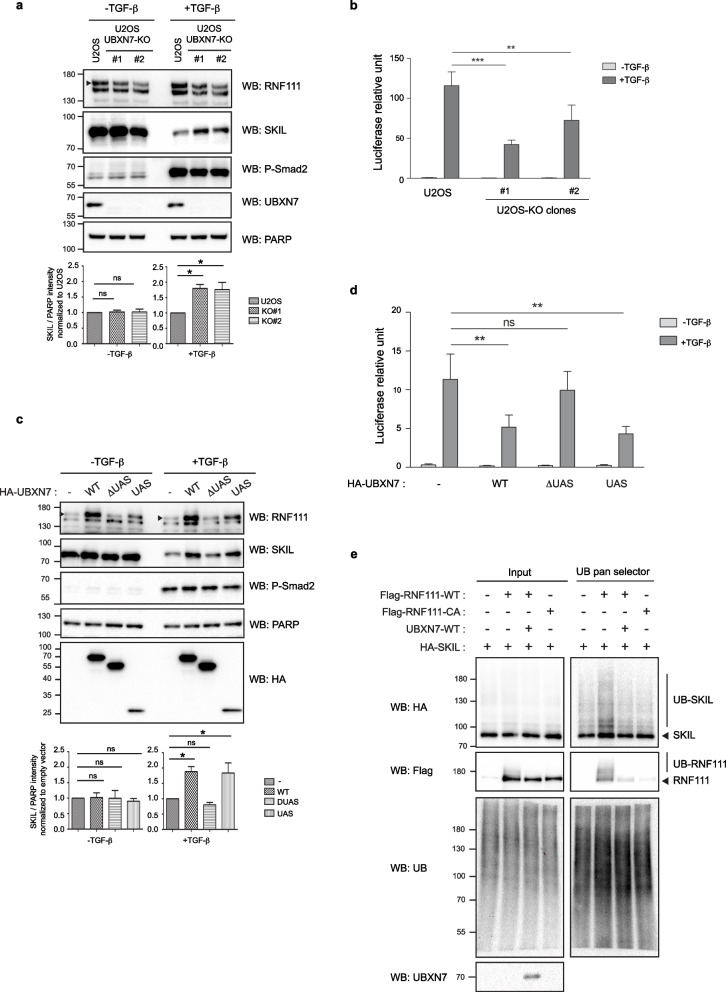
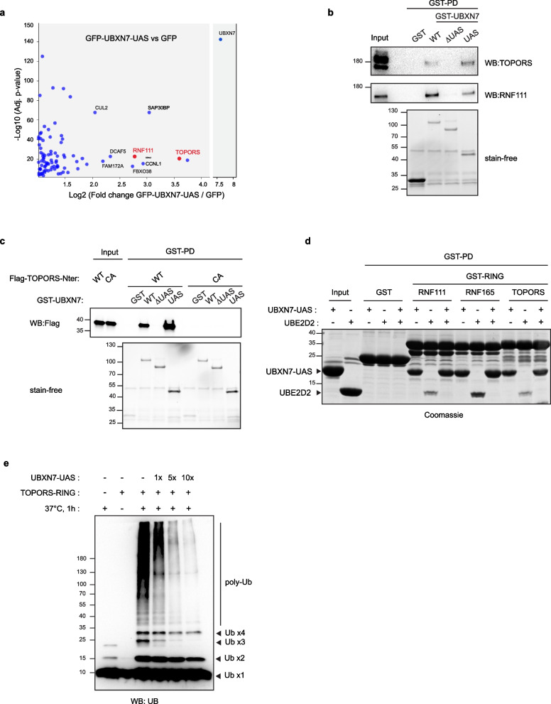
Results: We found that UBXN7, a member of the UBA-UBX family, directly interacts with the RING domain of RNF111 or its related E3 RNF165/ARK2C that shares high sequence homology with RNF111. We showed that UBXN7 docks on RNF111 or RNF165 RING domain through its UAS thioredoxin-like domain. Overexpression of UBXN7 or its UAS domain increases endogenous RNF111, while an UBXN7 mutant devoid of UAS domain has no effect. Conversely, depletion of UBXN7 decreases RNF111 protein level. As a consequence, we found that UBXN7 can modulate degradation of the RNF111 substrate SKIL in response to TGF-β signaling. We further unveiled this mechanism of regulation by showing that docking of the UAS domain of UBXN7 inhibits RNF111 ubiquitylation by preventing interaction of the RING domain with the E2 conjugating enzymes. By analyzing the interactome of the UAS domain of UBXN7, we identified that it also interacts with the RING domain of the E3 TOPORS and similarly regulates its E3 ubiquitin ligase activity by impairing E2 binding.

Conclusions: Taken together, our results demonstrate that UBXN7 acts as a direct regulator for the E3 ubiquitin ligases RNF111, RNF165, and TOPORS and reveal that a thioredoxin-like domain can dock on specific RING domains to regulate their E3 ubiquitin ligase activity.

Keywords: E3 ubiquitin ligase; RING; RNF111; RNF165; SKIL; TGF-β; TOPORS; Thioredoxin; UBXN7.


Conflict of interest statement

The authors declare that they have no competing interests.

Figures

Fig. 1
Fig. 1
UBXN7 interacts with RNF111 and RNF165 Arkadia-like RING domains. a Schematic representation of RNF111 showing its characterized domains: the three SUMO-interacting motifs (SIM) and C-terminal RING domain. The sequence of the RING core is indicated below with the cysteines and histidines of the RING-H2 domain in bold. Mutation of the first cysteine of the RING domain is annotated in red. A schematic representation of the “cross-brace” zinc finger structure of the RING domain is shown [12]. b Workflow for the identification of RING specific RNF111 partners. HEK-293 cells were transfected with GFP, GFP-RNF111-WT, or GFP-RNF111-CA before GFP-Trap affinity purification and proteomics mass spectrometry analysis. The number of peptides identified for UBXN7 in each condition is shown below. c Endogenous UBXN7 interacts with RNF111-WT but not with RNF111-CA. Whole-cell lysates (Input) and GFP-Trapped lysates (GFP-Trap) from HEK-293 cells transfected with GFP, GFP-RNF111-WT, or GFP-RNF111-CA were analyzed by western blotting with anti-GFP and anti-UBXN7 antibodies. The arrows indicate the protein of interest. d Schematic representation of Flag-tagged constructs expressing RNF111 (human) or RNF165 (rat) used in this study. The sequence alignment of the RING core is indicated below. Identical amino acids are highlighted in yellow. The RING core is 80% identical between RNF111 and RNF165. Note that the RING core sequence of RNF165 is identical in rat and human. The sequence alignment with human PIRH2, another RING-H2, is also indicated. e, f UBXN7 specifically interacts with RNF111 and RNF165 RING domains. Lysates from HEK-293 cells transfected with HA-UBXN7-WT and the indicated Flag-tagged RNF111, RNF165, or PIRH2 constructs were immunoprecipitated with Flag antibody and analyzed by western blotting with anti-Flag and anti-HA antibodies. The corresponding whole-cell lysates (Input) were analyzed with anti-HA antibody. The arrows indicate unmodified RNF111 and RNF165 proteins
Fig. 2
Fig. 2
UBXN7 interacts directly with RNF111 RING domain through its UAS domain. a Schematic representation of the domain organization of UBXN7 and the different mutants generated, together with their binding capacity to RNF111, N8-CUL2, and VCP observed in b. b, c UBXN7 binds RNF111 via its UAS domain. Whole-cell Lysates (input) and corresponding GFP immunoprecipitated lysates (GFP-Trap) from HEK-293 cells transfected with the different GFP-UBXN7 deletion mutants were analyzed by western blotting with the indicated antibodies. The arrows indicate RNF111 and CUL2. The asterisk indicates neddylated CUL2. d The UAS domain of UBXN7 binds directly to RNF111. UBXN7-WT, UBXN7-∆UAS, and UBXN7-UAS recombinant proteins were pulled down with GST, GST-RNF111-Cter-WT, or GST-RNF111-Cter-CA. UBXN7 proteins were revealed by western blotting with anti-UBXN7 antibody. The Ponceau staining shows GST proteins. The input shows the initial amount of each recombinant UBXN7 proteins before pull-down. e The RING domain of RNF111 has a binding affinity in the low micromolar range for UBXN7-WT and its UAS domain (Kd = 2.3 and 0.9 µM, respectively) and a stoichiometry of 1:1 (N = 1). ITC experiments were performed by titration of 200 µM of RNF111 RING domain into 10 µM of UBXN7-WT or UBXN7-UAS domain, or into 15 µM of UBXN7-∆UAS at 25 °C. ND not determined. The Kd values correspond to the average of replicate experiments (UAS, n = 3; UBXN7-WT, n = 2). ITC data values obtained for each experiments are indicated in Additional file 3: Table S1. f Schematic representation of the domain organization of the UAS containing UBA-UBX proteins UBXN7, FAF1, and FAF2, together with the matrix of percent identity of their UAS domain obtained with Clustal Omega multiple sequence alignments. g The RNF111 RING domain binds specifically to the UAS domain of UBXN7. HEK-293 lysates transfected with constructs expressing HA-tagged UAS domains from UBXN7, FAF1, or FAF2 were pulled down with GST-RNF111-Cter-WT or GST-RNF111-Cter-CA and subsequently analyzed by western blotting along with the corresponding whole-cell lysates (input). GST proteins were detected with stain-free as a control
Fig. 3
Fig. 3
UBXN7 stabilizes RNF111 in a RING and UAS dependent-manner. a Endogenous RNF111 is degraded by the proteasome in a RING-dependent manner. Western blotting of lysates from U2OS cells and CRISPR U2OS RNF111-RING-KO clones #1 and #2 treated or not with MG132 for 4 h. Quantification of variations in RNF111 protein level is indicated below the blots panel. Values represent the mean ± SEM of RNF111 intensity normalized to GAPDH loading control intensity and to the indicated control condition from three independent experiments. Statistical analysis was performed using a paired t-test. **p < 0.01, ns not significant. Arrows on western blots indicate the band corresponding to RNF111. b UBXN7 stabilizes RNF111 in a RING and UAS-dependent manner. Lysates from U2OS cells or U2OS RNF111-RING-KO clones #1 or #2 transiently transfected with HA-tagged empty vector (-), UBXN7-WT (WT), UBXN7-∆UAS (∆UAS), or UBXN7-UAS domain (UAS) were analyzed by western blotting with the indicated antibodies. Quantification of variations in RNF111 protein level was made as in a except it was normalized to the empty vector. Statistical analysis was performed using one-way ANOVA with Dunnett’s post test. *p < 0.05, **p < 0.01, ***p < 0.001, ns not significant. c Whole-cell extracts of U2OS cells transfected with a control siRNA or 2 independent siRNA #1 and #2 targeting UBXN7 were analyzed by western blotting as indicated. Quantification and statistical analysis as in b except that values were normalized to siRNA control. d Western blotting analysis of lysates from U2OS cells and U2OS UBXN7-KO clones #1 and #2 treated or not with MG132. Quantification and statistical analysis as in b except that values were normalized to U2OS. e Western blotting analysis of lysates from U2OS UBXN7-KO clone #1 transfected with the indicated plasmids and treated or not with MG132. Quantification and statistical analysis as in b. Western blot quantifications values obtained for each experiments are shown in Additional file 3: Table S1
Fig. 4
Fig. 4
UBXN7 inhibits RNF111 auto-ubiquitylation in a UAS-dependent manner. a UBXN7 inhibits RNF111 auto-ubiquitylation in cells. Lysates (input) from U2OS UBXN7-KO clone #1 transiently transfected with Flag-RNF111-WT or Flag-RNF111-CA together with HA-tagged empty vector ( −), UBXN7-WT (WT), UBXN7-∆UAS (∆UAS), or UBXN7-UAS (UAS) were immunoprecipitated with the UB pan selector resin and subsequently analyzed by western blotting. b UBXN7 inhibits RNF111 auto-ubiquitylation in vitro. Recombinant RNF111-Cter-CA or WT were incubated at 37 °C for 1 h in the presence of UBE1 (E1), UBE2D2 (E2), ubiquitin (UB), and recombinant UBXN7-WT (WT), UBXN7-∆UAS (∆UAS), or UBXN7-UAS (UAS). RNF111 ubiquitylation was revealed by western blotting using an antibody targeting the RNF111 C-terminal domain. c The UAS domain inhibits free ubiquitin chains formation induced by RNF111-RING domain in vitro. Recombinant RNF111-RING was incubated at 37 °C for 10 min in the presence of UBE1 (E1), UBE2D2 (E2), ubiquitin (UB) and 1 to 10 molar excess of recombinant UBXN7-UAS. Ubiquitin chains were revealed by western blotting using an anti-UB antibody
Fig. 5
Fig. 5
Binding of the UAS domain impedes RING-E2 interaction. a The UAS domain competes with RING-E2 but not with RING-UB interaction. GST pull-down (GST-PD) of GST, GST-UB (upper panel) or GST-UBE2D2 (lower panel) with recombinant RNF111 RING, in the presence or not of 10 × molar excess of recombinant UBXN7-UAS. The proteins were revealed on acrylamide gel by Coomassie staining. The unbound panel shows the unbound proteins in the flow-through. Recombinant UB (in the upper panel) and UBE2D2 (in the lower panel) were used as a positive control for binding competition. b UBE2D2 binding to GST-RNF111-RING in the presence of increasing amount of UBXN7-UAS. GST pull-down (GST-PD) of GST-RNF111-RING-WT with equimolar concentration of UBE2D2 in the presence of 1 to 5 × molar excess of UBXN7-UAS. The UBE2D2 input and the unbound proteins in the flow-through are shown. c The UAS domain inhibits the binding of RNF111-RING with different known interacting E2s. GST pull-down of different GST-E2s (UBE2D1-3, UBE2E1-3, UBE2K, UBE2N, and UBE2W2) with RNF111-RING-WT in the presence or not of 2 × molar excess of UBXN7-UAS. The unbound proteins in the flow-through are shown
Fig. 6
Fig. 6
UBXN7 regulates SKIL degradation in a UAS-dependent manner. a SKIL increases in the absence of UBXN7. Nuclear extracts from parental U2OS and U2OS UBXN7-KO clones #1 and #2 treated or not with TGF-β for 1 h were analyzed by western blotting. Quantification of SKIL protein level variations is indicated below the blots panel. Values represent the mean ± SEM of SKIL intensity normalized to PARP loading control intensity and to the U2OS control condition in three independent experiments. Statistical analysis was performed using one-way ANOVA with Dunnett’s post test. ns not significant; *p < 0.05. The arrow indicates the band corresponding to RNF111. b TGF-β-induced SMAD-dependent transcription is attenuated in the absence of UBXN7. Parental U2OS and U2OS UBXN7-KO clones #1 and #2 were co-transfected with the CAGA12-Luc and pRL-TK reporters and treated or not with TGF-β for 8 h before lysis. Data represent means ± SEM of luciferase activities normalized to Renilla in three independent experiments. Statistical analysis was performed using two-way ANOVA with Bonferroni post test on three independent experiments. **p < 0.01, ***p < 0.001. c UBXN7 overexpression impairs SKIL degradation in response to TGF-β. Nuclear extracts from U2OS UBXN7-KO clone #1 cells transfected with HA-tagged empty vector (-), UBXN7-WT, UBXN7-∆UAS, or UBXN7-UAS and treated or not for 1 h with TGF-β before extraction were analyzed by western blotting. Quantification and statistics as in a except that values were normalized to the empty vector condition. d UBXN7 significantly attenuates TGF-β-induced SMAD-dependent transcription in a UAS-dependent manner. U2OS UBXN7-KO clone #1 cells were co-transfected with CAGA12-Luc, pRL-TK, and HA-tagged empty vector ( −) or the indicated HA-UBXN7 constructs. Luciferase assays were performed as in be UBXN7 inhibits SKIL ubiquitylation by RNF111. U2OS UBXN7-KO clone #1 cells were transiently transfected as indicated with HA-SKIL, Flag-RNF111-WT, Flag-RNF111-CA, and untagged UBXN7-WT. Lysates (input) were immunoprecipitated with the UB pan selector resin and subsequently analyzed by western blotting. Western blot quantification and luciferase assay values obtained for each experiments are shown in Additional file 3: Table S1
Fig. 7
Fig. 7
TOPORS is another E3 regulated by the UAS domain of UBXN7. a GFP-UBXN7-UAS versus GFP Interactome. Cell lysate from HEK-293 cells transfected with GFP or GFP-UBXN7-UAS were subjected to GFP-Trap affinity purification and co-immunoprecipitated proteins were subsequently identified using quantitative label-free mass spectrometry. Volcano plot illustrating the distribution of the proteins significantly enriched (fold change ≥ 2, p value ≤ 0.05, n = 5) in GFP-UBXN7-UAS compared to GFP (-Log10 (adj p value), y-axis; log2 fold change GFP-UBXN7-UAS/GFP, x-axis). b, c TOPORS binds to UBXN7-UAS. HEK-293 cellular extracts (b) or HEK-293 cellular extracts from cells transfected with Flag-TOPORS-Nter-WT or Flag-TOPORS-Nter-C103A (c) were pulled down with the indicated GST-UBXN7 constructs and analyzed by western blotting along with the corresponding cellular extracts (input). The amount of GST proteins in each condition was revealed by stain-free as a control. d The UAS domain competes with UBE2D2 for binding to TOPORS RING domain. GST pull-down (GST-PD) of the indicated GST-RING domains with UBE2D2, in the presence or not of UBXN7-UAS. The proteins were revealed on acrylamide gel by Coomassie staining. e The UAS domain inhibits free ubiquitin chain formation induced by TOPORS RING domain in vitro. Recombinant TOPORS RING domain was incubated at 37 °C for 1 h in the presence of UBE1 (E1), UBE2D2 (E2), ubiquitin (UB), and 1 × , 5 × , or 10 × molar excess of recombinant UBXN7-UAS (UAS). Free ubiquitin chain formation was revealed by western blotting using an anti-ubiquitin (anti-UB) antibody

References

    1. Yau R, Rape M. The increasing complexity of the ubiquitin code. Nat Cell Biol. 2016;18:579–586. doi: 10.1038/ncb3358. - DOI - PubMed
    1. Hänzelmann P, Schindelin H. The interplay of cofactor interactions and post-translational modifications in the regulation of the AAA+ ATPase p97. Front Mol Biosci. 2017;4:21. doi: 10.3389/fmolb.2017.00021. - DOI - PMC - PubMed
    1. Alexandru G, Graumann J, Smith GT, Kolawa NJ, Fang R, Deshaies RJ. UBXD7 binds multiple ubiquitin ligases and implicates p97 in HIF1alpha turnover. Cell. 2008;134:804–816. doi: 10.1016/j.cell.2008.06.048. - DOI - PMC - PubMed
    1. den Besten W, Verma R, Kleiger G, Oania RS, Deshaies RJ. NEDD8 links cullin-RING ubiquitin ligase function to the p97 pathway. Nat Struct Mol Biol. 2012;19(511–6):S1. - PMC - PubMed
    1. Bandau S, Knebel A, Gage ZO, Wood NT, Alexandru G. UBXN7 docks on neddylated cullin complexes using its UIM motif and causes HIF1α accumulation. BMC Biol. 2012;10:36. doi: 10.1186/1741-7007-10-36. - DOI - PMC - PubMed

Publication types

MeSH terms