. 2023 Apr 3;18(16):e1307-e1327.
doi: 10.4244/EIJ-D-22-00776.

Computed tomographic angiography in coronary artery disease

Affiliations

Computed tomographic angiography in coronary artery disease

Patrick W Serruys et al. EuroIntervention. .

Abstract

Coronary computed tomographic angiography (CCTA) is becoming the first-line investigation for establishing the presence of coronary artery disease and, with fractional flow reserve (FFRCT), its haemodynamic significance. In patients without significant epicardial obstruction, its role is either to rule out atherosclerosis or to detect subclinical plaque that should be monitored for plaque progression/regression following prevention therapy and provide risk classification. Ischaemic non-obstructive coronary arteries are also expected to be assessed by non-invasive imaging, including CCTA. In patients with significant epicardial obstruction, CCTA can assist in planning revascularisation by determining the disease complexity, vessel size, lesion length and tissue composition of the atherosclerotic plaque, as well as the best fluoroscopic viewing angle; it may also help in selecting adjunctive percutaneous devices (e.g., rotational atherectomy) and in determining the best landing zone for stents or bypass grafts.


Conflict of interest statement

P.W. Serruys has received consultancy fees from Philips/Volcano, SMT, Novartis, Xeltis, and Meril Life, outside of the submitted work. N. Kotoku has received a grant for studying overseas from the Fukuda Foundation for Medical Technology. B.L. Nørgaard has received an unrestricted institutional research grant from HeartFlow, outside of the submitted work. K. Nieman reports research support from the National Institutes of Health (NIH R01- HL141712; NIH R01 - HL146754); unrestricted institutional research support from Siemens Healthineers, Bayer, and HeartFlow, unrelated to this work; has consulted for Siemens Medical Solutions USA and Novartis; and has equity in Lumen Therapeutics. M.R. Dweck is supported by the British Heart Foundation (FS/SCRF/21/32010); is the recipient of the Sir Jules Thorn Award for Biomedical Research 2015 (15/JTA); has received speaker fees from Pfizer and Novartis; and reports consultancy fees from Novartis, Jupiter Bioventures, Beren, and Silence Therapeutics. J.J. Bax has received speaker fees from Abbott Vascular and Edwards Lifesciences; and his department has received unrestricted research grants from Abbott and Edwards Lifesciences. J. Knuuti has receivedconsultancy fees from GE Healthcare and AstraZeneca; and has received speaker fees from GE Healthcare, Bayer, Lundbeck, Merck and Pfizer, outside of the submitted work. C.A. Taylor is an employee and shareholder of HeartFlow. J.A. Leipsic has served as a consultant to Circle CVI and HeartFlow; holds stock options in Circle CVI and HeartFlow; and served on the speaker bureau of Philips. K. Kitagawa has received support from Bayer Yakuhin, Siemens Healthcare K.K., FUJIFILM Medical Co., and Pfizer; and speaker fees from GE Healthcare Japan, HeartFlow Japan, Ono Pharmaceutical Co., Otsuka Pharmaceutical Co., Eisai Co., FUJIFILM Toyama Chemical Co., Bayer Yakuhin, Plusman LLC, and Amgen. B. De Bruyne reports receiving consultancy fees from Boston Scientific and Abbott Vascular; research grants from Coroventis Research, Pie Medical Imaging, CathWorks, Boston Scientific, Siemens, HeartFlow, and Abbott Vascular; and owns equity in Siemens, GE Healthcare, Philips, HeartFlow, Edwards Lifesciences, Bayer, Sanofi, and Celyad. C. Collet has received research grants from GE Healthcare, Siemens, Coroventis Research, Medis Medical Imaging, Pie Medical Imaging, CathWorks, Boston Scientific, HeartFlow, and Abbott Vascular; and consultancy fees from HeartFlow, CryoTherapeutics, and Abbott Vascular. A. Zlahoda-Huzior is a former employee of MedApp. S. Tu reports research grants and consultancy fees from Pulse Medical outside of this submitted work. W. Wijns is supported by the Science Foundation Ireland Research Professorship Award (15/RP/2765). M.J. Budoff has received grant support from General Electric and the National Institutes of Health. The other authors have no conflicts of interest to declare.

Figures
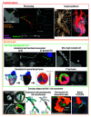
Central illustration
Central illustration. The role of CCTA in coronary artery disease: a diagnostic tool, decision maker and treatment planner.
APS: axial plaque stress; CCTA: coronary computed tomographic angiography; CT: computed tomography; FFR: fractional flow reserve; LAD: left anterior descending; LM: left main; PCI: percutaneous coronary intervention; PET: positron emission tomography; WSS: wall shear stress
Figure 1
Figure 1. Comparison between ultra-high resolution CT and conventional CT.
A) In ultra-high resolution CT in a patient with a calcium score >2,000, the presence of a significant stenosis in the proximal LAD can be ruled out despite extensive calcifications. B) A significant lesion cannot be ruled out on conventional CT due to the substantial blooming artefacts. C) ICA confirmed the absence of significant stenosis. Reproduced with permission from. CT: computed tomography; ICA: invasive coronary angiography; LAD: left anterior descending
Figure 2
Figure 2. Troponin-guided CCTA. Reproduced with permission from.
ACS: acute coronary syndrome; CAD: coronary artery disease; CCTA: coronary computed tomographic angiography; ECG: electrocardiogram; hs-cTnI: high sensitivity cardiac troponin I concentrations; NOCA: non-obstructive coronary artery
Figure 3
Figure 3. Comparison between full-order and on-site CT-derived FFR.
Functional diagnostic performance of full-order and on-site CT-derived FFR is shown in Supplementary Table 2. Modified and reproduced with permission from. 3D: three-dimensional; cFFR: computed fractional flow reserve: CT: computed tomography; FFR: fractional flow reserve; FFRCT: fractional flow reserve derived from coronary computed tomographic angiography; ML: machine learning; QFR: quantitative flow ratio
Figure 4
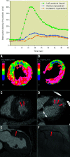
Figure 4. Dynamic myocardial perfusion imaging using dual-source CT and TAC.
By dynamic CTP, absolute myocardial blood flow (MBF) can be calculated from the time-attenuation curves (TAC). Dynamic CTP showed reduced MBF in the LAD territory in both the (A) short-axis and (B) long-axis view (C,D) CT-delayed enhancement revealed a subendocardial infarction in the anterior wall within the reduced MBF area (red arrows). E) CCTA showed a high-grade stenosis in the LAD just proximal to the stent (red arrow). F) ICA revealed >90% stenosis (red arrow). Upper graph reproduced with permission from. A-F) reproduced with permission from. CT: computed tomography; CCTA: coronary computed tomographic angiography; CTP: computed tomography perfusion; ICA: invasive coronary angiography; LAD: left anterior descending artery
Figure 5
Figure 5. Simulation of myocardial perfusion and coronary lumen volume to myocardial mass ratio.
A) Approach to extend non-invasive physiological assessment from epicardial coronary arteries to microcirculation. B) Coronary lumen volume to myocardial mass ratio (V/M). Reproduced with permission from. CCTA: coronary computed tomographic angiography; FFRCT: fractional flow reserve derived from coronary computed tomographic angiography; PET: positron emission tomography
Figure 6
Figure 6. CT Leaman score and Leiden CT risk score.
A) Both are calculated by weighting for plaque localisation according to proximality or distality in the coronary circulation×type of plaque×stenosis severity. B) Kaplan-Meier survival curves for hard cardiac events stratified by obstructive versus non-obstructive CAD and CT Leaman score (CT-LeSc). Reproduced with permission from. CAD: coronary artery disease; CT: computed tomography; LAD: left anterior descending artery; LCx: left circumflex; PDA: posterior descending artery; PL: postero-lateral; RCA: right coronary artery
Figure 7
Figure 7. Advanced assessments of plaque type, plaque thrombosis and disease activity with CT and hybrid PET/CT.
A) CCTA with regions of LAP (<30 HU, orange areas) B) CCTA showing a low-density area in the lumen of RCA in a patient with inferior STEMI. Hybrid PET/CT images demonstrate 18F-GP1 activity as acute thrombus in this region (yellow area). C) Hybrid PET/CT image after administration of 18F-fluoride. Coloured areas represent regions of increased calcification activity. CT: computed tomography; CCTA: coronary computed tomographic angiography; HU: Hounsfield units; LAP: low-attenuation plaque; PET: positron emission tomography; RCA: right coronary artery; STEMI: ST-elevation myocardial infarction
Figure 8
Figure 8. Anatomical and haemodynamic plaque characteristics derived from CCTA.
A) Probability of having ACS according to adverse plaque characteristics (APC). B) Adverse haemodynamic characteristics (AHC) based on 4 haemodynamic parameters derived from CCTA. C) Lesions with both APC and AHC showed significantly higher risk compared with those without. Reproduced/modified with permission from (A), (B), and (C). ACS: acute coronary syndrome; APSCT: axial plaque stress derived from computed tomography; CCTA: coronary computed tomographic angiography; CI: confidence interval; FFRCT: fractional flow reserve derived from coronary computed tomographic angiography; HU: Hounsfield units; RR: relative risk; WSSCT: wall shear stress derived from computed tomography
Figure 9
Figure 9. Optimal fluoroscopic viewing angle for ostial, stem, and bifurcation of LM.
A,B) The optimal fluoroscopic angle for ostial left main (LM) is obtained by the intersection between the aortic annulus and the ostial LM optimal projection curves. A,C) The optimal fluoroscopic angle for the LM stem is obtained by the intersection between the ostial LM and proximal LM optimal projection curves. D) FFRCT view matching the optimal angle of panel C. E,F) The optimal fluoroscopic angle for LM bifurcation is obtained as the perpendicular angle to the “en face” view created by placing 3 dots in the LM, left descending artery (LAD), and left circumflex artery (LCx) 5 mm from the point of the bifurcation. G) FFRCT view matching the optimal angle of panel F. FFRCT: fractional flow reserve derived from coronary computed tomographic angiography
Figure 10
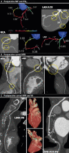
Figure 10. Anatomical and physiological assessment based on non-invasive imaging for planning and follow-up of CABG.
A) Preoperative maximum intensity projection (MIP) images and FFRCT. B) Preoperative curved multiplanar reconstruction (MPR) images: yellow circles indicate significant stenoses. FFRCT indicates flow-limiting lesions at the proximal and distal RCA and proximal LAD, and total occlusion at the proximal LCx. C) Postoperative curved MPR and volume rendering (VR) images at 30-day follow-up. Left internal mammary artery (LIMA)-#8 and saphenous vein graft (SVG)-#14-#4 were patent. CABG: coronary artery bypass graft; FFRCT: fractional flow reserve derived from coronary computed tomographic angiography; LAD: left anterior descending artery; LCx: left circumflex artery; RCA: right coronary artery

Similar articles

Cited by

References

    1. Serruys PW, Hara H, Garg S, Kawashima H, Nørgaard BL, Dweck MR, Bax JJ, Knuuti J, Nieman K, Leipsic JA, Mushtaq S, Andreini D, Onuma Y. Coronary Computed Tomographic Angiography for Complete Assessment of Coronary Artery Disease: JACC State-of-the-Art Review. J Am Coll Cardiol. 2021;78:713–36. - PubMed
    1. Collet C, Onuma Y, Andreini D, Sonck J, Pompilio G, Mushtaq S, La Meir M, Miyazaki Y, de Mey J, Gaemperli O, Ouda A, Maureira JP, Mandry D, Camenzind E, Macron L, Doenst T, Teichgräber U, Sigusch H, Asano T, Katagiri Y, Morel MA, Lindeboom W, Pontone G, Lüscher TF, Bartorelli AL, Serruys PW. Coronary computed tomography angiography for heart team decision-making in multivessel coronary artery disease. Eur Heart J. 2018;39:3689–98. - PMC - PubMed
    1. Serruys PW, Chichareon P, Modolo R, Leaman DM, Reiber JHC, Emanuelsson H, Di Mario, Pijls NHJ, Morel MA, Valgimigli M, Farooq V, van Klaveren, Capodanno D, Andreini D, Bourantas CV, Davies J, Banning AP, Escaned J, Piek JJ, Echavarría-Pinto M, Taylor CA, Thomsen B, Collet C, Pompilio G, Bartorelli AL, Glocker B, Dressler O, Stone GW, Onuma Y. The SYNTAX score on its way out or … towards artificial intelligence: part II. EuroIntervention. 2020;16:60–75. - PubMed
    1. Kunadian V, Chieffo A, Camici PG, Berry C, Escaned J, Maas AHEM, Prescott E, Karam N, Appelman Y, Fraccaro C, Louise Buchanan, Manzo-Silberman S, Al-Lamee R, Regar E, Lansky A, Abbott JD, Badimon L, Duncker DJ, Mehran R, Capodanno D, Baumbach A. An EAPCI Expert Consensus Document on Ischaemia with Non-Obstructive Coronary Arteries in Collaboration with European Society of Cardiology Working Group on Coronary Pathophysiology & Microcirculation Endorsed by Coronary Vasomotor Disorders International Study Group. Eur Heart J. 2020;41:3504–20. - PMC - PubMed
    1. Knuuti J, Wijns W, Saraste A, Capodanno D, Barbato E, Funck-Brentano C, Prescott E, Storey RF, Deaton C, Cuisset T, Agewall S, Dickstein K, Edvardsen T, Escaned J, Gersh BJ, Svitil P, Gilard M, Hasdai D, Hatala R, Mahfoud F, Masip J, Muneretto C, Valgimigli M, Achenbach S, Bax JJ ESC Scientific Document Group. 2019 ESC Guidelines for the diagnosis and management of chronic coronary syndromes. Eur Heart J. 2020;41:407–77. - PubMed

MeSH terms