. 2023 Mar 21:11:1069428.
doi: 10.3389/fped.2023.1069428. eCollection 2023.

The epifascial cap: A typical imaging sign for subcutaneous granuloma annulare in children

Affiliations

The epifascial cap: A typical imaging sign for subcutaneous granuloma annulare in children

Besiana P Beqo et al. Front Pediatr. .

Abstract

Objectives: Subcutaneous granuloma annulare (SGA) is a rare, self-limiting granulomatous disease in children, commonly diagnosed by histopathology following biopsy or surgical excision. This study aimed to identify imaging clues for SGA that could expedite accurate diagnosis and avoid the need for biopsy in children.

Methods: We retrospectively analyzed complete hospital records of all children diagnosed with SGA at our institution from January 2001 to December 2020. Detailed disease history, imaging findings, management, and outcome were evaluated.

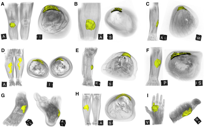
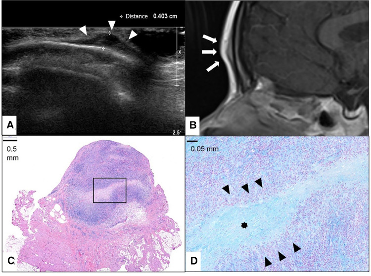
Results: We identified 28 patients (20 girls) at a median age of 3.75 (range 1-12.5 years). Ten patients presented with multiple lesions. Most lesions were located on the lower extremities (n = 26/41). Ultrasound examinations were performed on all patients, and 12 (43%) patients also received an MRI. Surgical intervention was conducted in 18 (64%) patients either by incisional biopsy (n = 6) or total excision of the lump (n = 12). In all patients who did not undergo surgery, SGA resolved spontaneously. A careful review of the MRIs led to the discovery of a characteristic imaging shape of SGA lesions: the epifascial cap with a typical broad circular base laying on the fascia, extending towards the subdermal/dermal tissue. This distinctive shape was evident in every patient in our cohort.

Conclusions: The "Epifascial Cap Sign" is a specific imaging sign for SGA, which to the best of our knowledge, helps distinguish this disease from other subcutaneous lesions. Recognition of this novel diagnostic sign combined with the historical and physical findings should enable clinicians to establish SGA diagnosis easily and diminish the need for further invasive diagnostic procedures.

Keywords: benign lesion; children; granuloma annulare; lumps and bumps; self limiting disease; subcutaneous lesion; treatment.


Conflict of interest statement

The authors declare that the research was conducted in the absence of any commercial or financial relationships that could be construed as a potential conflict of interest.

Figures

Figure 1
Figure 1
Representative MRI sections and 3D-reconstructions of subcutaneous granuloma annulare lesion (colored blue) on the right lower leg of a 2.5-year-old girl (A–D). A T2-weighted slice in axial orientation (A) shows the cap-shaped epifascial lesion with high signal intensity (bright). The corresponding 3D reconstruction is given in (B), visualizing the lesion in blue color and the remaining other tissues by overlaying a volume rendering in a skin-like color. (C) shows the lesion in a T1-weighted sagittal slice with low signal intensity (dark). The corresponding 3D rendering (D) demonstrates the cap-shaped morphology, extending from the deep fascia to the subcutaneous layer.
Figure 2
Figure 2
This composite figure shows 3D-reconstructions of magnetic resonance imaging (MRI) of all 9 patients with subcutaneous granuloma annulare (SGA) on the extremities who were evaluated by MRI. The 3D reconstructions of SGA (yellow) are overlaid by volume renderings of the respective extremity regions (gray) for reference. Note that all SGA lesions characteristically demonstrate a round or oval area with cap-shaped morphology, extending from the deep fascia into the subcutaneous layer. (A) right knee; (B) left lower leg; (C) left forearm; (D) right and left lower legs; (E) right lower leg; (F) left forearm; (G) left foot; (H) right lower leg; (I) left hand.
Figure 3
Figure 3
This composite figure shows representative T1-weighted magnetic resonance images (MRI) in sagittal or coronal orientation through the extremities of 4 children with subcutaneous granuloma annulare (SGA). Note the cap-shaped appearance of SGA lesions and that in T1-weighted MRIs they present as homogenous lesions (arrows) isointense relative to the muscles. (A) left lower leg, (B) left forearm, (C) right lower leg, (D) left forearm.
Figure 4
Figure 4
This composite figure shows representative slices of magnetic resonance imaging (MRI) through the subcutaneous granuloma annulare (SGA) on the extremities of 9 patients evaluated by MRI. The large boxes depict T2-weighted sequences with fat suppression (“T2 FS”). The small boxes depict T1-weighted sequences with fat suppression and intravenous contrast (“T1 FS C+”). Asterisks mark the locations of the SGA. A common finding in all presented cases is the cap-shaped lesion extending from the deep fascia into the subcutaneous fatty tissue. These lesions show heterogeneously hyperintense signal relative to the muscles in T2 FS images and marked contrast enhancement in T1 FS C+ . (A) right knee; (B) left lower leg; (C) left forearm; (D) right lower leg; (E) right lower leg; (F) left forearm; (G) left foot; (H) right lower leg; (I) left hand.
Figure 5
Figure 5
This composite figure shows a frontal location of the subcutaneous granuloma annulare (SGA). Note the epifascial cap sign in the ultrasound image marked with white arrowheads (A) and in the representative slice of magnetic resonance imaging (MRI) marked with white arrows. (B) The completely excised SGA lesion is localized in the subcutis overlying the fascia and is characterized by areas of necrobiotic granulomas shown here in hematoxylin and eosin staining. (C) The area marked with a box in (C) is enlarged in (D), showing alcian blue stain highlighting the mucin within the central zone of necrobiosis (star) surrounded by palisading histiocytes and lymphocytes (black arrowheads).
Figure 6
Figure 6
This composite figure shows representative slices of ultrasound imaging through the subcutaneous granuloma annulare (SGA) in 6 patients who had their final diagnosis by histopathology after excisional or incisional biopsy. A common finding in all presented cases is the cap-shaped SGA lesion extending from the deep fascia to the subcutaneous fatty tissue. The epifascial border is marked with white arrows. These lesions show heterogeneously hypoechoic signal in B-mode (A–C). The lesions stay hypoechoic and show mild perfusion in color mode US (D–F). (A)—right lower leg, (B)—scalp, (C)—right lower leg, (D)—right lower leg, (E)—left foot dorsum, (F)—left lower leg.
Figure 7
Figure 7
This composite figure shows various imaging methods used to evaluate a firm, immobile, indolent subcutaneous lesion in the mid-pretibial region of the right lower leg of a 4-years old girl. The lateral x-ray view of the lower leg depicts a thickened pretibial subcutaneous tissue (arrow), but no osseous abnormalities. The ultrasound image (US) shows a hypoechoic lesion in the typical epifascial cap shape (2 arrows) characteristic of subcutaneous granuloma annulare (SGA), confirmed by histopathological examination. Power Doppler US indicates slight hyperperfusion (2 arrows) in the area of SGA as compared to the surrounding tissue. In T1-weighted magnetic resonance images (MRI T1 C-), SGA presents as a homogenous lesion (1 arrow) isointense relative to the muscles. In T2-weighted images with fat suppression (MRI T2 FS), SGA shows a heterogeneously hyperintense signal relative to the muscles (2 arrows) extending from the muscular fascia into the adjacent subcutaneous tissue in a cap shape. Finally, in T1-weighted images with fat suppression and intravenous contrast (MRI T1 C+) SGA shows a marked contrast enhancement (1 arrow).

Similar articles

References

    1. Keimig EL. Granuloma annulare. Dermatol Clin. (2015) 33:315–29. 10.1016/j.det.2015.03.001 - DOI - PubMed
    1. Piette EW, Rosenbach M. Granuloma annulare: clinical and histologic variants, epidemiology, and genetics. J Am Acad Dermatol. (2016) 75:457–65. 10.1016/j.jaad.2015.03.054 - DOI - PubMed
    1. Thornsberry LA, English JC. Etiology, diagnosis, and therapeutic management of granuloma annulare: an update. Am J Clin Dermatol. (2013) 14:279–90. 10.1007/s40257-013-0029-5 - DOI - PubMed
    1. Joshi TP, Duvic M. Granuloma annulare: an updated review of epidemiology, pathogenesis, and treatment options. Am J Clin Dermatol. (2022) 23(1):37–50. 10.1007/s40257-021-00636-1 - DOI - PMC - PubMed
    1. Patrizi A, Gurioli C, Neri I. Childhood granuloma annulare: a review. G Ital Dermatol Venereol. (2014) 149:663–74. PMID: . - PubMed