Review
. 2023 Mar 21:14:1144069.
doi: 10.3389/fphys.2023.1144069. eCollection 2023.

Pathophysiology of Cav1.3 L-type calcium channels in the heart

Affiliations
Review

Pathophysiology of Cav1.3 L-type calcium channels in the heart

Sahil Zaveri et al. Front Physiol. .

Abstract

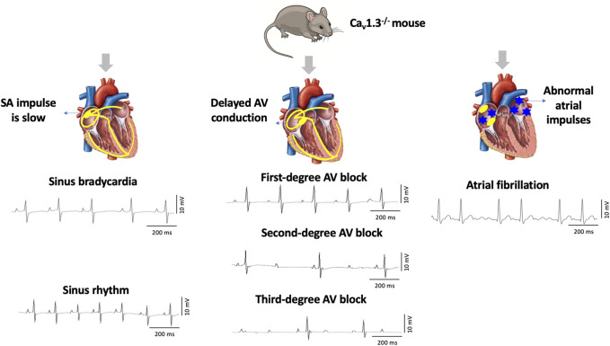
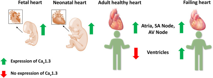
Ca2+ plays a crucial role in excitation-contraction coupling in cardiac myocytes. Dysfunctional Ca2+ regulation alters the force of contraction and causes cardiac arrhythmias. Ca2+ entry into cardiomyocytes is mediated mainly through L-type Ca2+ channels, leading to the subsequent Ca2+ release from the sarcoplasmic reticulum. L-type Ca2+ channels are composed of the conventional Cav1.2, ubiquitously expressed in all heart chambers, and the developmentally regulated Cav1.3, exclusively expressed in the atria, sinoatrial node, and atrioventricular node in the adult heart. As such, Cav1.3 is implicated in the pathogenesis of sinoatrial and atrioventricular node dysfunction as well as atrial fibrillation. More recently, Cav1.3 de novo expression was suggested in heart failure. Here, we review the functional role, expression levels, and regulation of Cav1.3 in the heart, including in the context of cardiac diseases. We believe that the elucidation of the functional and molecular pathways regulating Cav1.3 in the heart will assist in developing novel targeted therapeutic interventions for the aforementioned arrhythmias.

Keywords: atrial fibrillation; calcium channel; heart failure; protein kinase regulation; sinoatrial node dysfunction.


Conflict of interest statement

The authors declare that the research was conducted in the absence of any commercial or financial relationships that could be construed as a potential conflict of interest.

Figures

FIGURE 1
FIGURE 1
Deletion of Cav1.3 in mice leads to electrographic abnormalities. The absence of Cav1.3 in mice leads to the development of sinoatrial (SA) and atrioventricular (AV) node dysfunction leading to sinus bradycardia and first-degree, second-degree, and third-degree AV block as well as atrial fibrillation (AF).
FIGURE 2
FIGURE 2
Alternative splicing at the C-terminus of Cav1.3. Alternative splicing of exon 41 that removes the IQ motif resulted in a truncated Cav1.3 protein with diminished inactivation. Splicing of exon 43 which causes a frameshift variant and is susceptible to accelerating the inactivation similar to Cav1.342A. Ca2+ current through Cav1.342A channels increased at negative voltages, and inactivation was faster because of Ca2+-dependent inactivation. Splicing of exons 44 and 48 was an in-frame variant and caused disruption of the binding of distal modulator to the IQ domain. PCRD (proximal C-terminal regulatory domain), DCRD (distal C-terminal regulatory domain). Current tracing are from reference Tan et al., 2011.
FIGURE 3
FIGURE 3
Regulation of Cav1.3 L-type Ca2+ channel by protein kinase A, protein kinase C, and calmodulin-dependent protein kinase II. Panel (A) Schematic representation of the four homologous domains (I-IV) of the Cav1.3 α1-subunit with 6 transmembrane segments (S1-S6) and N- and C- termini. Phosphorylation of the channel by PKA is at serine residues located at positions 1743 and 1816 of the C-terminus. PKC phosphorylates at the N-terminal domain at serine residue located at position 81. Calmodulin binding site is on the proximal C-terminus. Missense variant A376V and the founder variant G403_V404insG, as well as heterozygous non-synonymous variant R930H in CACNA1D gene have been associated with sinoatrial node dysfunction (Liaqat et al., 2019; Rinné et al., 2022). The missense variants G403R and I770M has been found in patients with atrioventricular node dysfunction (Scholl et al., 2013). Panel (B) The sketch summarizes the regulation of atrial Cav1.3 channel by the different kinases, including PKA, PKC and CaMKII. Ca2+ entry through Cav1.3 channel and subsequent Ca2+ release from RyR contributes to pacemaking, while Ca2+ entry through Cav1.2 contributes to excitation-contraction coupling. PKA (protein kinase A), PKC (protein kinase C), SR (sarcoplasmic reticulum), SERCA (sarcoendoplasmic reticulum calcium ATPase), RyR (ryanodine receptor), P (phosphorylation site), CaMKII (calmodulin-dependent protein kinase II).
FIGURE 4
FIGURE 4
Expression of Cav1.3 L-type Ca2+ channel in the fetal, neonatal, adult, and failing heart. Cav1.3 is expressed in the supraventricular and ventricular tissue of the fetal and neonatal hearts. However in adult hearts, it is expressed only in the atria, sinoatrial (SA) node, and atrioventricular (AV) node, but not in the ventricles. Recent evidence suggests a Cav1.3 de novo expression in the ventricles of adult failing hearts.

Similar articles

Cited by

References

    1. Baig S. M., Koschak A., Lieb A., Gebhart M., Dafinger C., Nürnberg G., et al. (2011). Loss of Ca(v)1.3 (CACNA1D) function in a human channelopathy with bradycardia and congenital deafness. Nat. Neurosci. 14 (1), 77–84. 10.1038/nn.2694 - DOI - PubMed
    1. Banerjee R., Yoder J. B., Yue D. T., Amzel L. M., Tomaselli G. F., Gabelli S. B., et al. (2018). Bilobal architecture is a requirement for calmodulin signaling to Ca(V)1.3 channels. Proc. Natl. Acad. Sci. U. S. A. 115 (13), E3026–e3035. 10.1073/pnas.1716381115 - DOI - PMC - PubMed
    1. Baroudi G., Qu Y., Ramadan O., Chahine M., Boutjdir M. (2006). Protein kinase C activation inhibits Cav1.3 calcium channel at NH2-terminal serine 81 phosphorylation site. Am. J. Physiol. Heart Circ. Physiol. 291 (4), H1614–H1622. 10.1152/ajpheart.00095.2006 - DOI - PubMed
    1. Ben Johny M., Yang P. S., Bazzazi H., Yue D. T. (2013). Dynamic switching of calmodulin interactions underlies Ca2+ regulation of CaV1.3 channels. Nat. Commun. 4, 1717. 10.1038/ncomms2727 - DOI - PMC - PubMed
    1. Benitah J. P., Alvarez J. L., Gómez A. M. (2010). L-type Ca(2+) current in ventricular cardiomyocytes. J. Mol. Cell Cardiol. 48 (1), 26–36. 10.1016/j.yjmcc.2009.07.026 - DOI - PubMed