

Clinical utility of positron emission tomography leading to rapid and accurate diagnosis of intravascular large B-cell lymphoma presenting with the central nervous system symptoms alone: A case report and review of the literature
- PMID: 37025518
- PMCID: PMC10070256
- DOI: 10.25259/SNI_1175_2022
Clinical utility of positron emission tomography leading to rapid and accurate diagnosis of intravascular large B-cell lymphoma presenting with the central nervous system symptoms alone: A case report and review of the literature
Abstract
Background: Intravascular large B-cell lymphoma (IVLBCL) is a rare entity among large B-cell non-Hodgkin lymphomas and is often difficult to diagnose. We report the case of a patient with IVLBCL who presented with central nervous system (CNS) symptoms alone, in which positron emission tomography (PET) enabled a rapid and accurate diagnosis.
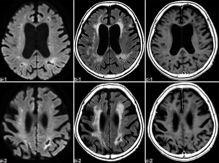
Case description: An 81-year-old woman was admitted to our hospital with a 3-month history of gradually progressive dementia and declining spontaneity. Magnetic resonance imaging revealed multiple hyperintense lesions bilaterally on diffusion-weighted imaging without enhancement on gadolinium-enhanced T1-weighted imaging. Laboratory findings showed elevated serum lactate dehydrogenase (626 U/L) and soluble interleukin-2 receptor (sIL-2R) (4692 U/mL). Cerebrospinal fluid (CSF) analysis showed slightly elevated levels of protein (166 mg/dL) and lymphocytic cells (29/μL), and β2-microglobulin (β2-MG) (4.6 mg/L) was highly elevated. Whole-body computed tomography revealed faint ground-glass opacities in the upper and middle lung fields and diffuse enlargement of both kidneys without lymph node swelling. 18F-fluorodeoxyglucose (FDG)-PET showed diffuse and remarkably high FDG uptake in both upper lungs and kidneys without uptake by lymph nodes, suggesting a malignant hematological disease. IVLBCL was confirmed histologically by incisional random skin biopsy from the abdomen. Chemotherapy using R-CHOP regimen in combination with intrathecal methotrexate injection was started on day 5 after admission and follow-up neuroimaging showed no signs of recurrence.
Conclusion: IVLBCL presenting with CNS symptoms alone is rare and often has a poor prognosis associated with delayed diagnosis, and various evaluations (including systemic analysis) are therefore necessary for early diagnosis. FDG-PET, in addition to identification of clinical symptoms and evaluation of serum sIL-2R and CSF β2-MG, enables rapid therapeutic intervention in IVLBCL presenting with CNS symptoms.
Keywords: 18F-fluorodeoxyglucose; Intravascular large B-cell lymphoma; Positron emission tomography; R-CHOP; Random skin biopsy.
Copyright: © 2023 Surgical Neurology International.
Conflict of interest statement
There are no conflicts of interest.
Figures


Similar articles
-
Early detection of intravascular large B-cell lymphoma by (18)FDG-PET/CT with diffuse FDG uptake in the lung without respiratory symptoms or chest CT abnormalities.Asia Ocean J Nucl Med Biol. 2014 Spring;2(1):65-8. Asia Ocean J Nucl Med Biol. 2014. PMID: 27408860 Free PMC article.
-
A case of lung intravascular large B cell lymphoma developed with respiratory failure rescued by corticosteroid prior to definite diagnosis.Respir Med Case Rep. 2022 Mar 11;37:101625. doi: 10.1016/j.rmcr.2022.101625. eCollection 2022. Respir Med Case Rep. 2022. PMID: 35309974 Free PMC article.
-
CT-Imaging Manifestations and Diagnostic Insights in Pulmonary Intravascular Large B-Cell Lymphoma: A Case Series and Literature Review.Cancer Rep (Hoboken). 2025 Mar;8(3):e70159. doi: 10.1002/cnr2.70159. Cancer Rep (Hoboken). 2025. PMID: 40019334 Free PMC article. Review.
-
Rapid deterioration of intravascular large B-cell lymphoma with mass formation in the trigeminal nerve and multiple organ infiltration: An autopsy case report.J Clin Exp Hematop. 2022 Mar 9;62(1):41-45. doi: 10.3960/jslrt.21013. Epub 2021 Nov 26. J Clin Exp Hematop. 2022. PMID: 34840206 Free PMC article.
-
Case report: Intravascular large B cell lymphoma mimicking acute demyelinating encephalomyelitis after SARS-CoV-2 reinfection: diagnostic value of advanced MRI techniques and the literature review with the assistance of ChatGPT.Front Immunol. 2024 Nov 14;15:1478163. doi: 10.3389/fimmu.2024.1478163. eCollection 2024. Front Immunol. 2024. PMID: 39611151 Free PMC article. Review.
Cited by
-
Intravascular Large B-Cell Lymphoma Diagnosed After Recurrent Stroke: Case Report and Literature Review.Neurol Int. 2025 Apr 27;17(5):68. doi: 10.3390/neurolint17050068. Neurol Int. 2025. PMID: 40423224 Free PMC article.
References
-
- Chapin JE, Davis LE, Kornfeld M, Mandler RN. Neurologic manifestations of intravascular lymphomatosis. Acta Neurol Scand. 1995;91:494–9. - PubMed
-
- Enzan N, Kitadate A, Tanaka A, Matsue K. Incisional random skin biopsy, not punch biopsy, is an appropriate method for diagnosis of intravascular large B-cell lymphoma: A clinicopathological study of 25 patients. Br J Dermatol. 2019;181:200–1. - PubMed
-
- Glass J, Hochberg FH, Miller DC. Intravascular lymphomatosis. A systemic disease with neurologic manifestations. Cancer. 1993;71:3156–64. - PubMed
-
- Hans CP, Weisenburger DD, Greiner TC, Gascoyne RD, Delabie J, Ott G, et al. Confirmation of the molecular classification of diffuse large B-cell lymphoma by immunohistochemistry using a tissue microarray. Blood. 2004;103:275–82. - PubMed
Publication types
LinkOut - more resources
Full Text Sources
Research Materials