

Microneurosurgical anatomy of the basal cisterns: A brief review for cisternostomy
- PMID: 37025519
- PMCID: PMC10070334
- DOI: 10.25259/SNI_1095_2022
Microneurosurgical anatomy of the basal cisterns: A brief review for cisternostomy
Abstract
Background: Cisternostomy is a surgical technique thought of and developed as an option for severe brain trauma treatment. It demands a particular knowledge and skill to microsurgically approach basal cisterns and effectively manipulate their contents. To perform this procedure safely, the anatomy and pathophysiology must be clearly understood.
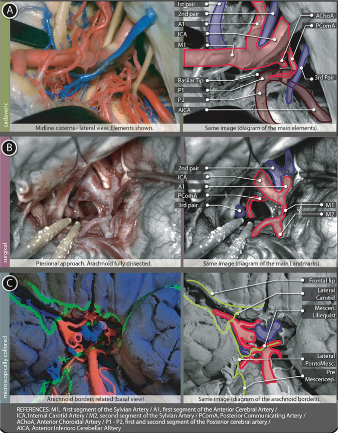
Methods: Detailed microscopic dissection and anatomical review were done, after a detailed reading of facts and recent publications about cisternostomy. Cisternal pathways and landmark planning are described and augmented using a new method to show de arachnoid borders. Finally, a brief discussion is written as a synopsis.
Results: Cisternostomy requires thorough microscopic knowledge and microsurgical skills. This paper intends to provide information to understand better the anatomy related, thus, easing the learning curve. The technique used to show arachnoid borders, complementing cadaveric and surgical images, was useful for this purpose.
Conclusion: To perform this procedure safely, it is mandatory to handle microscopic details of cistern anatomy. Reaching a core cistern is necessary to assure effectiveness. This procedure needs, as well, surgical step-by-step landmark planning and performing. Cisternostomy could be a life-saving procedure and a new powerful tool for severe brain trauma treatment. Evidence is being collected to support its indications.
Keywords: Basal cisterns anatomy; Cisternostomy; Microsurgical technique; Severe brain trauma.
Copyright: © 2023 Surgical Neurology International.
Conflict of interest statement
There are no conflicts of interest.
Figures
Similar articles
-
Basal Cisternostomy for Severe TBI: Surgical Technique and Cadaveric Dissection.Front Surg. 2022 May 6;9:915818. doi: 10.3389/fsurg.2022.915818. eCollection 2022. Front Surg. 2022. PMID: 35599786 Free PMC article.
-
Basal Cisternostomy - A Microsurgical Cerebro Spinal Fluid Let Out Procedure and Treatment Option in the Management of Traumatic Brain Injury. Analysis of 40 Consecutive Head Injury Patients Operated with and without Bone Flap Replacement Following Cisternostomy in a Tertiary Care Centre in India.Neurol India. 2021 Mar-Apr;69(2):328-333. doi: 10.4103/0028-3886.314535. Neurol India. 2021. PMID: 33904445
-
Anatomy and physiology of cisternostomy.Chin J Traumatol. 2016;19(1):7-10. doi: 10.1016/j.cjtee.2016.01.003. Chin J Traumatol. 2016. PMID: 27033265 Free PMC article.
-
Arachnoid membrane: the first and probably the last piece of the roadmap.Surg Radiol Anat. 2015 Mar;37(2):127-38. doi: 10.1007/s00276-014-1361-z. Epub 2014 Aug 19. Surg Radiol Anat. 2015. PMID: 25135312 Review.
-
The Sylvian fissure, cistern and arachnoid membrane.Br J Neurosurg. 2014 Jan;28(1):98-106. doi: 10.3109/02688697.2013.815324. Epub 2013 Jul 19. Br J Neurosurg. 2014. PMID: 23869573 Review.
Cited by
-
Basal cisternostomy as an adjunct to decompressive hemicraniectomy in moderate to severe traumatic brain injury: a systematic review and meta-analysis.Neurosurg Rev. 2024 Oct 2;47(1):717. doi: 10.1007/s10143-024-02954-4. Neurosurg Rev. 2024. PMID: 39354191 Free PMC article.
-
Morphology and Variations of the Posterior Cerebral Artery: A Literature Review.Cureus. 2025 Mar 25;17(3):e81205. doi: 10.7759/cureus.81205. eCollection 2025 Mar. Cureus. 2025. PMID: 40291304 Free PMC article. Review.
-
The basal cisternostomy for management of severe traumatic brain injury: A retrospective study.Chin J Traumatol. 2025 Mar;28(2):118-123. doi: 10.1016/j.cjtee.2024.09.007. Epub 2024 Nov 20. Chin J Traumatol. 2025. PMID: 39632242 Free PMC article.
-
Posterior vascular anatomy of the encephalon: a comprehensive review.Surg Radiol Anat. 2024 Jun;46(6):843-857. doi: 10.1007/s00276-024-03358-1. Epub 2024 Apr 23. Surg Radiol Anat. 2024. PMID: 38652250 Free PMC article. Review.
References
-
- Adib SD, Herlan S, Ebner FH, Hirt B, Tatagiba M, Honegger J. Interoptic, trans-lamina terminalis, opticocarotid triangle, and caroticosylvian windows from mini-supraorbital, frontomedial, and pterional perspectives: A comparative cadaver study with artificial lesions. Front Surg. 2019;6:40. - PMC - PubMed
-
- Altafulla J, Bordes S, Jenkins S, Litvack Z, Iwanaga J, Loukas M, et al. The basal subarachnoid cisterns: Surgical and anatomical considerations. World Neurosurg. 2019;129:190–9. - PubMed
LinkOut - more resources
Full Text Sources