

Proper ophthalmic artery aneurysms
- PMID: 37025535
- PMCID: PMC10070312
- DOI: 10.25259/SNI_1151_2022
Proper ophthalmic artery aneurysms
Abstract
Background: The ophthalmic segment of the internal carotid artery (ICA) represents a common site for cerebral aneurysms. However, aneurysms of the ophthalmic artery (OphA) itself represent rare lesions and have been associated with trauma and flow-related lesions such as arteriovenous fistulas or malformations. Here, we explore clinical and radiological features of four patients managed for five proper ophthalmic artery aneurysms (POAAs).
Methods: Patients undergoing diagnostic cerebral angiogram (DCA) between January 2018 and November 2021 with newly or previously identified POAA were retrospectively reviewed. Clinical and radiological data were analyzed to identify common and unique features.
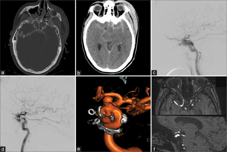
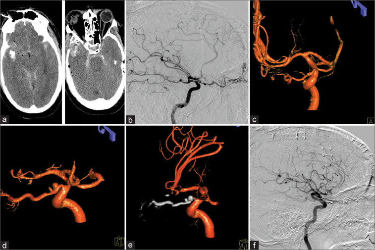
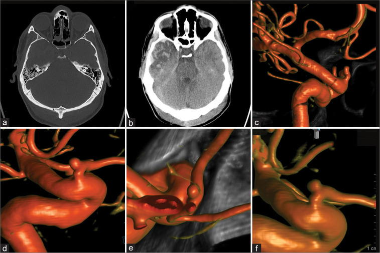
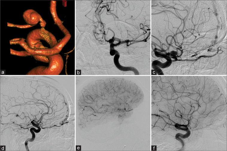
Results: Four patients with identification of five POAA were identified. Three patients suffered traumatic brain injury with subsequent identification of POAA on DCA. Patient 1 presented with a traumatic carotid-cavernous-sinus fistula requiring transvenous coil embolization and second stage flow diversion of the ICA. Patient 2 suffered a gunshot wound with ICA compromise, ethmoidal dural arteriovenous fistula (dAVF) development with rapid growth of two POAAs eventually requiring Onyx embolization. Patient 3 was assaulted and DCA showed a POAA without any other cerebrovascular pathology. Patient 4 had undergone N-butyl cyanoacrylate embolization of an ethmoidal dAVF 13 years ago with the feeding OphA carrying a large POAA. Re-DCADCA was performed for a newly developed and unrelated transverse-sigmoid-sinus dAVF.
Conclusion: Management of POAAs poses a challenge to neurovascular surgeons since POAAs inherit a risk for visual deterioration or hemorrhage. DCA facilitates identification of coexisting cerebrovascular pathology. If clinically silent and not accompanied by cerebrovascular disease, observation appears reasonable.
Keywords: Aneurysm; Dural arteriovenous fistula; Ophthalmic artery.
Copyright: © 2023 Surgical Neurology International.
Conflict of interest statement
There are no conflicts of interest.
Figures
References
-
- Cagnazzo F, Peluso A, Vannozzi R, Brinjikji W, Lanzino G, Perrini P. Arterial aneurysms associated with intracranial dural arteriovenous fistulas: Epidemiology, natural history, and management. A systematic review. Neurosurg Rev. 2019;42:277–85. - PubMed
-
- Chun HJ, Yi HJ. Traumatic extracranial pseudoaneurysm on the peripheral ophthalmic artery presenting as delayed intraparenchymal hematoma: Case report. Surg Neurol. 2009;71:701–4. - PubMed
-
- Dehdashti AR, Safran AB, Martin JB, Rüfenacht DA, de Tribolet N. Intraorbital ophthalmic artery aneurysm associated with basilar tip saccular aneurysm. Neuroradiology. 2002;44:600–3. - PubMed
-
- Foreman PM, Hendrix P, Harrigan MR, Fisher WS, 3rd, Vyas NA, Lipsky RH, et al. PHASES score applied to a prospective cohort of aneurysmal subarachnoid hemorrhage patients. J Clin Neurosci. 2018;53:69–73. - PubMed
LinkOut - more resources
Full Text Sources
Miscellaneous