A challenging recurrent thoracic disc herniation
- PMID: 37025536
- PMCID: PMC10070332
- DOI: 10.25259/SNI_139_2023
A challenging recurrent thoracic disc herniation
Abstract
Background: Thoracic disc herniations are rare and occur at the rate of 1/1,000,000/year. Surgical approach must be individually tailored to the size, location, and consistency of the herniated disc. Notably, here, we report the unusual recurrence of a thoracic herniated disc.
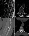
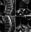
Case description: In 2014, a 53-year-old female presented with thoracic back pain, and paraparesis, attributed to an magnetic resonance imaging/computed tomography (CT)-documented left paramedian T8-T9 calcific disc herniation. She underwent a left hemilaminectomy/costotrasversectomy following which she experienced complete regression of her symptoms. Notably, the postoperative radiological studies at that time demonstrated some residual although asymptomatic calcific disc herniation. Eight years later, she again presented, but now with the chief complaint of difficulty breathing. The new CT scan showed a new calcified herniated disc fragment superimposed on the previously documented residual disc. Through a posterolateral transfacet approach, she underwent resection of the disc complex. An intraoperative CT scan confirmed complete removal of the recurrent calcified disc herniation. Following the second surgery, the patient fully recovered and remains asymptomatic.
Conclusion: A 53-year-old female first presented with a left-sided T8/T9 thoracic calcified disc herniation that was initially partially resected). When another larger fragment appeared 8 years later, superimposed on the previously documented residual disc, it was successfully removed through a posterolateral transfacet approach completed with CT guidance and neuronavigation.
Keywords: Residual thoracic hernia; Thoracic disc herniation; Thoracic herniation recurrence; Thoracic myelopathy.
Copyright: © 2023 Surgical Neurology International.
Conflict of interest statement
There are no conflicts of interest.
Figures


References
-
- Arts MP, Bartels RH. Anterior or posterior approach of thoracic disc herniation? A comparative cohort of mini-transthoracic versus transpedicular discectomies. Spine J. 2014;14:1654–62. - PubMed
-
- Bouthors C, Benzakour A, Court C. Surgical treatment of thoracic disc herniation: An overview. Int Orthop. 2019;43:807–16. - PubMed
-
- Court C, Mansour E, Bouthors C. Thoracic disc herniation: Surgical treatment. Orthop Traumatol Surg Res. 2018;104:S31–40. - PubMed
-
- Dickman CA, Rosenthal D, Regan JJ. Reoperation for herniated thoracic discs. J Neurosurg. 1999;91(Suppl 2):157–62. - PubMed
Publication types
LinkOut - more resources
Full Text Sources
