

Lumbar paraspinal intramuscular myxoma: A case report
- PMID: 37025538
- PMCID: PMC10070254
- DOI: 10.25259/SNI_141_2023
Lumbar paraspinal intramuscular myxoma: A case report
Abstract
Background: With an estimated incidence of about 1 case/million patients, paravertebral intramuscular myxomas represent a rare cause of lumbar pain. Rather, they typically occur in the heart and in bone tissues.
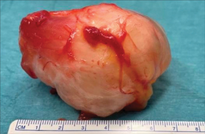
Case description: A 64-year-old female presented with a protracted course of nocturnal lumbar pain that radiated to the anterior aspect of the right thigh accompanied by numbness. She reported a slow-growing right paramedian lumbar mass in the previous months. The magnetic resonance (MR) showed a right lumbar paravertebral intramuscular mass at the L3 level (i.e., 70 × 50 mm) that had well-defined margins, and markedly enhanced with gadolinium. Following gross total "en bloc" tumor resection, the patient fully recovered. Pathologically, the myofibroblastic lesion proved to be an intramuscular myxoma without malignant changes.
Conclusion: A 64-year-old female presented with a slow-growing MR-documented right paramedian lumbar L3 mass responsible for proximal right-thigh numbness. Following "en bloc" gross total removal of the benign intramuscular myxoma, the patient was asymptomatic.
Keywords: Intramuscular myxoma; Lumbar pain; Paraspinal tumors.
Copyright: © 2023 Surgical Neurology International.
Conflict of interest statement
There are no conflicts of interest.
Figures


References
-
- Al Awadhi A, Benichi S, Lot G, Rogers A. A case of intramuscular lumbar myxoma: Uncertainty in the preoperative diagnosis of a spinal soft tissue tumour. Neurochirurgie. 2022;68:530–4. - PubMed
-
- Guppy KH, Wagner F, Tawk R, Gallagher L. Intramuscular myxoma causing lumbar radiculopathy. Case report and review of the literature. J Neurosurg. 2001;95:260–3. - PubMed
Publication types
LinkOut - more resources
Full Text Sources
Research Materials