. 2023 Mar 22;9(4):e14741.
doi: 10.1016/j.heliyon.2023.e14741. eCollection 2023 Apr.

Why the clock ticks differently in Parkinson's disease: Insights from motor imagery and resting-state functional magnetic resonance imaging

Affiliations

Why the clock ticks differently in Parkinson's disease: Insights from motor imagery and resting-state functional magnetic resonance imaging

Marina Christine Ruppert-Junck et al. Heliyon. .

Abstract

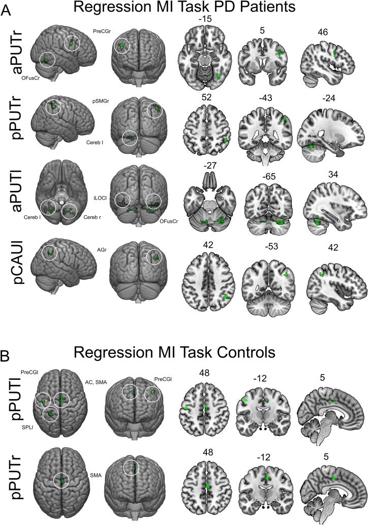
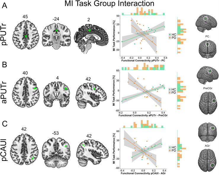
In Parkinson's disease (PD), an impaired perception of suprasecond time intervals has been reported. From a neurobiological perspective, dopamine is thought to be an important mediator of timing. Nevertheless, it is still unclear whether timing deficits in PD occur mainly in the motor context and are associated with corresponding striatocortical loops. This study attempted to fill this gap by investigating time reproduction in the context of a motor imagery task, and its neurobiological correlates in resting-state networks of basal ganglia substructures in PD. Nineteen PD patients and 10 healthy controls therefore underwent two time reproduction tasks. In a motor imagery task, subjects were asked to walk down a corridor for 10 s and reproduce the time spent walking during motor imagery afterwards. In an auditory task, the subjects had to reproduce an acoustically presented time interval of 10 s. Subsequently, resting-state functional magnetic resonance imaging was performed and voxel-wise regressions were conducted between striatal functional connectivity and performance in the individual task at group level and compared between groups. Patients significantly misjudged the time interval in the motor imagery task and an auditory task in comparison to controls. Seed-to-voxel functional connectivity analysis of basal ganglia substructures revealed a significant association between striatocortical connectivity and motor imagery performance. PD patients showed a different pattern of associated striatocortical connections as indicated by significantly different regression slopes for connections of the right putamen and left caudate nucleus. In accordance with previous findings, our data confirm an impaired time reproduction of suprasecond time intervals in PD patients. Our data imply that deficits in time reproduction tasks are not specific to motor context but reflect a general time reproduction deficit. According to our findings, impaired performance in context of motor imagery is accompanied by a different configuration of striatocortical resting-state networks responsible for timing.

Keywords: Internal clock; Motor imagery; Parkinson's disease; Resting-state fMRI; Time reproduction.


Conflict of interest statement

The contributing authors declare no conflicts of interest related to the content.

Figures

Fig. 1
Fig. 1
Group comparison of performance in A) Motor Imagery Task and B) Auditory Control Task. Between-group differences in numeric variables were assessed by Student's t-test or Mann-Whitney U test. C) Direct relation between performance in motor imagery task and auditory control task for each individual. Abbreviations: HC = healthy controls; PD = Parkinson's disease; MI = motor imagery.
Fig. 2
Fig. 2
Striatocortical functional connectivity networks with significant association to motor imagery task performance. Voxel-wise regressions were performed for all striatal subcompartments with motor imagery task performance (percentage deviation to 10 s interval) as variable of interest and age as covariate of no interest in A) PD patients and B) healthy controls in CONN. For all presented analyses, thresholds were set at pFWE < 0.05 (two-tailed). Results are shown in render view and axial, coronal and sagittal slices with numbers above representing corresponding x-,y- or z-coordinates. Abbreviations: AC = anterior cingulum; AGr = right angular gyrus; aPUTl = left anterior putamen; aPUTr = right anterior putamen; Cereb = Cerebellum; HC = healthy controls; iLOCl = left inferior lateral occipital cortex; OFusCr = right occipital fusiform cortex; pCAUl = left posterior caudate; PC = posterior cingulate gyrus; PD = Parkinson's disease; pPUTl = left posterior putamen; pPUTr = right posterior putamen; PreCGl = left precentral gyrus; PreCGr = right precentral gyrus; SMA = supplementary motor area; pSMGr = right supramarginal gyrus posterior division; SPLl = left superior parietal lobe.
Fig. 3
Fig. 3
Striatocortical functional connectivity networks with group differences in relation to motor imagery. Voxels with significant group differences in regression slopes between motor imagery task performance and seed-based connectivity of A) the right posterior putamen, B) the right anterior putamen and C) the left posterior caudate are shown in the left column. The right column shows regression plots for both groups for motor imagery task performance and the extracted connectivity values for voxels with significant group-differences obtained by voxel-wise comparison of regression slopes. Marginal histograms represent the frequency of the individual data sections in the respective group. Significant group differences in regression slopes were tested voxel-wise for all striatal seeds in CONN to analyze group:task interactions. For all presented analysis, thresholds were set at pFWE < 0.05 (two-tailed). Results are shown in render view and axial, coronal and sagittal slices with numbers above representing corresponding x-, y- or z-coordinates. Abbreviations: AGr = right angular gyrus; aPUTr = right anterior putamen; HC = healthy controls; pCAUl = posterior caudate left; PC = posterior cingulate gyrus; PD = Parkinson's disease; pPUTr = right posterior putamen; PreCGr = right precentral gyrus.

References

    1. Hayashi M.J., Ivry R.B. Duration-selectivity in right parietal cortex reflects the subjective experience of time. J. Neurosci. 2020 doi: 10.1523/JNEUROSCI.0078-20.2020. - DOI - PMC - PubMed
    1. Pastor M.A., Artieda J., Jahanshahi M., Obeso J.A. Time estimation and reproduction IS abnormal in Parkinson's DISease. Brain. 1992:211–225. - PubMed
    1. Gibbon J., Church R.M., Meck W.H. Scalar timing in memory. Ann. N. Y. Acad. Sci. 1984;423:52–77. doi: 10.1111/j.1749-6632.1984.tb23417.x. - DOI - PubMed
    1. Wiener M., Turkeltaub P., Coslett H.B. The image of time: a voxel-wise meta-analysis. Neuroimage. 2010;49:1728–1740. doi: 10.1016/j.neuroimage.2009.09.064. - DOI - PubMed
    1. Kosillo P., Smith A.T. The role of the human anterior insular cortex in time processing. Brain Struct. Funct. 2010;214:623–628. doi: 10.1007/s00429-010-0267-8. - DOI - PubMed

LinkOut - more resources