

A richer and more diverse future for microglia phenotypes
- PMID: 37025898
- PMCID: PMC10070543
- DOI: 10.1016/j.heliyon.2023.e14713
A richer and more diverse future for microglia phenotypes
Abstract
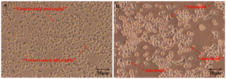
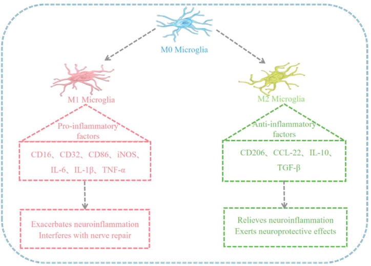
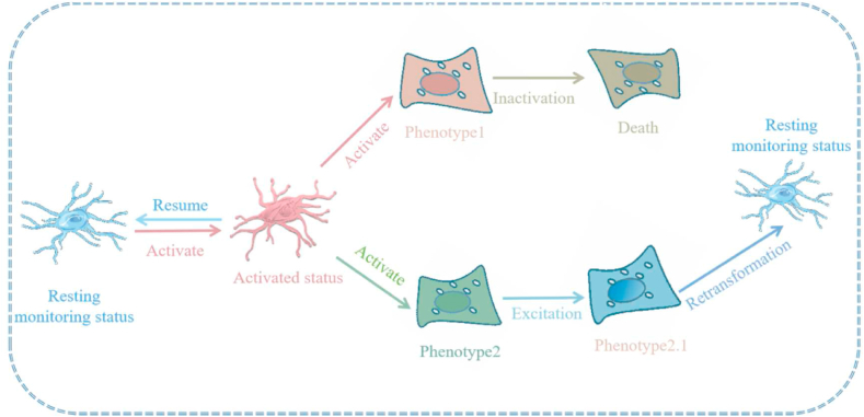
Microglia are the only resident innate immune cells derived from the mesoderm in the nerve tissue. They play a role in the development and maturation of the central nervous system (CNS). Microglia mediate the repair of CNS injury and participate in endogenous immune response induced by various diseases by exerting neuroprotective or neurotoxic effects. Traditionally, microglia are considered to be in a resting state, the M0 type, under physiological conditions. In this state, they perform immune surveillance by constantly monitoring pathological responses in the CNS. In the pathological state, microglia undergo a series of morphological and functional changes from the M0 state and eventually polarize into classically activated microglia (M1) and alternatively activated microglia (M2). M1 microglia release inflammatory factors and toxic substances to inhibit pathogens, while M2 microglia exert neuroprotective effects by promoting nerve repair and regeneration. However, in recent years, the view regarding M1/M2 polarization of microglia has gradually changed. According to some researchers, the phenomenon of microglia polarization is not yet confirmed. The M1/M2 polarization term is used for a simplified description of its phenotype and function. Other researchers believe that the microglia polarization process is rich and diverse, and consequently, the classification method of M1/M2 has limitations. This conflict hinders the academic community from establishing more meaningful microglia polarization pathways and terms, and therefore, a careful revision of the concept of microglia polarization is required. The present article briefly reviews the current consensus and controversy regarding microglial polarization typing to provide supporting materials for a more objective understanding of the functional phenotype of microglia.
Keywords: M1/M2 subtype; Microglia; Multiple subtypes; Polarization.
© 2023 The Authors.
Conflict of interest statement
6The author(s) declared no potential conflicts of interest with respect to the research, authorship, and/or publication of this article..
Figures
Similar articles
-
Dehydrocorydaline attenuates bone cancer pain by shifting microglial M1/M2 polarization toward the M2 phenotype.Mol Pain. 2018 Jan-Dec;14:1744806918781733. doi: 10.1177/1744806918781733. Mol Pain. 2018. PMID: 29882480 Free PMC article.
-
Cyclic AMP is a key regulator of M1 to M2a phenotypic conversion of microglia in the presence of Th2 cytokines.J Neuroinflammation. 2016 Jan 13;13:9. doi: 10.1186/s12974-015-0463-9. J Neuroinflammation. 2016. PMID: 26757726 Free PMC article.
-
Targeting MAPK Pathways by Naringenin Modulates Microglia M1/M2 Polarization in Lipopolysaccharide-Stimulated Cultures.Front Cell Neurosci. 2019 Jan 11;12:531. doi: 10.3389/fncel.2018.00531. eCollection 2018. Front Cell Neurosci. 2019. PMID: 30687017 Free PMC article.
-
Diversity and plasticity of microglial cells in psychiatric and neurological disorders.Pharmacol Ther. 2015 Oct;154:21-35. doi: 10.1016/j.pharmthera.2015.06.010. Epub 2015 Jun 27. Pharmacol Ther. 2015. PMID: 26129625 Review.
-
Selective modulation of microglia polarization to M2 phenotype for stroke treatment.Int Immunopharmacol. 2015 Apr;25(2):377-82. doi: 10.1016/j.intimp.2015.02.019. Epub 2015 Feb 20. Int Immunopharmacol. 2015. PMID: 25704852 Review.
Cited by
-
Age-dependent Powassan virus lethality is linked to glial cell activation and divergent neuroinflammatory cytokine responses in a murine model.J Virol. 2024 Aug 20;98(8):e0056024. doi: 10.1128/jvi.00560-24. Epub 2024 Aug 1. J Virol. 2024. PMID: 39087762 Free PMC article.
-
Extracellular domain of TREM2 possess two distinct ligand recognition sites: Insights from machine-learning guided docking and all-atoms molecular dynamics simulations.Heliyon. 2024 Dec 20;11(1):e41414. doi: 10.1016/j.heliyon.2024.e41414. eCollection 2025 Jan 15. Heliyon. 2024. PMID: 39866401 Free PMC article.
-
Role of Glial Cells and Receptors in Schizophrenia Pathogenesis.Neurochem Res. 2025 Jan 27;50(2):85. doi: 10.1007/s11064-025-04336-8. Neurochem Res. 2025. PMID: 39869278 Review.
-
Microglia-induced neuroinflammation in hippocampal neurogenesis following traumatic brain injury.Heliyon. 2024 Aug 8;10(16):e35869. doi: 10.1016/j.heliyon.2024.e35869. eCollection 2024 Aug 30. Heliyon. 2024. PMID: 39220913 Free PMC article. Review.
-
Unraveling the molecular complexity: Wtap/Ythdf1 and Lcn2 in novel traumatic brain injury secondary injury mechanisms.Cell Biol Toxicol. 2024 Aug 7;40(1):65. doi: 10.1007/s10565-024-09909-x. Cell Biol Toxicol. 2024. PMID: 39110292 Free PMC article.
References
-
- Deckert-Schlüter M. [Rudolf-Virchow Prize 1998. Award lecture. Toxoplasmosis: a model infection for studying systemic and intracerebral immune reactions] Verh. Dtsch. Ges. Pathol. 1998;82:9–22. - PubMed
-
- Sierra A., de Castro F., Del Río-Hortega J., Rafael Iglesias-Rozas J., Garrosa M., Kettenmann H. The “Big-Bang” for modern glial biology: translation and comments on Pío del Río-Hortega 1919 series of papers on microglia. Glia. 2016;64:1801–1840. - PubMed
-
- Borst K., Dumas A.A., Prinz M. Microglia: immune and non-immune functions. Immunity. 2021;54:2194–2208. - PubMed
Publication types
LinkOut - more resources
Full Text Sources
